. 2009 Dec;119(12):3774-86.
doi: 10.1172/JCI39692. Epub 2009 Nov 9.

PKCtheta is required for alloreactivity and GVHD but not for immune responses toward leukemia and infection in mice

Affiliations

PKCtheta is required for alloreactivity and GVHD but not for immune responses toward leukemia and infection in mice

Javier O Valenzuela et al. J Clin Invest. 2009 Dec.

Abstract

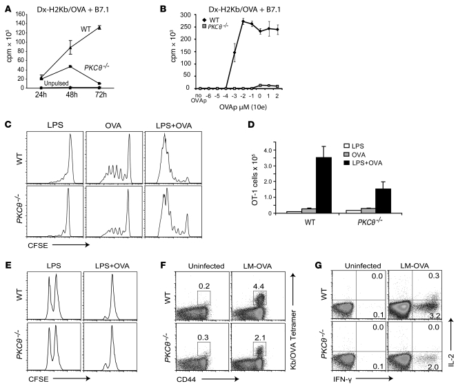
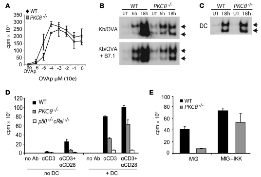
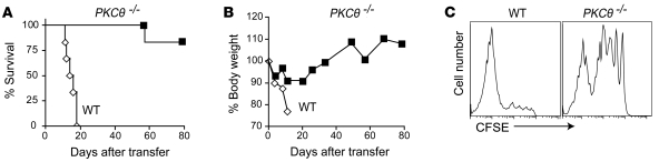
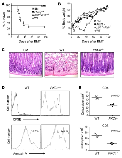
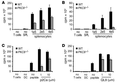
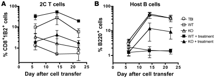
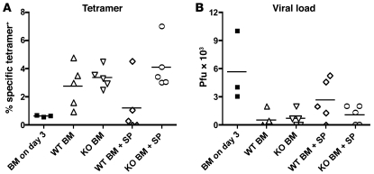
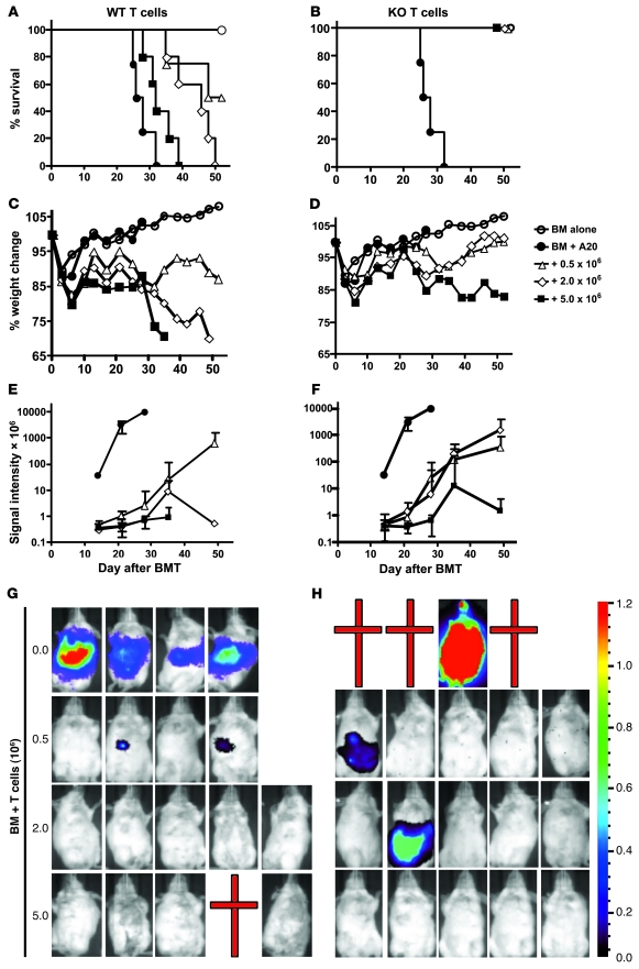
When used as therapy for hematopoietic malignancies, allogeneic BM transplantation (BMT) relies on the graft-versus-leukemia (GVL) effect to eradicate residual tumor cells through immunologic mechanisms. However, graft-versus-host disease (GVHD), which is initiated by alloreactive donor T cells that recognize mismatched major and/or minor histocompatibility antigens and cause severe damage to hematopoietic and epithelial tissues, is a potentially lethal complication of allogeneic BMT. To enhance the therapeutic potential of BMT, we sought to find therapeutic targets that could inhibit GVHD while preserving GVL and immune responses to infectious agents. We show here that T cell responses triggered in mice by either Listeria monocytogenes or administration of antigen and adjuvant were relatively well preserved in the absence of PKC isoform theta (PKCtheta), a key regulator of TCR signaling. In contrast, PKCtheta was required for alloreactivity and GVHD induction. Furthermore, absence of PKCtheta raised the threshold for T cell activation, which selectively affected alloresponses. Most importantly, PKCtheta-deficient T cells retained the ability to respond to virus infection and to induce GVL effect after BMT. These findings suggest PKCtheta is a potentially unique therapeutic target required for GVHD induction but not for GVL or protective responses to infectious agents.


Figures

Figure 1
Figure 1. PKCθ is required for efficient proliferation of CD8 T cells in vitro but not in vivo.
WT and PKCθ–/– OT-1 CD8 T cells were activated in vitro with microspheres bearing H-2Kb:Ig, B7.1:Fc, and 0.1 μM of OVA257–264 (OVAp) (A) or indicated OVA257–264 amounts (B). Cell proliferation (A and B) was measured by 3H-thymidine incorporation. SD within triplicate samples is indicated. For in vivo studies, CFSE-labeled WT and PKCθ–/– OT-1 T cells were transferred into C57BL/6 CD45.1 congenic recipients prior to immunization with 25 μg LPS with or without 10 μg OVA. Cell proliferation (C) and clonal expansion (D) of donor T cells was measured in draining lymph nodes by CFSE dilution. SD within triplicate samples is indicated in D. (E) WT and PKCθ–/– OT-1 T cells were transferred and mice immunized as in C. 3 days later, splenocytes pulsed with OVA257–264 (CFSE 0.25 μM) or left unpulsed (CFSE 2.5 μM) were injected and monitored 10–12 hours later in the spleen by FACS. The T cell response to the OVA-expressing L. monocytogenes (LM-OVA) was measured by immunizing mice i.v. with 2 × 10³ CFUs. 7 days later, clonal expansion (F) and intracellular expression of IL-2 and IFN-γ (G) of activated OVA-specific CD8 T cells were measured in spleens by staining for CD8, CD44, Kb/OVA tetramer (F), and CD8, IL-2, and IFN-γ after a 4-hour in vitro restimulation with 100 nM OVA257–264 in the presence of brefeldin A (G) followed by FACS. Numbers in F and G represent percentages within spleen CD8 T cells.
Figure 2
Figure 2. Crucial role of NF-κB in mediating PKCθ-dependent and -independent responses.
(A) 1 × 10⁵ MACS-purified WT and PKCθ–/– OT-1 CD8 T cells were stimulated with 1 × 10⁴ BM-derived DCs in the presence of increasing amounts of OVAp ranging from 0 μM to 10 μM, as indicated. (B) 1 × 10⁵ MACS-purified WT and PKCθ–/– OT-1 CD8 T cells were activated with microspheres bearing DimerX H-2Kb:Ig (pulsed with 0.1 μM OVAp) and B7.1:Fc or (C) 1 × 10⁴ BM-derived DCs in the presence of 1 μM OVAp. Untreated (UT) samples received no stimulation. NF-κB nuclear localization was measured at the indicated times by EMSA. Prior to generating EMSA extracts, DCs were removed by positive selection of CD11c+ cells on a MACS column. (D) 1 × 10⁵ MACS-purified WT, PKCθ–/–, and p50–/–cRel–/– CD8 T cells were activated with 0.1 μg anti-TCR ± anti-CD28 agonistic antibodies (coated on plates) in the presence or absence of 1 × 10⁴ BM-derived DCs. T cell proliferation was measured after 3 days by 3H-thymidine incorporation. (E) 1 × 10⁵ MACS-purified WT and PKCθ–/– OT-1 CD8 T cells were activated with microspheres coated with H-2Kb/OVA plus B7.1:Fc as described above. After 24 and 48 hours of activation, the cells were infected with the GFP-expressing retroviral vectors MIG or MIG-IKK. GFP+ cells were sorted by FACS and restimulated with Kb/OVA/B7 microspheres for an additional 48 hours. Proliferation was measured by 3H-thymidine incorporation during the last 10 hours of restimulation. Error bars indicate SD in triplicate samples.
Figure 3
Figure 3. PKCθ plays a critical role in CD8 T cell–induced GVHD.
1 × 10⁶ MACS-purified WT and PKCθ–/– CD8 T cells were transferred into sublethally irradiated (500 cGy) bm1 recipients, and the recipients were monitored for survival (A) and weight loss (B). (C) 5–8 × 10⁶ MACS-purified and CFSE-labeled B6 WT or PKCθ–/– CD8 donor T cells were injected i.v. into sublethally irradiated bm1 recipients, and their proliferation was monitored in the spleen 4 days after transfer. Representative results from 2 separate experiments are shown.
Figure 4
Figure 4. PKCθ is required for GVHD development in a B6→BALB/c BMT model.
2 × 10⁶ MACS-purified B6 WT, PKCθ–/–, or NF-κB p50–/–cRel–/– CD4+ and CD8+ donor T cells were injected i.v. into lethally irradiated (800–900 cGy) BALB/c recipient mice along with T cell–depleted BM (BM). Recipients were monitored 2–3 times weekly for clinical signs of GVHD and survival (A) and weight loss (B). Animals that were moribund were sacrificed and counted as GVHD mortality. H&E staining of the gut epithelium was performed approximately 20 days after T cell transfer (C). Original magnification, ×200. The initial proliferation and survival of donor CD8 T cells was measured by transferring CFSE-labeled cells and staining for CFSE+ CD8+ H-2b+ annexin V+ cells 4 days after transplantation (D). Donor T cell expansion was measured by staining for CD4+ H-2b+ and CD8+ H-2b+ cells in the spleen 4 days after transplantation (E). Horizontal bars indicate the averages of 4 mice in each group.
Figure 5
Figure 5. Strength of TCR stimulation determines the requirement for PKCθ in T cell activation.
1 × 10⁵ MACS-purified Tg WT OT-1 and PKCθ–/– OT-1 CD8 T cells (A) or WT and PKCθ–/– CD8 T cells (B) were stimulated with increasing amounts of whole irradiated splenocytes from syngeneic C57BL/6 (A) or allogeneic BALB/c (B) mice. Syngeneic APCs were cocultured with OT-1 responders in the presence of 1 μM OVA257–264 peptide. To measure proliferation induced by different affinity peptides, CB6 BM-derived DCs (1 × 10⁴) were cultured with WT or PKCθ–/– 2C CD8 T cell responders in the presence of indicated amounts of p2Ca (C) or QL9 (D) peptides. T cell proliferation was determined 3 days later by 3H-thymidine incorporation. Representative results from 2 separate experiments are shown. Error bars indicate SD in triplicate samples.
Figure 6
Figure 6. Essential requirement for PKCθ in 2C T cell expansion and cytotoxicity in vivo.
(A) 5 × 10⁶ WT and PKCθ–/– (KO) 2C T cells were injected into sublethally irradiated CB6 recipient mice (3–4 mice per group). Percentages of 2C T cells in peripheral blood 6, 14, and 22 days after T cell transfer are shown. Some mice also received 50 μg anti-CD40 and 100 μg poly(I:C) after irradiation as indicated (treatment). (B) Host B cell reconstitution was evaluated on different days after T cell transfer as indicated. TBI, total body irradiation. Error bars indicate the SD of 3–4 mice in each group at each time point.
Figure 7
Figure 7. PKCθ–/– T cells can mount an effective anti-MCMV response after BMT.
Lethally irradiated CB6 recipients were transplanted with WT or PKCθ–/– (KO) TCD BM alone or with 5 × 10⁶ splenocytes from WT or KO donors, respectively. 60 days after BMT, recipients were infected with MCMV, and MCMV tetramer–positive CD8 T cell numbers were determined after 10 days (A). Virus titers (B) were measured in recipient livers 10 days after infection. A few mice receiving BM alone were sacrificed on day 3 after infection to determine both tetramer-positive cells and virus titer in livers as indicated (BM on day 3). Horizontal bars indicate the averages of 3–5 mice in each group.
Figure 8
Figure 8. Preservation of GVL responses in the absence of PKCθ in T cells.
Lethally irradiated BALB/c mice were transplanted with B6 TCD BM alone or with purified T cells (0.5, 2.0, or 5.0 × 10⁶) from WT or PKCθ–/– (KO) B6 mice. All recipients, except those of TCD BM alone, were also given luciferase-transduced A20 cells (2 × 10³) at the same time as BM cells. The TCD BM recipients are shown in both WT and KO groups (AF) for direct comparison. Survival and body weight of the recipients that were transplanted with WT (A and C) or KO T cells (B and D) was followed over time after BMT. The summary of BLI signal intensity in WT (E) or KO (F) recipients are shown at multiple time points after BMT. The data present geometric mean ± 1 SEM. The BLI signal of individual recipients of WT (G) or KO (H) T cells is shown 28 days after BMT, except that the images in the top row (G) were obtained from recipients of BM alone on day 21. The numbers (0.0, 0.5, 2.0, and 5.0) on the left (G) indicate the number of T cells (× 106) transplanted per recipient. The pseudo color indicates the relative signal strength for tumor growth, with strongest in red and weakest in purple. Number of recipients was 5 in all the groups, except 4 in the groups of TCD BM alone and TCD BM plus 0.5 × 10⁶ WT T cells. The data represent 1 of 2 replicate experiments. Crosses indicate death of mice.

Similar articles

Cited by

References

    1. Greenwald R.J., Freeman G.J., Sharpe A.H. The B7 family revisited. Annu. Rev. Immunol. 2005;23:515–548. doi: 10.1146/annurev.immunol.23.021704.115611. - DOI - PubMed
    1. Watts T.H. TNF/TNFR family members in costimulation of T cell responses. Annu. Rev. Immunol. 2005;23:23–68. doi: 10.1146/annurev.immunol.23.021704.115839. - DOI - PubMed
    1. Curtsinger J.M., Gerner M.Y., Lins D.C., Mescher M.F. Signal 3 availability limits the CD8 T cell response to a solid tumor. J. Immunol. 2007;178:6752–6760. - PubMed
    1. Samelson L.E. Signal transduction mediated by the T cell antigen receptor: the role of adapter proteins. Annu. Rev. Immunol. 2002;20:371–394. doi: 10.1146/annurev.immunol.20.092601.111357. - DOI - PubMed
    1. Spitaler M., Cantrell D.A. Protein kinase C and beyond. Nat. Immunol. 2004;5:785–790. doi: 10.1038/ni1097. - DOI - PubMed

Publication types

MeSH terms