Role of HIV membrane in neutralization by two broadly neutralizing antibodies
- PMID: 19906992
- PMCID: PMC2787149
- DOI: 10.1073/pnas.0908713106
Role of HIV membrane in neutralization by two broadly neutralizing antibodies
Abstract
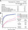
Induction of effective antibody responses against HIV-1 infection remains an elusive goal for vaccine development. Progress may require in-depth understanding of the molecular mechanisms of neutralization by monoclonal antibodies. We have analyzed the molecular actions of two rare, broadly neutralizing, human monoclonal antibodies, 4E10 and 2F5, which target the transiently exposed epitopes in the membrane proximal external region (MPER) of HIV-1 gp41 envelope during viral entry. Both have long CDR H3 loops with a hydrophobic surface facing away from the peptide epitope. We find that the hydrophobic residues of 4E10 mediate a reversible attachment to the viral membrane and that they are essential for neutralization, but not for interaction with gp41. We propose that these antibodies associate with the viral membrane in a required first step and are thereby poised to capture the transient gp41 fusion intermediate. These results bear directly on strategies for rational design of HIV-1 envelope immunogens.
Conflict of interest statement
The authors declare no conflict of interest.
Figures


Similar articles
-
N-terminal residues of an HIV-1 gp41 membrane-proximal external region antigen influence broadly neutralizing 2F5-like antibodies.Virol Sin. 2015 Dec;30(6):449-56. doi: 10.1007/s12250-015-3664-6. Epub 2015 Dec 21. Virol Sin. 2015. PMID: 26715302 Free PMC article.
-
Mechanism of HIV-1 neutralization by antibodies targeting a membrane-proximal region of gp41.J Virol. 2014 Jan;88(2):1249-58. doi: 10.1128/JVI.02664-13. Epub 2013 Nov 13. J Virol. 2014. PMID: 24227838 Free PMC article.
-
An affinity-enhanced neutralizing antibody against the membrane-proximal external region of human immunodeficiency virus type 1 gp41 recognizes an epitope between those of 2F5 and 4E10.J Virol. 2007 Apr;81(8):4033-43. doi: 10.1128/JVI.02588-06. Epub 2007 Feb 7. J Virol. 2007. PMID: 17287272 Free PMC article.
-
Antigp41 membrane proximal external region antibodies and the art of using the membrane for neutralization.Curr Opin HIV AIDS. 2017 May;12(3):250-256. doi: 10.1097/COH.0000000000000364. Curr Opin HIV AIDS. 2017. PMID: 28422789 Review.
-
Progress towards the development of a HIV-1 gp41-directed vaccine.Curr HIV Res. 2004 Apr;2(2):193-204. doi: 10.2174/1570162043484933. Curr HIV Res. 2004. PMID: 15078183 Review.
Cited by
-
Rational design of membrane proximal external region lipopeptides containing chemical modifications for HIV-1 vaccination.Clin Vaccine Immunol. 2013 Jan;20(1):39-45. doi: 10.1128/CVI.00615-12. Epub 2012 Oct 31. Clin Vaccine Immunol. 2013. PMID: 23114698 Free PMC article.
-
HIV-1 Entry and Membrane Fusion Inhibitors.Viruses. 2021 Apr 23;13(5):735. doi: 10.3390/v13050735. Viruses. 2021. PMID: 33922579 Free PMC article. Review.
-
Transmembrane domain membrane proximal external region but not surface unit-directed broadly neutralizing HIV-1 antibodies can restrict dendritic cell-mediated HIV-1 trans-infection.J Infect Dis. 2012 Apr 15;205(8):1248-57. doi: 10.1093/infdis/jis183. Epub 2012 Mar 6. J Infect Dis. 2012. PMID: 22396600 Free PMC article.
-
Elicitation of structure-specific antibodies by epitope scaffolds.Proc Natl Acad Sci U S A. 2010 Oct 19;107(42):17880-7. doi: 10.1073/pnas.1004728107. Epub 2010 Sep 27. Proc Natl Acad Sci U S A. 2010. PMID: 20876137 Free PMC article.
-
Strategies for eliciting HIV-1 inhibitory antibodies.Curr Opin HIV AIDS. 2010 Sep;5(5):421-7. doi: 10.1097/COH.0b013e32833d2d45. Curr Opin HIV AIDS. 2010. PMID: 20978384 Free PMC article. Review.
References
-
- Wei X, et al. Antibody neutralization and escape by HIV-1. Nature. 2003;422:307–312. - PubMed
-
- Kwong PD, et al. HIV-1 evades antibody-mediated neutralization through conformational masking of receptor-binding sites. Nature. 2002;420:678–682. - PubMed
-
- Haynes BF, et al. Cardiolipin polyspecific autoreactivity in two broadly neutralizing HIV-1 antibodies. Science. 2005;308:1906–1908. - PubMed
Publication types
MeSH terms
Substances
Grants and funding
LinkOut - more resources
Full Text Sources
Other Literature Sources
Medical

