. 2009 Dec;70(6):596-607.
doi: 10.1111/j.1365-3083.2009.02332.x.

Immunization with Cry1Ac from Bacillus thuringiensis increases intestinal IgG response and induces the expression of FcRn in the intestinal epithelium of adult mice

Affiliations

Immunization with Cry1Ac from Bacillus thuringiensis increases intestinal IgG response and induces the expression of FcRn in the intestinal epithelium of adult mice

S L Verdin-Terán et al. Scand J Immunol. 2009 Dec.

Abstract

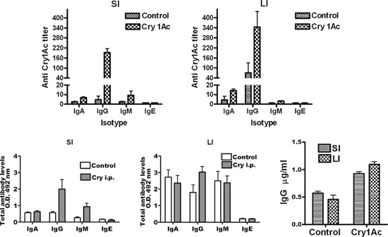
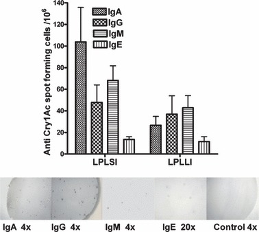
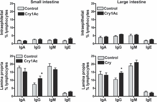
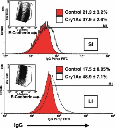
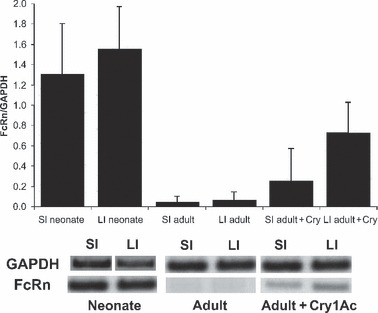
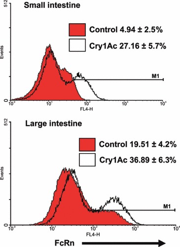
We have shown that Cry1Ac protoxin from Bacillus thuringiensis is a potent mucosal and systemic immunogen with adjuvant properties. Interestingly, we have observed that Cry1Ac preferentially induces high specific IgG responses in intestinal fluid when it is intraperitoneally administered to mice; therefore, in the present study, we used this protocol, as a model to address the influence of systemic immunization on the induction of the intestinal IgG response. The data shown indicate that upon intraperitoneal immunization with Cry1Ac, significant intestinal specific IgG cell responses were produced in the lamina propria, accompanied by an increased frequency of intestinal IgG+ lymphocytes and epithelial cells containing IgG. Considering that FcRn is the receptor responsible for the transport of IgG in neonatal intestinal epithelia, but it is developmentally downregulated in the rodent intestine, we analysed whether upon intestinal IgG induction, FcRn mRNA expression was induced in intestinal epithelial cells, of adult mice. Whereas in intestinal epithelia of unimmunized adult mice FcRn mRNA was not detected, in Cry1Ac immunized mice it was expressed, although the level was lower in comparison with that found in neonatal epithelia. Then using flow cytometry and immunofluorescence we confirmed that the expression of the protein FcRn was induced in the intestines of adult immunized mice especially in the large intestine. Finally, we found that Cry1Ac also increased FcRn expression in isolated intestinal epithelial cells stimulated in vitro. The outcomes suggest that the expression of FcRn in intestinal epithelium might be reactivated upon immunization, and possibly facilitate IgG transport.


Figures

Figure 1
Figure 1
Anti Cry1Ac titers and total antibody levels in the fluids from the small and large intestines of control and immunized mice. Cry1Ac: immunized mice that received three intraperitoneal doses of 50 μg of Cry1Ac; control: untreated mice. Above: anti Cry1Ac antibody titers. Below panel: total antibody levels. SI: small intestine, LI: large intestine. Mucosal specific antibody titers and antibody levels were determined by ELISA as described in the Methods. Intestinal fluids were collected with 5 and 3 ml volumes, for the large and small intestines respectively. Anti Cry1Ac titers are expressed as the last dilution giving an OD 492 value of 0.2 (twofold higher than the mean background value). Total antibody levels are shown for a mucosal sample dilution of 1:2. Bottom left: Concentration of IgG values were determined in samples diluted 1:2. Results are expressed as the arithmetic mean ± SD. n = 5 animals per experimental group. P < 0.001.
Figure 2
Figure 2
Intraperitoneal immunization with Cry1Ac induces specific antibody cell responses in the lamina propria of the small and large intestine in Balb/c mice. Lamina propria lymphocytes were isolated from the small (SI) and large intestine (LI), and the elicited IgA, IgG, IgM and IgE anti Cry1Ac antibody cell responses were evaluated by ELISPOT as described in the Methods in control and immunized mice. Upper panel: Data shown are from Cry1Ac immunized mice and are expressed as the arithmetic mean ± SD. (n = 5). In control mice were not detected significant anti Cry1Ac antibody cell responses, nor in the small neither in the large intestines; as the values recorded in this group for any isotype analysed were <2 spots per million of cells added (data not shown). Bottom panel: representative images of the spots recorded in immunized mice indicating the isotype and magnification. Control image corresponds to anti Cry1Ac IgG response in an un‐immunized mouse.
Figure 3
Figure 3
FACS analysis of intestinal B lymphocytes expressing surface immunoglobulins from control and mice i.p. immunized with Cry1Ac. Single cell suspensions of isolated intestinal lymphocytes (1 × 106 cells/sample) were stained with either FITC or Percp ‐anti‐CD3, and either Percp or PE‐anti‐B220, and PE anti‐IgG or IgM, and FITC anti IgA and IgE. Left: small intestine, Right: large intestine, upper panel: intraepithelial lymphocytes, Bottom panel: lamina propria. Data represent the mean percentage of cells expressing Igs among gated B lymphocytes ± SD from five independent experiments using cells of a given tissue from three mice.
Figure 4
Figure 4
Immunohistochemical staining for detection of IgG in paraffin sections from the small and large intestine, of unimmunized and Cry1Ac immunized mice. Above: small intestine (SI), Bottom: large intestine (LI). Left: unimmunized, middle: i.p. immunized with Cry1Ac (Cry1Ac) and right: isotype control. In the small intestine IgG staining was detected in some cells located in the lamina propria as well as in several epithelial cells. The number of IgG positive cells observed in immunized mice was higher than in unimmunized mice. In the epithelia of the large intestine, IgG reaction was slight. Five micron thickness sections were counter stained with Harris’s Haematoxilin. Each section represents consensus staining profiles from five independent animals. Arrow indicates an IgG + intraephitelial lymphocyte.
Figure 5
Figure 5
Flow cytometry analysis of the intestinal epithelial cells displaying IgG. Cells were analysed within the epithelial gate and E‐cadherin was used an epithelial cell marker. The frequencies of epithelial cells displaying IgG were higher in both the small and large intestines of immunized mice than in control mice, although the difference was markedly in the large intestine. Numbers represent the percentage of IgG positive cells ± SD (from the marker to the right M1) within the epithelial gate. Eight independent experiments were performed using two mice per group and analysing the cells of a given tissue from individual mice. A representative histogram is shown.
Figure 6
Figure 6
Immunization with Cry1Ac increases FcRn mRNA abundance in intestinal epithelial cells of adult mice. Epithelial cells from small and large intestine (SI and LI) from untreated neonate or adult mice; and adult Cry1Ac immunized mice (adult+Cry), were isolated and FcRn mRNA expression was analized by RT‐PCR. Data represent mean ± SEM of relative expression of FcRn regarding GAPDH in three independent experiments.
Figure 7
Figure 7
Flow cytometry analysis of the intestinal epithelial cells expressing FcRn from control and immunized adult mice. Cells were analysed within the epithelial gate and E‐cadherin was used an epithelial cell marker. The frequencies of epithelial cells displaying FcRn were higher in both the small and large intestines of immunized mice (Cry1Ac) than in control unimmunized mice (Control), although the expression was higher in the large than in the small intestine. Numbers represent the mean percentage of cells expressing FcRn ± SD (from the marker to the right M1), from three independent experiments using two mice per group and analysing the cells of a given tissue from individual mice. A representative histogram is shown.
Figure 8
Figure 8
Localization of IgG and FcRn in intestines from neonatal and adult unimmunized and immunized mice. Immunofluorescence in labelled intestinal sections from neonatal (neonate), adult control (unimmunized) and adult immunized mice (Cry1Ac i.p.), was visualized using a confocal microscope. Intestinal tissue sections were double labelled for; Left: IgG (green) and Middle: FcRn (blue). Right: merged images (clear blue). Double staining (Merge) shows the bright and wide co‐localization of IgG and FcRn observed in neonatal intestines (especially in the small intestine), in comparison with that found in tissues from adult mice. In immunized mice the localization of both IgG and FcRn is increased in the epithelia in relation to adult unimmunized mice, which presented scarce FcRn epithelial mark and moderate IgG staining. Each figure is representative of three similar experiments. Control staining with isotype resulted in no detectable staining (not shown).
Figure 9
Figure 9
Flow cytometry analysis of FcRn expression in intestinal epithelial cells stimulated in vitro with Cry1Ac. Epithelial cells were isolated from the small (SI) and large (LI) intestines from three control adult Balb/c mice and cultured in supplemented RPMI 1640 medium. Cells were cultured in 24 well plates (2 × 106 cells/ml) in culture medium with or without Cry1Ac (20 μg/ml) at 37 °C in 5% CO2 for 3 h in freshly isolated cells (left) and after the last 24 h in cells cultivated for 7 days (right). The cells were harvested and stained with anti‐FcRn and E‐cadherin antibodies and analysed by flow cytometry as described in Fig. 7. Above: small intestine (SI), Bottom: large intestine (LI). Numbers represent the mean percentage of cells expressing FcRn ± SD (from the marker to the right M1), from two independent experiments using the isolated cells of a given tissue from individual mice (n = 3). Representative histograms are shown.

Similar articles

Cited by

References

    1. Woof JM, Mestecky J. Mucosal immunoglobulins. Immunol Rev 2005;206:64–82. - PubMed
    1. Woof JM, Kerr MA. The function of immunoglobulin A in immunity. J Pathol 2006;208:270–82. - PubMed
    1. Robert‐Guroff M. IgG surfaces as an important component in mucosal protection. Nat Med 2000;6:129–30. - PubMed
    1. Baba TW, Liska V, Hofmann‐Lehmann R et al. Human neutralizing monoclonal antibodies of the IgG1 subtype protect against mucosal simian‐human immunodeficiency virus infection. Nat Med 2000;6:200–6. - PubMed
    1. Mbawuike IN, Pacheco S, Acuna CL, Switzer KC, Zhang Y, Harriman GR. Mucosal immunity to influenza without IgA: an IgA knockout mouse model. J Immunol 1999;162:2530–7. - PubMed

Publication types

MeSH terms