

ALK1 signaling regulates early postnatal lymphatic vessel development
- PMID: 19903896
- PMCID: PMC2830767
- DOI: 10.1182/blood-2009-07-235655
ALK1 signaling regulates early postnatal lymphatic vessel development
Abstract
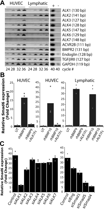
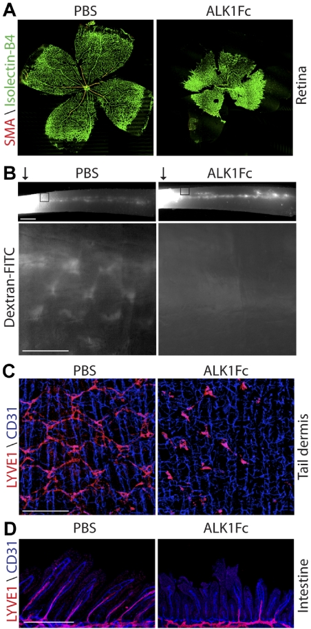
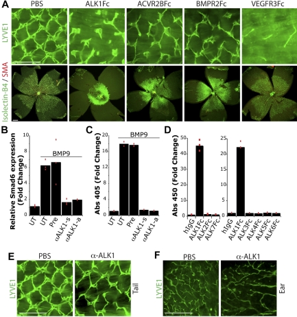
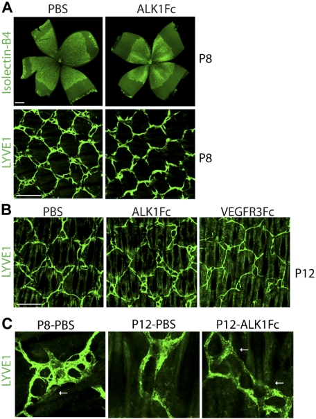
In vertebrates, endothelial cells form 2 hierarchical tubular networks, the blood vessels and the lymphatic vessels. Despite the difference in their structure and function and genetic programs that dictate their morphogenesis, common signaling pathways have been recognized that regulate both vascular systems. ALK1 is a member of the transforming growth factor-beta type I family of receptors, and compelling genetic evidence suggests its essential role in regulating blood vascular development. Here we report that ALK1 signaling is intimately involved in lymphatic development. Lymphatic endothelial cells express key components of the ALK1 pathway and respond robustly to ALK1 ligand stimulation in vitro. Blockade of ALK1 signaling results in defective lymphatic development in multiple organs of neonatal mice. We find that ALK1 signaling regulates the differentiation of lymphatic endothelial cells to influence the lymphatic vascular development and remodeling. Furthermore, simultaneous inhibition of ALK1 pathway increases apoptosis in lymphatic vessels caused by blockade of VEGFR3 signaling. Thus, our study reveals a novel aspect of ALK1 signaling in regulating lymphatic development and suggests that targeting ALK1 pathway might provide additional control of lymphangiogenesis in human diseases.
Figures

Comment in
-
Jazzing up vessel growth.Blood. 2010 Feb 25;115(8):1479. doi: 10.1182/blood-2009-11-254995. Blood. 2010. PMID: 20185593 No abstract available.
Similar articles
-
BMP9 (Bone Morphogenetic Protein-9)/Alk1 (Activin-Like Kinase Receptor Type I) Signaling Prevents Hyperglycemia-Induced Vascular Permeability.Arterioscler Thromb Vasc Biol. 2018 Aug;38(8):1821-1836. doi: 10.1161/ATVBAHA.118.310733. Arterioscler Thromb Vasc Biol. 2018. PMID: 29880487
-
Bone Morphogenetic Protein 9 Regulates Early Lymphatic-Specified Endothelial Cell Expansion during Mouse Embryonic Stem Cell Differentiation.Stem Cell Reports. 2019 Jan 8;12(1):98-111. doi: 10.1016/j.stemcr.2018.11.024. Epub 2018 Dec 27. Stem Cell Reports. 2019. PMID: 30595547 Free PMC article.
-
Tumour necrosis factor superfamily member 15 (Tnfsf15) facilitates lymphangiogenesis via up-regulation of Vegfr3 gene expression in lymphatic endothelial cells.J Pathol. 2015 Nov;237(3):307-18. doi: 10.1002/path.4577. Epub 2015 Aug 6. J Pathol. 2015. PMID: 26096340
-
VEGFR signaling during lymphatic vascular development: From progenitor cells to functional vessels.Dev Dyn. 2015 Mar;244(3):323-31. doi: 10.1002/dvdy.24227. Epub 2014 Dec 4. Dev Dyn. 2015. PMID: 25399804 Review.
-
Two Birds, One Stone: Double Hits on Tumor Growth and Lymphangiogenesis by Targeting Vascular Endothelial Growth Factor Receptor 3.Cells. 2019 Mar 21;8(3):270. doi: 10.3390/cells8030270. Cells. 2019. PMID: 30901976 Free PMC article. Review.
Cited by
-
Tumor lymphangiogenesis as a potential therapeutic target.J Oncol. 2012;2012:204946. doi: 10.1155/2012/204946. Epub 2012 Feb 16. J Oncol. 2012. PMID: 22481918 Free PMC article.
-
TGF-β Signaling in Control of Cardiovascular Function.Cold Spring Harb Perspect Biol. 2018 Feb 1;10(2):a022210. doi: 10.1101/cshperspect.a022210. Cold Spring Harb Perspect Biol. 2018. PMID: 28348036 Free PMC article. Review.
-
Molecular regulation of lymphangiogenesis in development and tumor microenvironment.Cancer Microenviron. 2012 Dec;5(3):249-60. doi: 10.1007/s12307-012-0119-6. Epub 2012 Aug 4. Cancer Microenviron. 2012. PMID: 22864800 Free PMC article.
-
Anti-human activin receptor-like kinase 1 (ALK1) antibody attenuates bone morphogenetic protein 9 (BMP9)-induced ALK1 signaling and interferes with endothelial cell sprouting.J Biol Chem. 2012 May 25;287(22):18551-61. doi: 10.1074/jbc.M111.338103. Epub 2012 Apr 5. J Biol Chem. 2012. PMID: 22493445 Free PMC article.
-
Bone Morphogenetic Proteins in Vascular Homeostasis and Disease.Cold Spring Harb Perspect Biol. 2018 Feb 1;10(2):a031989. doi: 10.1101/cshperspect.a031989. Cold Spring Harb Perspect Biol. 2018. PMID: 28348038 Free PMC article. Review.
References
-
- François M, Caprini A, Hosking B, et al. Sox18 induces development of the lymphatic vasculature in mice. Nature. 2008;456(7222):643–647. - PubMed
-
- Wigle JT, Oliver G. Prox1 function is required for the development of the murine lymphatic system. Cell. 1999;98(6):769–778. - PubMed
-
- Oliver G, Alitalo K. The lymphatic vasculature: recent progress and paradigms. Annu Rev Cell Dev Biol. 2005;21:457–483. - PubMed
-
- Karkkainen MJ, Haiko P, Sainio K, et al. Vascular endothelial growth factor C is required for sprouting of the first lymphatic vessels from embryonic veins. Nat Immunol. 2004;5(1):74–80. - PubMed
-
- Mäkinen T, Jussila L, Veikkola T, et al. Inhibition of lymphangiogenesis with resulting lymphedema in transgenic mice expressing soluble VEGF receptor-3. Nat Med. 2001;7(2):199–205. - PubMed
MeSH terms
Substances
LinkOut - more resources
Full Text Sources
Other Literature Sources
Molecular Biology Databases
Miscellaneous
