

Integration of BMP and Wnt signaling via vertebrate Smad1/5/8 and Drosophila Mad
- PMID: 19896409
- PMCID: PMC2810204
- DOI: 10.1016/j.cytogfr.2009.10.017
Integration of BMP and Wnt signaling via vertebrate Smad1/5/8 and Drosophila Mad
Abstract
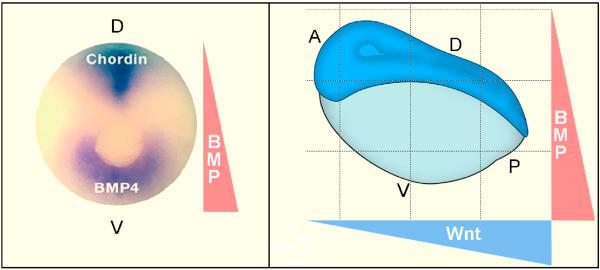
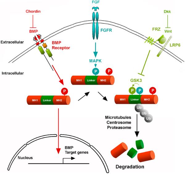
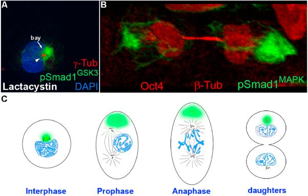
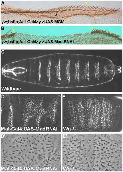
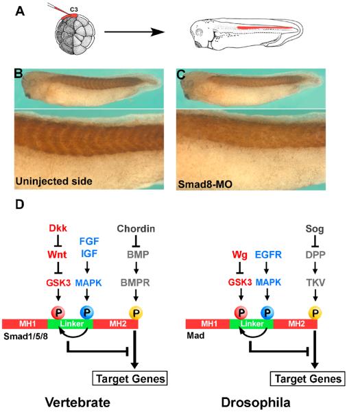
BMPs pattern the dorsal-ventral axis of vertebrate embryos. Smad1/5/8 transduces the BMP signal, and receives phosphorylation inputs from both MAPK and GSK3. Phosphorylation of Smad1 by MAPK and GSK3 result in its polyubiquitination and transport to the centrosome where it is degraded by the proteasome. These linker phosphorylations inhibit BMP/Smad1signaling by shortening its duration. Wnt, which negatively regulates GSK3 activity, prolongs the BMP/Smad1 signal. Remarkably, linker-phosphorylated Smad1 has been shown to be inherited asymmetrically during cell division. Drosophila contains a single Smad1/5/8 homologue, Mad, and is stabilized by phosphorylation-resistant mutations at GSK3 sites, causing Wingless-like effects. We summarize here the significance of linker-phosphorylated Smad1/Mad in relation to signal intensity and duration, and how this integrates the Wnt and BMP pathways during cell differentiation.
Figures
Similar articles
-
Integrating positional information at the level of Smad1/5/8.Curr Opin Genet Dev. 2008 Aug;18(4):304-10. doi: 10.1016/j.gde.2008.06.001. Epub 2008 Jul 14. Curr Opin Genet Dev. 2008. PMID: 18590818 Free PMC article. Review.
-
BMP canonical Smad signaling through Smad1 and Smad5 is required for endochondral bone formation.Development. 2009 Apr;136(7):1093-104. doi: 10.1242/dev.029926. Epub 2009 Feb 18. Development. 2009. PMID: 19224984 Free PMC article.
-
Integrating patterning signals: Wnt/GSK3 regulates the duration of the BMP/Smad1 signal.Cell. 2007 Nov 30;131(5):980-93. doi: 10.1016/j.cell.2007.09.027. Cell. 2007. PMID: 18045539 Free PMC article.
-
Dose-dependent Smad1, Smad5 and Smad8 signaling in the early mouse embryo.Dev Biol. 2006 Aug 1;296(1):104-18. doi: 10.1016/j.ydbio.2006.04.442. Dev Biol. 2006. PMID: 16765933 Free PMC article.
-
BMP-Smad 1/5/8 signalling in the development of the nervous system.Prog Neurobiol. 2013 Oct;109:28-41. doi: 10.1016/j.pneurobio.2013.07.002. Epub 2013 Jul 24. Prog Neurobiol. 2013. PMID: 23891815 Review.
Cited by
-
Wnt signaling in axial patterning and regeneration: lessons from planaria.Sci Signal. 2010 Jun 22;3(127):pe21. doi: 10.1126/scisignal.3127pe21. Sci Signal. 2010. PMID: 20571126 Free PMC article.
-
Novel Pathways for Targeting Tumor Angiogenesis in Metastatic Breast Cancer.Front Oncol. 2021 Dec 3;11:772305. doi: 10.3389/fonc.2021.772305. eCollection 2021. Front Oncol. 2021. PMID: 34926282 Free PMC article. Review.
-
BRASSINOSTEROID-INSENSITIVE2 Negatively Regulates the Stability of Transcription Factor ICE1 in Response to Cold Stress in Arabidopsis.Plant Cell. 2019 Nov;31(11):2682-2696. doi: 10.1105/tpc.19.00058. Epub 2019 Aug 13. Plant Cell. 2019. PMID: 31409630 Free PMC article.
-
bantam microRNA is a negative regulator of the Drosophila decapentaplegic pathway.Fly (Austin). 2018;12(2):105-117. doi: 10.1080/19336934.2018.1499370. Epub 2018 Aug 19. Fly (Austin). 2018. PMID: 30015555 Free PMC article.
-
Signaling crosstalk between TGFβ and Dishevelled/Par1b.Cell Death Differ. 2012 Oct;19(10):1689-97. doi: 10.1038/cdd.2012.50. Epub 2012 May 11. Cell Death Differ. 2012. PMID: 22576663 Free PMC article.
References
-
- Niehrs C. Regionally specific induction by the Spemann-Mangold organizer. Nat Rev Genet. 2004;6:425–434. - PubMed
Publication types
MeSH terms
Substances
Grants and funding
LinkOut - more resources
Full Text Sources
Other Literature Sources
Molecular Biology Databases
