. 2010 Jan;30(1):131-45.
doi: 10.1128/MCB.01000-09.

Dephosphorylation of gamma H2A by Glc7/protein phosphatase 1 promotes recovery from inhibition of DNA replication

Affiliations

Dephosphorylation of gamma H2A by Glc7/protein phosphatase 1 promotes recovery from inhibition of DNA replication

Marco Bazzi et al. Mol Cell Biol. 2010 Jan.

Abstract

Replication fork stalling caused by deoxynucleotide depletion triggers Rad53 phosphorylation and subsequent checkpoint activation, which in turn play a crucial role in maintaining functional DNA replication forks. How cells regulate checkpoint deactivation after inhibition of DNA replication is poorly understood. Here, we show that the budding yeast protein phosphatase Glc7/protein phosphatase 1 (PP1) promotes disappearance of phosphorylated Rad53 and recovery from replication fork stalling caused by the deoxynucleoside triphosphate (dNTP) synthesis inhibitor hydroxyurea (HU). Glc7 is also required for recovery from a double-strand break-induced checkpoint, while it is dispensable for checkpoint inactivation during methylmethane sulfonate exposure, which instead requires the protein phosphatases Pph3, Ptc2, and Ptc3. Furthermore, Glc7 counteracts in vivo histone H2A phosphorylation on serine 129 (gamma H2A) and dephosphorylates gamma H2A in vitro. Finally, the replication recovery defects of HU-treated glc7 mutants are partially rescued by Rad53 inactivation or lack of gamma H2A formation, and the latter also counteracts hyperphosphorylated Rad53 accumulation. We therefore propose that Glc7 activity promotes recovery from replication fork stalling caused by dNTP depletion and that gamma H2A dephosphorylation is a critical Glc7 function in this process.


Figures
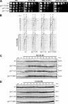
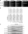
FIG. 1.
FIG. 1.
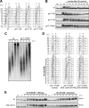
DNA replication in HU-treated glc7 mutants. (A) Serial dilutions (1:10) of exponentially growing cultures of cells with the indicated genotypes were spotted onto YPD plates with or without MMS, HU, and phleomycin (phleo) and incubated at 25°C for 3 days. wt, wild type. (B to D) Exponentially growing wild-type, glc7-129, glc7-132, and glc7-T152K cells were arrested in G1 with α-factor (αf) and released from the pheromone block in YPD or YPD containing 0.03 M HU. Aliquots of each culture were harvested at the indicated times after α-factor release to determine DNA content by fluorescence-activated cell sorting (FACS) analysis (B) and to detect Rad53 (C) and Ddc2 (D) by Western blot analysis with anti-Rad53 and anti-HA antibodies, respectively. exp, exponentially growing cells.
FIG. 2.
FIG. 2.
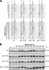
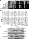
Restart of a stalled replication fork is defective in glc7 mutants, and this defect is suppressed by antagonizing Rad53 activity. (A and B) Wild-type (wt), glc7-129, glc7-132, and glc7-T152K cells were released from G1 arrest (αf) into YPD medium containing 0.2 M HU and incubated for 210 min (HU). Then, cultures were released into fresh medium, and samples were taken at the indicated times after HU removal to determine DNA content by FACS analysis (A) and to detect Rad53 by Western blot analysis with anti-Rad53 antibodies (B). (C) Immunodetection of BrdU-pulsed DNA. G1-arrested cells were released into YPgal containing 0.2 M HU plus 25 mM BrdU. After 1 h (HU), cells were chased with 2 mM thymidine into fresh medium, and DNA from samples taken at the indicated times after chase was prepared to detect BrdU-labeled DNA with anti-BrdU antibody. (D) glc7-T152K cells containing a URA3 centromeric plasmid, either empty or carrying the GAL-RAD53 or the GAL-rad53-K227A allele, were blocked in G1 with α-factor (αf) in SCraf-Ura and released into YPgal in the absence (top) or presence (bottom) of 0.05 M HU. Cell samples were collected at the indicated times after α-factor release to determine DNA content by FACS analysis. (E) Wild-type and GAL-GLC7 cells were released from G1 arrest (αf) in SCraf-Ura into YPraf or YPgal with 0.05 M HU. Cell samples were collected at the indicated times after α-factor release to detect Rad53 protein by Western blot analysis.
FIG. 3.
FIG. 3.
Glc7 is not involved in the recovery from MMS-induced DNA damage. Exponentially growing wild-type (wt), glc7-129, glc7-132, and glc7-T152K cells were arrested in G1 with α-factor (αf) and released from the pheromone block in YPD or YPD containing 0.005% MMS. Aliquots of each culture were harvested at the indicated times after α-factor release to determine DNA content by FACS analysis (A) and to detect Rad53 by Western blot analysis with anti-Rad53 antibodies (B).
FIG. 4.
FIG. 4.
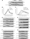
Recovery from a DSB-induced checkpoint involves Glc7. (A) Nocodazole-arrested (noc) wild-type (wt) cells containing URA3 centromeric plasmids, either empty or carrying the GAL-GLC7 allele, were incubated with 10 μg/ml phleomycin (phleo) in YPraf. After 40 min, two samples of each cell culture were transferred to either YPraf or YPgal medium, both lacking phleomycin but still containing nocodazole. Cell samples were collected at the indicated times after phleomycin removal to detect Rad53 by Western blot analysis. exp, exponentially growing cells. (B and C) Nocodazole-arrested wild-type, glc7-129, glc7-132, and glc7-T152K cells were incubated in YPD or YPD with 5 μg/ml phleomycin (+ phleo). After 15 min, cells were released into YPD lacking both phleomycin and nocodazole (time zero) and the percentages of binucleate cells in untreated (B) and phleomycin-treated (C) cultures were determined at the indicated times. (D to G) Wild-type, glc7-129, glc7-132, and glc7-T152K cells arrested in G2 with nocodazole (noc) were incubated with 5 μg/ml (D to F) or 10 μg/ml (G) phleomycin. After 15 min (phleo), cells were transferred to medium lacking phleomycin but still containing nocodazole. Aliquots of each culture were harvested at the indicated times after phleomycin removal to detect Rad53 (D), Rad9 (E), Ddc2-HA (F), and Mre11-HA (G) by Western blot analysis. Asterisks in panel E indicate hyperphosphorylated Rad9.
FIG. 5.
FIG. 5.
DSB formation and repair in glc7 mutant cells. Exponentially growing wild-type (wt), glc7-129, glc7-132, and glc7-T152K SCraf-Ura cell cultures (raf), all carrying the MATa allele and expressing the HO gene from the GAL1 promoter, were transferred to YPgal to induce HO expression. After 1 h, cells were transferred to medium containing glucose to allow cells to repair the HO-induced break (time zero). StyI-BamHI-digested genomic DNA prepared from cell samples collected at the indicated time points after galactose removal was subjected to Southern blot analysis with a MAT probe that detects 0.9-kb fragments (MATa) in the absence of HO-cut, while HO-induced DSB formation results in generation of HO-cut (0.7-kb fragment), which can be eventually repaired by HR with donor sequence HMR or HML, generating MATa (0.9-kb) and MATα (1.8-kb) repair products, respectively.
FIG. 6.
FIG. 6.
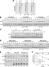
Glc7 regulates γH2A formation. (A and B) α-Factor-arrested (αf) wild-type (wt), glc7-T152K, and glc7-132 cells were released in YPD with 0.03 M HU. Aliquots of each culture were harvested at the indicated times after α-factor release to determine the DNA content by FACS (A) and to detect γH2A and H2A by Western blot analysis with anti-γH2A and anti-H2A antibodies, respectively (B). Specificity of the latter was checked with protein extracts from hta1Δ cells expressing the H2A-S129A variant (hta2-S129A), which were treated with 0.03 M HU for 120 min. (C) Cell cultures arrested in G2 with nocodazole (noc) were incubated with 5 μg/ml phleomycin. After 15 min (phleo), cells were transferred to medium lacking phleomycin but still containing nocodazole. (D) α-Factor-arrested (αf) cell cultures were released in YPD with 0.005% MMS. In panels C and D, aliquots of each culture were harvested at the indicated times after α-factor release to detect γH2A and H2A by Western blot analysis with anti-γH2A and anti-H2A antibodies, respectively. (E) Protein extracts from cells carrying either untagged GLC7 (Glc7) or fully functional HA-tagged GLC7 (Glc7-HA) at the GLC7 chromosomal locus were immunoprecipitated with anti-HA antibody. Immunoprecipitates were assayed for phosphatase activity toward purified histones at 30°C (lanes 1 to 6). The Glc7-HA immunoprecipitate was also preincubated for 15 min with inhibitor 2 (I-2) prior to phosphatase assay (lanes 7 to 9). At the indicated time points after histone addition, samples of each reaction mixture were subjected to Western blot analysis with anti-γH2A, anti-H2A, and anti-HA antibodies. (F) Densitometric analysis. Plotted values are the mean values ± standard deviations (SD) from three independent experiments as in panel E. The amount of γH2A was determined as the ratio between γH2A and total H2A band intensities.
FIG. 7.
FIG. 7.
Lack of γH2A formation alleviates the recovery defects of HU-treated glc7 mutants. (A) Serial dilutions (1:10) of exponentially growing cultures of wild-type (wt), glc7-T152K, hta2-S129A, and glc7-T152K hta2-S129A cells were spotted onto YPD plates with or without HU or phleomycin at the indicated concentrations and incubated at 25°C for 3 days. (B and C) Cells were released from G1 arrest (αf) in YPD medium with 0.03 M HU. Aliquots were harvested at the indicated times after release to determine DNA content by FACS analysis (B) and to detect Rad53 by Western blot analysis (C). The hta2-S129A and glc7-T152K hta2-S129A strains also carry the HTA1 deletion.
FIG. 8.
FIG. 8.
Epistatic relationships between GLC7, PPH3, PTC2, and PTC3. (A) Serial dilutions (1:10) of exponentially growing cells with the indicated genotypes were spotted onto YPD plates with or without MMS, HU, and phleomycin at the indicated concentrations and incubated at 25°C for 3 days. wt, wild type. (B and C) Exponentially growing cells with the indicated genotypes were arrested in G1 with α-factor (αf) and released from the pheromone block in YPD or in YPD containing 0.03 M HU. Aliquots of each culture were harvested at the indicated times after α-factor release to determine DNA content by FACS analysis (B) and to detect Rad53 by Western blot analysis with anti-Rad53 antibodies (C).

Similar articles

Cited by

References

    1. Altaf, M., A. Auger, M. Covic, and J. Côté. 2009. Connection between histone H2A variants and chromatin remodeling complexes. Biochem. Cell Biol. 87:35-50. - PubMed
    1. Bailis, J. M., and G. S. Roeder. 2000. Pachytene exit is controlled by reversal of Mek1-dependent phosphorylation. Cell 101:211-221. - PubMed
    1. Baker, S. H., D. L. Frederick, A. Bloecher, and K. Tatchell. 1997. Alanine-scanning mutagenesis of protein phosphatase type 1 in the yeast Saccharomyces cerevisiae. Genetics 145:615-626. - PMC - PubMed
    1. Chowdhury, D., X. Xu, X. Zhong, F. Ahmed, J. Zhong, J. Liao, D. M. Dykxhoorn, D. M. Weinstock, G. P. Pfeifer, and J. Lieberman. 2008. A PP4-phosphatase complex dephosphorylates γ-H2AX generated during DNA replication. Mol. Cell 31:33-46. - PMC - PubMed
    1. Clerici, M., V. Baldo, D. Mantiero, F. Lottersberger, G. Lucchini, and M. P. Longhese. 2004. A Tel1/MRX-dependent checkpoint inhibits the metaphase-to-anaphase transition after UV irradiation in the absence of Mec1. Mol. Cell. Biol. 24:10126-10144. - PMC - PubMed

Publication types

MeSH terms