. 2009 Oct 26;206(11):2557-72.
doi: 10.1084/jem.20091010.

KIR2DS4 is a product of gene conversion with KIR3DL2 that introduced specificity for HLA-A*11 while diminishing avidity for HLA-C

Affiliations

KIR2DS4 is a product of gene conversion with KIR3DL2 that introduced specificity for HLA-A*11 while diminishing avidity for HLA-C

Thorsten Graef et al. J Exp Med. .

Abstract

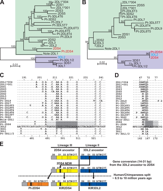
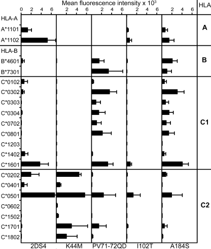
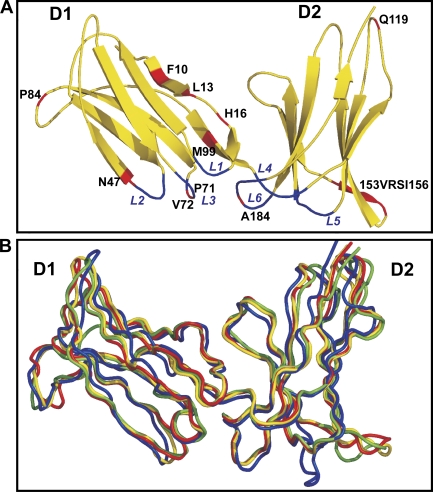
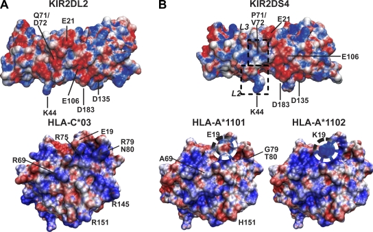
Human killer cell immunoglobulin-like receptors (KIRs) are distinguished by expansion of activating KIR2DS, whose ligands and functions remain poorly understood. The oldest, most prevalent KIR2DS is KIR2DS4, which is represented by a variable balance between "full-length" and "deleted" forms. We find that full-length 2DS4 is a human histocompatibility leukocyte antigen (HLA) class I receptor that binds specifically to subsets of C1+ and C2+ HLA-C and to HLA-A*11, whereas deleted 2DS4 is nonfunctional. Activation of 2DS4+ NKL cells was achieved with A*1102 as ligand, which differs from A*1101 by unique substitution of lysine 19 for glutamate, but not with A*1101 or HLA-C. Distinguishing KIR2DS4 from other KIR2DS is the proline-valine motif at positions 71-72, which is shared with KIR3DL2 and was introduced by gene conversion before separation of the human and chimpanzee lineages. Site-directed swap mutagenesis shows that these two residues are largely responsible for the unique HLA class I specificity of KIR2DS4. Determination of the crystallographic structure of KIR2DS4 shows two major differences from KIR2DL: displacement of contact loop L2 and altered bonding potential because of the substitutions at positions 71 and 72. Correlation between the worldwide distributions of functional KIR2DS4 and HLA-A*11 points to the physiological importance of their mutual interaction.


Figures

Figure 1.
Figure 1.
KIR2DS4 binds to HLA-A*11 and subsets of C1+ and C2+ HLA-C allotypes. (A) Titration of KIR2D-Fc fusion proteins made from 2DL1, 2, and 3 and 2DS4 to a panel of beads individually coated with 1 out of 29 HLA-A, 50 HLA-B, 9 C1+ HLA-C, and 7 C2+ HLA-C allotypes. Informative reactions are shown individually; negatively reacting (NR) allotypes are collectively represented by their means. Mean values from three independent experiments are shown. (B) Comparison of the binding of the four KIR2D-Fc fusion proteins at a concentration of 100 µg/ml against the four HLA allotype groups. All binding data for KIR2D-Fc was normalized to the binding obtained with W6/32, an mAb recognizing a conserved epitope of HLA class I. Mean values from three independent experiments are shown.
Figure 2.
Figure 2.
Ligation of KIR2DS4 by HLA-A*1102 or a specific antibody induces NKL cell activation. (A) Redirected cytotoxicity of P815 target cells by G4-NKL and G4-NKL transfectants expressing KIR2DS4, KIR2DL3, and KIR2DS4-LT, a mutant of KIR2DS4 that has a long cytoplasmic tail with inhibitory function. Effector cells were preincubated with combinations of mAbs as indicated (1mg/ml each). Mean values from three independent experiments are shown for E/T ratios from 80:1 to 20:1. (B) Results of cytotoxicity with G4-NKL expressing KIR2DL3, KIR2DS4, KIR3DL2, or no KIR as effector cells and untransfected 221 cells (top) or 221 cells expressing HLA-A*1102 (bottom) as target cells. To reduce the basal level of 221 cell lysis by G4-NKL, mAbs against the activating receptors 2B4 and NKG2D were added to effectors (5 mg/ml; right) for blocking. E/T ratios from 50:1 to 12.5:1 are shown. (C) Killing relative to untransfected G4-NKL cells at a 25:1 E/T ratio against 221 cells or 221 cells expressing HLA-A*1102. Blocking antibodies added to effectors are indicated. Data points shown in B and C are mean values from four independent experiments.
Figure 3.
Figure 3.
Human and chimpanzee KIR2DS4 bind similarly to HLA-A*11 but have distinctive specificities for HLA-C. Shown is the binding of KIR-Fc fusion proteins (100 µg/ml) made from human 2DS4 and its chimpanzee orthologue, Pt-2DS4, to beads individually coated with 1 out of 95 HLA class I allotypes. Only data for positively reacting allotypes are shown individually. The mean for 27 negatively reacting (NR) HLA-A allotypes is shown. Mean fluorescence values from three independent experiments are shown.
Figure 4.
Figure 4.
Gene conversion with KIR3DL2 was a seminal event in the evolution of KIR2DS4. (A and B) Neighbor-joining tree topology for the genomic segment carrying the exon encoding the D1 domain (A) and for the segment of D1 targeted by the gene conversion between 3DL2 and 2DS4 (B); bootstrap support is indicated where ≥50%. Colored boxes denote KIR genes of lineage II (purple) or III (green). Nole, Nomascus leucogenys (white-cheeked gibbon); Pt, Pan troglodytes (common chimpanzee). (C) Alignment of nucleotide sequences encoding the region of D1 where gene conversion took place. KIR3DL2 is used as the reference sequence, and a dot indicates identity to the reference. Numbering for 2DS4 and other lineage III KIR genes is given at the top, and numbering for 3DL2 and other lineage II KIR genes is at the bottom. The minimal length for the gene conversion is shown by the dark gray shaded box, and the maximal length is shown by the light and dark gray shading. (D) Alignment of amino acid sequences encoded by the nucleotide sequences in C. (E) Model for the gene conversion in which a 3DL2 ancestor was the donor and a 2DS4 ancestor was the recipient. The gene conversion occurred before the speciation event separating humans and chimpanzees 6.5–10 million years ago (Benton and Donoghue, 2007).
Figure 5.
Figure 5.
Mutation at positions 44, 71+72, 102, and 184 alter the specificity and/or avidity of KIR2DS4 for HLA class I. The binding of Fc fusion proteins made from 2DS4 or 2DS4 mutants (K44M, PV71-72QD, I102T, or A184S) to 95 HLA class I allotypes was determined as described in the legend to Fig. 3. Mean values from four independent experiments are shown.
Figure 6.
Figure 6.
Comparison of the three-dimensional structure of KIR2DS4 to the structures of other lineage III KIRs. (A) An illustration of the structure of the D1 and D2 domains of 2DS4, with the β strands represented by arrows. Binding loops are indicated and shown in blue. 14 out of the 15 unique substitutions in the extracellular domains that distinguish 2DS4 from other human lineage III KIRs are labeled and shown in red; the 15th residue is glutamine at position 1 in D1, which was not included in the construct used to produce the protein for crystallization. (B) Cα trace superposition for 192 equivalent Cα atoms (residues 8–200) of 2DS4 (yellow), 2DL1 (red; 1IM9; Fan et al., 2001), 2DL2 (green; 1EFX; Boyington and Sun, 2002), and 2DS2 (blue; 1M4K; Saulquin et al., 2003).
Figure 7.
Figure 7.
Features distinguishing the KIR2DS4 structure. (A) The 2DS4 unique residue His16 of the D1 domain forms two novel interdomain bonds with the D2 domain. Shown are these bonds (dashed line) formed by His16 with Ala145 (3.3 Å) and the side chain of Glu147 (2.9 Å). The electron-density maps (gray) of the interdomain region are contoured at the 1.2-σ level to indicate the quality of the resolved structure. (B–F) Close-up views of key residues involved in KIR2DL/HLA-C recognition. The D1 and D2 domains of 2DS4 were superimposed on the respective D1 and D2 domains of the structures of 2DL1 bound to HLA-C*04 (Fan et al., 2001) and 2DL2 bound to HLA-C*03 (Boyington et al., 2000). KIRs are colored as in Fig. 6. HLA-C molecules are colored magenta (HLA-C*03) and light blue (HLA-C*04), the GAV peptide of the 2DL2–HLA-C*03 complex is colored in red, and the key water molecule 38 (w38) is shown as a red asterisk. (B) The close proximity (1.6 Å) of Lys44 in 2DL2/3 to Lys80 of HLA-C2 causes steric hindrance, which prevents functional interactions between the two lysine residues. With the backbone displacement of L2 in 2DS4, Lys44 Cα of 2DS4 is separated from that of 2DL2 by 2.5 Å (dashed line), increasing the minimum distance between their Nζ atoms to 4 Å. (C) Lys44 in 2DL2 forms hydrogen bonds with Asn80 and Arg79 of HLA-C*03. The backbone shift widens the distance between Lys44 in 2DS4 and Asn80 to 3.7 Å and eliminates contact with Arg79. Because the benzyl group of Phe45 is rotated 90° clockwise in 2DS4, it is unlikely to make the hydrophobic contacts reported for 2DL2 and HLA-C*03. (D) In both KIR–HLA-C structures, Gln71 and Asp72 in loop 3 form hydrogen and hydrophobic contacts with HLA-C. Gln71 of 2DL2 also forms hydrogen bonds with Ala8 of peptide and water molecule 38. These interactions are abrogated by the replacement of Gln71/Asp72 with Pro71/Val72 in 2DS4, although they do not alter the conformation of loop 3. (E and F) The replacement of Ser184 by alanine is unique to 2DS4. This substitution abolishes the interactions described between Ser184 and Lys146 in the 2DL2–HLA-C*03 structure (E), or between Ser184 and both Lys80 and Lys146 in the 2DL1–HLA-C*04 structure (F).
Figure 8.
Figure 8.
Possible effects of the difference between A*1101 and A*1102 on the interaction with KIR2DS4. Predicted effects of the single substitution between HLA-A*1101 (Glu19) and -A*1102 (Lys19) were modeled using the structure file for HLA-A*1101 (1QVO). Electrostatic potentials were calculated with APBS tools (Baker et al., 2001) for 2DS4, 2DL2 (1EFX), HLA-C*03 (1EFX), HLA-A*1101 (1QVO), and HLA-A*1102 and were used to color the molecular surface drawn on their Cα traces (red, negative [−4]; blue, positive [4]). (A) Characterizing the binding of HLA-C to KIR2DL is an electropositive surface on HLA-C (e.g., HLA-C*03; bottom) that interacts with a complementary electronegative surface on KIR2D (e.g., 2DL2; top). Key acidic residues for KIR2D and basic residues for HLA-C are indicated. (B) In 2DS4 the common electronegative surface is disrupted by small positive and uncharged patches in L2 and L3 (boxes; top). HLA-A*11 has four nonconservative changes in the predicted KIR–HLA-C contact region (A69R, G79R, T80N/K, and H151R), resulting in an uncharged altered electrostatic surface compared with HLA-C. As indicated in the black and white circles, the substitution of Glu19 for Lys19 in A*1102 restores the electropositive surface in this region, which could explain its stronger reaction with 2DS4 than A*1101.

Similar articles

Cited by

References

    1. Abi-Rached L., Parham P. 2005. Natural selection drives recurrent formation of activating killer cell immunoglobulin-like receptor and Ly49 from inhibitory homologues. J. Exp. Med. 201:1319–1332 10.1084/jem.20042558 - DOI - PMC - PubMed
    1. Adams E.J., Parham P. 2001. Species-specific evolution of MHC class I genes in the higher primates. Immunol. Rev. 183:41–64 10.1034/j.1600-065x.2001.1830104.x - DOI - PubMed
    1. Adams P.D., Grosse-Kunstleve R.W., Hung L.W., Ioerger T.R., McCoy A.J., Moriarty N.W., Read R.J., Sacchettini J.C., Sauter N.K., Terwilliger T.C. 2002. PHENIX: building new software for automated crystallographic structure determination. Acta Crystallogr. D Biol. Crystallogr. 58:1948–1954 10.1107/S0907444902016657 - DOI - PubMed
    1. Arase H., Lanier L.L. 2004. Specific recognition of virus-infected cells by paired NK receptors. Rev. Med. Virol. 14:83–93 10.1002/rmv.422 - DOI - PubMed
    1. Baker N.A., Sept D., Joseph S., Holst M.J., McCammon J.A. 2001. Electrostatics of nanosystems: application to microtubules and the ribosome. Proc. Natl. Acad. Sci. USA. 98:10037–10041 10.1073/pnas.181342398 - DOI - PMC - PubMed

Publication types

MeSH terms