. 2009 Nov;136(22):3801-10.
doi: 10.1242/dev.038109.

Wnt-Ror signaling to SIA and SIB neurons directs anterior axon guidance and nerve ring placement in C. elegans

Affiliations

Wnt-Ror signaling to SIA and SIB neurons directs anterior axon guidance and nerve ring placement in C. elegans

Jason R Kennerdell et al. Development. 2009 Nov.

Abstract

Wnt signaling through Frizzled proteins guides posterior cells and axons in C. elegans into different spatial domains. Here we demonstrate an essential role for Wnt signaling through Ror tyrosine kinase homologs in the most prominent anterior neuropil, the nerve ring. A genetic screen uncovered cwn-2, the C. elegans homolog of Wnt5, as a regulator of nerve ring placement. In cwn-2 mutants, all neuronal structures in and around the nerve ring are shifted to an abnormal anterior position. cwn-2 is required at the time of nerve ring formation; it is expressed by cells posterior of the nerve ring, but its precise site of expression is not critical for its function. In nerve ring development, cwn-2 acts primarily through the Wnt receptor CAM-1 (Ror), together with the Frizzled protein MIG-1, with parallel roles for the Frizzled protein CFZ-2. The identification of CAM-1 as a CWN-2 receptor contrasts with CAM-1 action as a non-receptor in other C. elegans Wnt pathways. Cell-specific rescue of cam-1 and cell ablation experiments reveal a crucial role for the SIA and SIB neurons in positioning the nerve ring, linking Wnt signaling to specific cells that organize the anterior nervous system.


Figures

Fig. 1.
Fig. 1.
Anterior displacement of the nerve ring in cwn-2 and sax-3 mutants. (A-C) Confocal images of wild-type (wt), cwn-2 and sax-3 C. elegans L1 larvae expressing ceh-23::GFP. Top row, bright field; middle row, GFP projection; bottom row, merged image. Arrows indicate the position of the nerve ring, and arrowheads indicate the posterior boundary of the metacorpus. Anterior is left and dorsal is up. Scale bar: 10 μm. (D) Diagram of wild-type pharynx and nerve ring. Dendrites extend anteriorly from the cell bodies. Sensory axons extend ventrally, then anteriorly, then dorsally into the nerve ring. (E) Diagram of an anteriorly displaced cwn-2 nerve ring circling the metacorpus of the pharynx. (F) Quantification of nerve ring defects at the L1 stage. An animal was scored as defective if the nerve ring was as far anterior as the metacorpus (arrowhead in A-E). (G) Diagram of the cwn-2 genomic region. Black boxes represent exons, white boxes represent untranslated regions, and lines represent introns. cwn-2 lesions are indicated.
Fig. 2.
Fig. 2.
Coherent mislocalization of the nervous system and associated structures in cwn-2 mutants. (A-D) Renderings of wild-type and cwn-2 C. elegans L1 larvae based on serial electron microscopy sections. (A,B,D) Lateral views; (C) ventral views. Off-white, perimeter of the worm; orange, neuronal nuclei; blue, epidermal nuclei; red, muscle; yellow, neuropil (nerve ring, ventral cords, amphid commissures); green, excretory canal and pore; gray, pharynx. (A) All tissues. (B) Renderings limited to nervous system and pharynx. (C) Renderings limited to neuropil, pharynx, excretory pore and select head muscles. Yellow arrowheads, amphid commissures; green arrow, excretory pore. (D) Renderings limited to neuropil, pharynx and select head muscles. Red arrowheads indicate termination points of muscle arms.
Fig. 3.
Fig. 3.
Expression pattern and rescue of cwn-2. (A-C) DIC images (top) and GFP images (bottom) showing expression of cwn-2::SL2::GFP. (A) Comma stage C. elegans embryo. Arrow indicates approximate location of the developing nerve ring. (B) Adult. Arrowhead, body wall muscle; arrows, intestine; P, pharynx. (C) High magnification of adult head. Arrow, SMD neuron; P, pharynx. Scale bars: 10 μm. (D) Transgenic rescue of cwn-2 nerve ring defects by promoter::cDNA fusions. Nerve ring defects in each strain were scored at the L4 stage and normalized to defects in control siblings without the transgene (see Materials and methods). (E) Transgenic rescue of cwn-2 nerve ring defects by embryonic heat shock-induced cwn-2 expression. Controls are heat shocked cwn-2 animals without the hs::cwn-2 transgene. In D and E, error bars represent the s.e. of proportion. Asterisks indicate values different from controls at P<0.01 by Student's t-test.
Fig. 4.
Fig. 4.
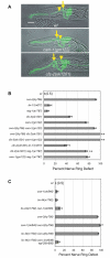
Nerve ring defects in Wnt ligand and receptor mutants. (A) Images of C. elegans L1 larvae illustrating anterior nerve rings in cam-1 and cfz-2 mutants. Images are confocal projections of ceh-23::GFP merged with a bright-field image. Arrows, nerve ring position; arrowheads, the posterior boundary of the metacorpus (as in Fig. 1). Anterior is left, dorsal is up. Scale bar: 10 μm. (B) Nerve ring defects in cwn-2, candidate receptor, and double mutants at the L1 stage. (C) Nerve ring defects in Wnt mutants and double mutants at the L1 stage. In B and C, error bars represent the s.e. of proportion. Asterisks indicate double mutants different from either single mutant by Student's t-test or Fisher's exact test with Bonferroni correction. *, P<0.05; **, P<0.01.
Fig. 5.
Fig. 5.
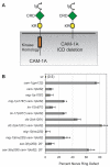
Double mutants uncover a function for the intracellular domain of CAM-1. (A) Schematic of CAM-1 and the CAM-1(ks52) protein with a deleted intracellular domain (ICD). Ig, immunoglobulin domain; CRD, cysteine-rich domain; KR, Kringle domain. (B) Nerve ring defects in cam-1(ks52) mutants and double mutants. Error bars represent the s.e. of proportion. Asterisks indicate double mutants different from either single mutant by Student's t-test or Fisher's exact test with Bonferroni correction. *, P<0.05; **, P<0.01.
Fig. 6.
Fig. 6.
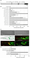
cam-1 functions in the SIA and/or SIB neurons to influence nerve ring placement. (A) Structure of the cam-1 locus, showing cam-1A, cam-1B and cam-1C transcriptional start sites. (B) Transgenic rescue of cam-1 nerve ring defects, scored at L4 stage. Defects in each strain were normalized to control siblings without the transgene (see Materials and methods). (C) Diagram and confocal projections of SIA, SIB and SMD neurons in wild type and cwn-2 mutants. SIA, SIB and SMD neurons express ceh-24::GFP (green), as does a neuron within the pharynx. AWC and AWB neurons (red) mark the position of the nerve ring. Diagram shows the trajectories of two SIA neurons; SIB and SMD trajectories are comparable. Green arrows, dorsal and ventral sublateral cords; green arrowheads, aberrant axons. Anterior is left, dorsal is up. (D) Partial rescue of mig-1 and sax-3 nerve ring defects by cDNA expression in SIA, SIB and SMD neurons. In B and D, error bars represent the s.e. of proportion. Asterisks indicate values different from controls by Student's t-test or Fisher's exact test. *, P<0.05; **, P<0.01.
Fig. 7.
Fig. 7.
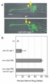
Nerve ring defects caused by ablation of SIA, SIB and SMD neurons. (A) C. elegans L1 larvae illustrating anterior nerve rings in animals carrying a ceh-24::egl-1 transgene. Images are confocal projections of ceh-23::GFP merged with a bright-field image. Arrows, nerve ring position; arrowheads, the posterior boundary of the metacorpus (as in Fig. 1). Anterior is left, dorsal is up. Scale bar: 10 μm. (B) Quantification of nerve ring defects in animals carrying ceh-24::egl-1 transgene in control and cwn-2 mutant backgrounds. All animals were ced-3/+ heterozygotes. Error bars represent the s.e. of proportion. Asterisks indicate values different from controls by Student's t-test or Fisher's exact test. **, P<0.01.
Fig. 8.
Fig. 8.
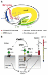
Cellular and molecular models for nerve ring placement. (A) Diagram of a C. elegans embryo near the time of nerve ring formation, showing cells and cues relevant for nerve ring guidance (this work) (Wadsworth et al., 1996; Hao et al., 2001; Pan et al., 2006; Whangbo and Kenyon, 1999). Gray neurons, which are negative for cam-1 rescue based on Fig. 6B, include BAG, ASK, ADL, ASI, AWC, ASH, AFD, ADF, AWB, AVE, AIM, AIY, AVK, AIZ, RIC, ADA, RMG, PHA, PHV, PVC and PVQ. (B) CAM-1 may be a receptor for CWN-2 in SIA and/or SIB neurons. MIG-1 (Frizzled) may be a co-receptor for CWN-2 in SIA and SIB. The SLT-1 receptor SAX-3 also affects nerve ring development, and can function in the same cells. CFZ-2 affects nerve ring development at an unknown site, and CWN-1 has mild effects on nerve ring development. Ig, immunoglobulin domain; CRD, cysteine-rich domain; KR, Kringle domain; FNIII, Fibronectin type III domain; LRR, leucine-rich repeat; EGF, Epidermal growth factor repeat.

Similar articles

Cited by

References

    1. Adler, C. E., Fetter, R. D. and Bargmann, C. I. (2006). UNC-6/Netrin induces neuronal asymmetry and defines the site of axon formation. Nat. Neurosci. 9, 511-518. - PMC - PubMed
    1. Aurelio, O., Hall, D. H. and Hobert, O. (2002). Immunoglobulin-domain proteins required for maintenance of ventral nerve cord organization. Science 295, 686-690. - PubMed
    1. Benard, C. Y., Boyanov, A., Hall, D. H. and Hobert, O. (2006). DIG-1, a novel giant protein, non-autonomously mediates maintenance of nervous system architecture. Development 133, 3329-3340. - PubMed
    1. Billiard, J., Way, D. S., Seestaller-Wehr, L. M., Moran, R. A., Mangine, A. and Bodine, P. V. (2005). The orphan receptor tyrosine kinase Ror2 modulates canonical Wnt signaling in osteoblastic cells. Mol. Endocrinol. 19, 90-101. - PubMed
    1. Brenner, S. (1974). The genetics of Caenorhabditis elegans. Genetics 77, 71-94. - PMC - PubMed

Publication types

MeSH terms

Substances