Review
. 2010:5:193-221.
doi: 10.1146/annurev.pathol.4.110807.092306.

Preinvasive breast cancer

Affiliations
Review

Preinvasive breast cancer

Dennis C Sgroi. Annu Rev Pathol. 2010.

Abstract

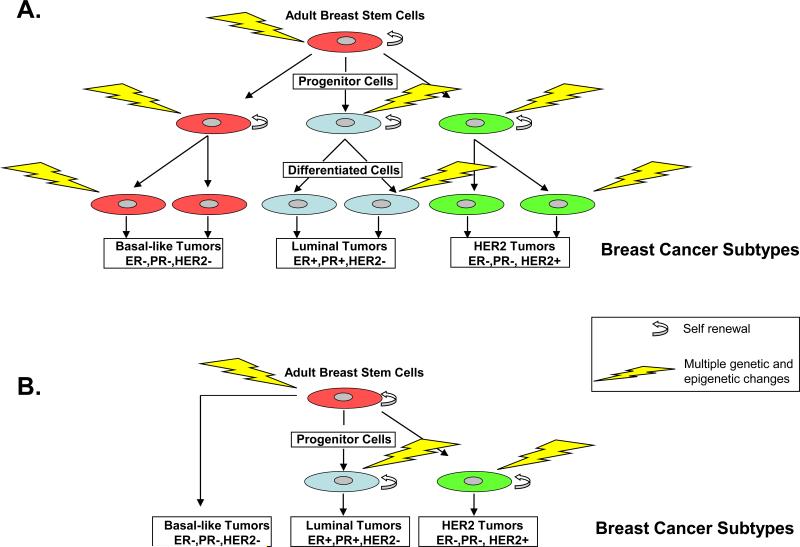
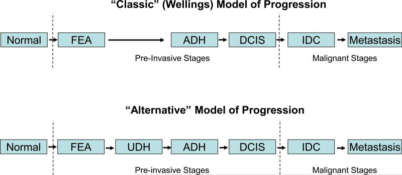
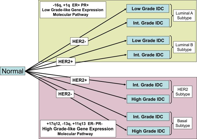
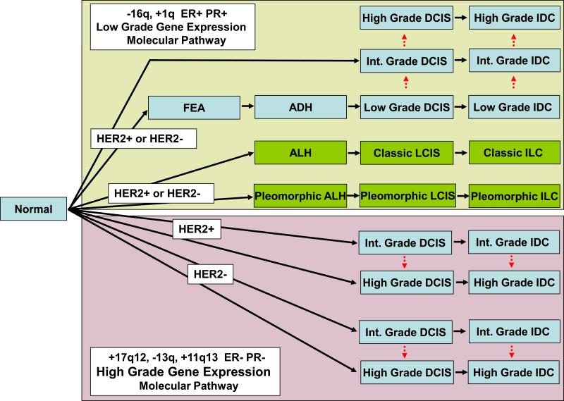
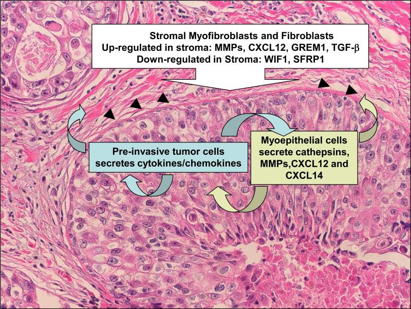
Preinvasive breast cancer accounts for approximately one-third of all newly diagnosed breast cancer cases in the United States and constitutes a spectrum of neoplastic lesions with varying degrees of differentiation and clinical behavior. High-throughput genetic, epigenetic, and gene-expression analyses have enhanced our understanding of the relationship of these early neoplastic lesions to normal breast tissue, and they strongly suggest that preinvasive breast cancer develops and evolves along two distinct molecular genetic and biological pathways that correlate with tumor grade. Although unique epigenetic and gene-expression changes are not observed in the tumor epithelial compartment during the transition from preinvasive to invasive disease, distinct molecular alterations are observed in the tumor-stromal and myoepithelial cells. This suggests that the stromal and myoepithelial microenvironment of preinvasive breast cancer actively participates in the transition from preinvasive to invasive disease. An improved understanding of the transition from preinvasive to invasive breast cancer will pave the way for novel preventative and therapeutic strategies.


Figures

Figure 1
Figure 1
Hypothetical models of the origin of human breast cancer. Based on the stochastic model of breast carcinogenesis (a), any epithelial cell type (e.g. stem cell, progenitor, or differentiated cell) may be the target of the initiating event. Each breast cancer subtype is initiated in a different cell type. In the cancer stem cell model (b), the cell of origin can be the same stem cell or progenitor cell for the different subtypes. The tumor phenotype is determined by a combination of genetic and epigenetic events. Abbreviations: ER+(−), presence (absence) of immunohistological expression or HER gene amplification; PR+(−), presence (absence) of immunohistochemical expression of progesterone receptor (PR) expression.
Figure 2
Figure 2
Microanatomy of the terminal duct lobular unit (TDLU). (a). Low-power photomicrograph of a hematoxylin and eosin (H&E)-stained tissue section of human breast with several TDLUs, each consisting of a cluster of acini (ductules) and a terminal duct (TD). High- power images of H&E-stained (b,c) and calponin-immunostained (d,e) tissue sections demonstrate the two cell layer anatomy of the acini (b,e) and the TD of the TDLU. Both the acini and TD display a continuous outer layer of myoepithelial cells (ME) that are immunoreactive for calponin (d,e;, brown elongate cells). The ME surround the cuboidal or the low columnar luminal epithelial cells (LE) of the acinus and TD, respectively. The basement membrane (BM) surrounds the ME. The TDLU is surrounded by the breast stromal (S) compartment.
Figure 3
Figure 3
Histomorphological classification of preinvasive and invasive breast cancer. The preinvasive stages of the lobular type consist of atypical lobular hyperplasia (ALH) and lobular carcinoma in situ (LCIS). Differentiation of ALH from LCIS is based upon the extent of proliferation and distension of the acini within the terminal duct lobular unit (TDLU). Pleomorphic LCIS differs from co-called classic LCIS in that the pleomorphic type consists of a proliferation of cells that are highly variable in size and shape and loosely cohesive, with or without necrosis, whereas the classic type consists of a proliferation of uniform, small, loosely cohesive, nonpolarized epithelial cells. The epithelial proliferation associated with preinvasive LCIS is confined to the ductal system, whereas invasive lobular carcinoma (ILC) infiltrates through the basement membrane into the surrounding stroma. The preinvasive stages of ductal type consists of flat epithelial atypia (FEA), atypical ductal hyperplasia (ADH), and ductal carcinoma in situ (DCIS). FEA is characterized by a minimal proliferation of native luminal cells of the TDLU by one to several layers of monomorphic cuboidal or columnar epithelial cells with low-grade cytological atypia. ADH differs from FEA in that in ADH the cells grow in a pseudostratified manner with secondary architectural atypia in the form of micropapillae, Roman arches (arrow) and trabecular bars. The differentiation of ADH from DCIS is based on the degree of architectural atypia and on the size and the extent of epithelial proliferation. Low-grade DCIS consists of small, cohesive, polarized, uniform cells of low proliferative capacity while high-grade DCIS consists of large, pleomorphic cells of high proliferative capacity with necrosis (asterisk). Intermediate-grade DCIS consists of small- to medium-sized, polarized cells with moderate nuclear pleomorphism and low proliferative capacity with or without necrosis (asterisk). Preinvasive carcinoma of the ductal type (e.g.; DCIS) consists of proliferation of medium- to large sized polarized cells confined to the ductal system whereas invasive ductal carcinoma (IDC) is characterized by the growth and invasion of neoplastic cells beyond the basement membrane and into the surrounding stroma.
Figure 4
Figure 4
Traditional linear models of breast cancer progression. (a) Classicand (b) alternative linear multistep models of human ductal breast cancer progression. The two models are identical, with the exception of the placement of usual ductal hyperplasia (UDH) as the precursor to atypical ductal hyperplasia (ADH). The classic model was initially based on histomorphological observations, whereas the alternative model was initially proposed based upon the epidemiological observations. Recently, multiple lines of evidence (histomorphological, immunohistochemical, and molecular genetic) support the classic model and contest the alternative model of progression. Molecular alterations occurring in normal breast epithelium result in flat epithelial atypia (FEA). FEA leads to additional changes give rise to ADH and ductal carcinoma in situ (DCIS), upon which subsequent genetic and epigenetic alterations in turn give rise to invasive ductal carcinoma (IDC).
Figure 5
Figure 5
Molecular classification of invasive ductalbreast cancer. Genetic and gene-expression data classify invasive ductal carcinomas into two distinct molecular biological and clinicopathological pathways. (a) The low grade-like gene-expression molecular pathway is characterized by chromosome 16q loss, predominant estrogen receptor (ER) and progesterone receptor (PR) expression (ER+, PR+) and a low-grade gene-expression profile populated with genes associated with ER positivity (ER+). (b) The high grade-like gene-expression molecular pathway is characterized by loss of chromosome 13q, gain of 11q13 and/or amplification of 17q12, infrequent expression of ER and PR, and a high grade-like gene-expression signature populated with genes associated with cell cycle, centrosomal function and DNA repair. The low-grade tumors express a unique set of genes that are rarely expressed in high-grade tumors, and vice versa. The luminal A and luminal B subtypes constitute the majority of invasive breast cancers in the low grade-like gene-expression molecular pathway and generally display indolent clinical behavior, whereas the human epidermal growth receptor 2 (HER2) and basal subtypes constitute the majority of cancers in the high grade-like pathway and generally display an aggressive clinical behavior. Light blue rectangles denote morphological ductal subtypes. Abbreviations: HER+(−), presence (absence) of immunohistological expression or HER gene amplification: IDC, invasive ductal carcinoma; –16q, loss of chromosome 16q; +1q, gain of chromosome 1q; –13q, loss of chromosome 13q; +11q13, gain of chromosomal region 11q13; +17q12, amplification of chromosomal region 17q12.
Figure 6
Figure 6
Contemporary multistep, two-dimensional model of human breast cancer progression derived from morphological, immunohistochemical, genetic and gene-expression data. Distinct molecular events occur in normal breast epithelium, giving rise to two distinct divergent molecular pathways within which linear pathological stage progression (horizontal black arrows) and intrastage heterogeneity (i.e., tumor-grade evolution; vertical red dashed arrows) occur. (a) The low grade-like gene-expression molecular pathway is characterized by chromosome 16q loss, predominant estrogen and progesterone receptor expression (ER+, PR+) and a low-grade gene-expression profile populated with genes associated with ER positivity (ER+). This low-grade pathway is observed in preinvasive lesions of both the ductal subtype (light blue rectangles) and lobular subtype (green rectangles). (b) The high grade-like gene-expression molecular pathway is characterized by loss of chromosome 13q; gain of 11q13 and/or amplification of 17q12; infrequent expression of ER and PR; and a high grade-like gene-expression signature populated with genes associated with cell cycle, centrosomal function, and DNA repair. Although pleomorphic atypical ductal hyperplasia (ALH), pleomorphic lobular carcinoma in situ (LCIS), and pleomorphic invasive lobular carcinoma (ILC), phenotypically resemble high grade tumors, immunohistochemical (ER positivity) and genetic (16q loss and 1q gain) data support an evolutionary association with the low grade-like gene-expression molecular pathway.. Recent immunohisto-chemical, morphological and genetic data support the concept of intrastage tumor-grade progression (red dashed arrows), which probably accounts for the observation of intratumoral heterogeneity. Light blue and green rectangles denote ductal and lobular morphological subtypes, respectively. Abbreviations: ALH, atypical lobular hyperplasia; DCIS, ductal carcinoma in situ; FEA, flat epithelial atypia; HER+(−), presence (absence) of immunohistochemical expression or HER gene amplification; IDC, invasive ductal carcinoma; −16q, loss of chromosome 16q; +1q, gain of chromosome 1q; −13q, loss of chromosome 13q; +11q13, gain of chromosomal region 11q13; +17q12, amplification of chromosomal region 17q12.
Figure 7
Figure 7
Tumor-stromal interactions during the preinvasive-to-invasive breast cancer transition. Multiple lines of evidence demonstrate marked genetic, epigenetic and gene-expression alterations in the neoplastic epithelium of preinvasive breast cancer as compared with normal breast epithelium. At the transition from preinvasive to invasive breast cancer, the neoplastic epithelium is not associated with additional qualitative molecular alterations. Instead, the nonneoplastic tumor microenvironment cells, in particular myoepithelial cells and stromal fibroblasts, myofibroblasts, and leukocytes, display significant molecular alterations that are associated with and may contribute to the transition from in-situ to invasive breast carcinoma. In preinvasive breast cancer (such as that illustrated in this hematoxylin and eosin-stained section), overexpression of cytokines and chemokines (as compared with normal breast epithelium) by neoplastic epithelium can induce an autocrine pathway that directly stimulates tumor growth. These tumor epithelial cell-derived cytokines can also induce several paracrine pathways. In the first paracrine pathway, tumor epithelial cell-derived cytokines and chemokines induce decreased stromal expression of growth pathway antagonists (i.e. WIF1 and SFRP1) that, in turn, favors tumor epithelial cell growth. Furthermore, the tumor epithelium can induce stromal cell expression of proteases [matrix metalloproteinases (MMPs) and cathepsins] that are involved in extracellular matrix remodeling, angiogenesis and basement membrane (black arrowheads) degradation. In the second paracrine pathway, epithelial cytokines induce myoepithelial cells to secrete proteases (MMPs) that degrade the basement membrane, thereby facilitating the transition from in situ to invasive carcinoma, and cytokines (CXCL12, CXCL14) that promote tumor epithelial cell growth, migration, and invasion. Abbreviation: TBF-β, transforming growth factor beta,

Similar articles

Cited by

References

    1. Jemal A, Siegel R, Ward E, Hao Y, Xu J, Murray T, Thun MJ. Cancer statistics, 2008. CA Cancer J Clin. 2008;58:71–96. - PubMed
    1. Harris JR, Lippman ME, Veronesi U, Willett W. Breast cancer (2). N Engl J Med. 1992;327:390–8. - PubMed
    1. Tavassoli FA, Devilee P. World Health Organization Classification of Tumours. Pathology and Genetics of Tumours of the Breast and Female Genital Organs. IARC Press; 2003.
    1. Wicha MS, Liu S, Dontu G. Cancer stem cells: an old idea--a paradigm shift. Cancer Res. 2006;66:1883–90. discussion 95-6. - PubMed
    1. Kakarala M, Wicha MS. Implications of the cancer stem-cell hypothesis for breast cancer prevention and therapy. J Clin Oncol. 2008;26:2813–20. - PMC - PubMed

Publication types