Progesterone and mifepristone regulate L-type amino acid transporter 2 and 4F2 heavy chain expression in uterine leiomyoma cells
- PMID: 19808856
- PMCID: PMC2775649
- DOI: 10.1210/jc.2009-1286
Progesterone and mifepristone regulate L-type amino acid transporter 2 and 4F2 heavy chain expression in uterine leiomyoma cells
Abstract
Context: Progesterone and its receptor (PR) play key roles in uterine leiomyoma growth. Previously, using chromatin immunoprecipitation-based cloning, we uncovered L-type amino acid transporter 2 (LAT2) as a novel PR target gene. LAT2 forms heterodimeric complexes with 4F2 heavy chain (4F2hc), a single transmembrane domain protein essential for LAT2 to exert its function in the plasma membrane. Until now, little is known about the roles of LAT2/4F2hc in the regulation of the growth of human uterine leiomyoma.
Objective: The aim of the study is to investigate the regulation of LAT2 and 4F2hc by progesterone and the antiprogestin mifepristone and their functions in primary human uterine leiomyoma smooth muscle (LSM) cells and tissues from 39 premenopausal women.
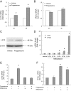
Results: In primary LSM cells, progesterone significantly induced LAT2 mRNA levels, and this was blocked by cotreatment with mifepristone. Progesterone did not alter 4F2hc mRNA levels, whereas mifepristone significantly induced 4F2hc mRNA expression. Small interfering RNA knockdown of LAT2 or 4F2hc markedly increased LSM cell proliferation. LAT2, PR-B, and PR-A levels were significantly higher in freshly isolated LSM cells vs. adjacent myometrial cells. In vivo, mRNA levels of LAT2 and PR but not 4F2hc were significantly higher in leiomyoma tissues compared with matched myometrial tissues.
Conclusion: We present evidence that progesterone and its antagonist mifepristone regulate the amino acid transporter system LAT2/4F2hc in leiomyoma tissues and cells. Our findings suggest that products of the LAT2/4F2hc genes may play important roles in leiomyoma cell proliferation. We speculate that critical ratios of LAT2 to 4F2hc regulate leiomyoma growth.
Figures


Similar articles
-
The Heavy Chain 4F2hc Modulates the Substrate Affinity and Specificity of the Light Chains LAT1 and LAT2.Int J Mol Sci. 2020 Oct 14;21(20):7573. doi: 10.3390/ijms21207573. Int J Mol Sci. 2020. PMID: 33066406 Free PMC article.
-
LAT1 regulates growth of uterine leiomyoma smooth muscle cells.Reprod Sci. 2010 Sep;17(9):791-7. doi: 10.1177/1933719110372419. Epub 2010 Jul 2. Reprod Sci. 2010. PMID: 20601542
-
Transcription factor KLF11 integrates progesterone receptor signaling and proliferation in uterine leiomyoma cells.Cancer Res. 2010 Feb 15;70(4):1722-30. doi: 10.1158/0008-5472.CAN-09-2612. Epub 2010 Feb 2. Cancer Res. 2010. PMID: 20124487 Free PMC article.
-
Function and structure of heterodimeric amino acid transporters.Am J Physiol Cell Physiol. 2001 Oct;281(4):C1077-93. doi: 10.1152/ajpcell.2001.281.4.C1077. Am J Physiol Cell Physiol. 2001. PMID: 11546643 Review.
-
Uterine Leiomyoma Stem Cells: Linking Progesterone to Growth.Semin Reprod Med. 2015 Sep;33(5):357-65. doi: 10.1055/s-0035-1558451. Epub 2015 Aug 6. Semin Reprod Med. 2015. PMID: 26251118 Review.
Cited by
-
Protein kinase C activation upregulates human L-type amino acid transporter 2 function.J Physiol Sci. 2021 Mar 31;71(1):11. doi: 10.1186/s12576-021-00795-0. J Physiol Sci. 2021. PMID: 33789576 Free PMC article.
-
The solute carrier SLC7A8 is a marker of favourable prognosis in ER-positive low proliferative invasive breast cancer.Breast Cancer Res Treat. 2020 May;181(1):1-12. doi: 10.1007/s10549-020-05586-6. Epub 2020 Mar 21. Breast Cancer Res Treat. 2020. PMID: 32200487 Free PMC article.
-
Comparison of the inhibitory effect of gonadotropin releasing hormone (GnRH) agonist, selective estrogen receptor modulator (SERM), antiprogesterone on myoma cell proliferation in vitro.Int J Med Sci. 2014 Jan 28;11(3):276-81. doi: 10.7150/ijms.7627. eCollection 2014. Int J Med Sci. 2014. PMID: 24516352 Free PMC article.
-
The selective progesterone receptor modulator CDB4124 inhibits proliferation and induces apoptosis in uterine leiomyoma cells.Fertil Steril. 2010 May 15;93(8):2668-73. doi: 10.1016/j.fertnstert.2009.11.031. Epub 2010 Jan 8. Fertil Steril. 2010. PMID: 20056218 Free PMC article.
-
Selective Progesterone Receptor Modulators-Mechanisms and Therapeutic Utility.Endocr Rev. 2020 Oct 1;41(5):bnaa012. doi: 10.1210/endrev/bnaa012. Endocr Rev. 2020. PMID: 32365199 Free PMC article. Review.
References
-
- Vollenhoven BJ, Lawrence AS, Healy DL 1990 Uterine fibroids: a clinical review. Br J Obstet Gynaecol 97:285–298 - PubMed
-
- Wilcox LS, Koonin LM, Pokras R, Strauss LT, Xia Z, Peterson HB 1994 Hysterectomy in the United States, 1988–1990. Obstet Gynecol 83:549–555 - PubMed
-
- Möller C, Hoffmann J, Kirkland TA, Schwede W 2008 Investigational developments for the treatment of progesterone-dependent diseases. Expert Opin Investig Drugs 17:469–479 - PubMed
-
- Marsh EE, Bulun SE 2006 Steroid hormones and leiomyomas. Obstet Gynecol Clin North Am 33:59–67 - PubMed
-
- Yin P, Lin Z, Cheng YH, Marsh EE, Utsunomiya H, Ishikawa H, Xue Q, Reierstad S, Innes J, Thung S, Kim JJ, Xu E, Bulun SE 2007 Progesterone receptor regulates Bcl-2 gene expression through direct binding to its promoter region in uterine leiomyoma cells. J Clin Endocrinol Metab 92:4459–4466 - PubMed
Publication types
MeSH terms
Substances
Grants and funding
LinkOut - more resources
Full Text Sources
Other Literature Sources
Medical
Research Materials
Miscellaneous

