. 2009 Nov;20(22):4826-37.
doi: 10.1091/mbc.e09-01-0034. Epub 2009 Sep 30.

The Cdc42 effectors Ste20, Cla4, and Skm1 down-regulate the expression of genes involved in sterol uptake by a mitogen-activated protein kinase-independent pathway

Affiliations

The Cdc42 effectors Ste20, Cla4, and Skm1 down-regulate the expression of genes involved in sterol uptake by a mitogen-activated protein kinase-independent pathway

Meng Lin et al. Mol Biol Cell. 2009 Nov.

Abstract

In Saccharomyces cerevisiae, the Rho-type GTPase Cdc42 regulates polarized growth through its effectors, including the p21-activated kinases (PAKs) Ste20, Cla4, and Skm1. Previously, we demonstrated that Ste20 interacts with several proteins involved in sterol synthesis that are crucial for cell polarization. Under anaerobic conditions, sterols cannot be synthesized and need to be imported into cells. Here, we show that Ste20, Cla4, and Skm1 form a complex with Sut1, a transcriptional regulator that promotes sterol uptake. All three PAKs can translocate into the nucleus and down-regulate the expression of genes involved in sterol uptake, including the Sut1 targets AUS1 and DAN1 by a novel mechanism. Consistently, deletion of either STE20, CLA4, or SKM1 results in an increased sterol influx and PAK overexpression inhibits sterol uptake. For Ste20, we demonstrate that the down-regulation of gene expression requires nuclear localization and kinase activity of Ste20. Furthermore, the Ste20-mediated control of expression of sterol uptake genes depends on SUT1 but is independent of a mitogen-activated protein kinase signaling cascade. Together, these observations suggest that PAKs translocate into the nucleus, where they modulate expression of sterol uptake genes via Sut1, thereby controlling sterol homeostasis.


Figures

Figure 1.
Figure 1.
Transcriptional regulation of genes mediating sterol uptake. In the presence of oxygen, yeast cells synthesize sterols and are unable to take up sterols from the extracellular medium. In contrast, under anaerobiosis cells rely on sterol uptake, because sterol synthesis requires oxygen. Under these conditions, the transcriptional regulators Sut1, Upc2, and Ecm22 promote the expression of AUS1, DAN1, and PDR11 and less well characterized genes (Y), which mediate sterol import. The underlying mechanisms of this transcriptional regulation are largely unknown. Here, we show that the PAKs Ste20, Cla4, and Skm1 can translocate into the nucleus, where they bind to the transcriptional regulator Sut1. The fact that the PAKs negatively regulate expression of AUS1, DAN1, and PDR11, suggests that PAKs inhibit Sut1 and/or Upc2 and Ecm22 by a novel mechanism. As demonstrated for Ste20, the down-regulation of gene expression requires its kinase activity. However, since there is no evidence for Sut1 phosphorylation by PAKs, an unknown protein (X), phosphorylated by PAKs, may be involved in the control of transcriptional regulators. Because sterol synthesis is crucial for cell polarization under aerobic conditions, it seems likely that sterol uptake plays an equally important role in the absence of oxygen.
Figure 2.
Figure 2.
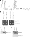
PAKs form a complex with Sut1. (A) The split-ubiquitin technique. See text for details. USPs, ubiquitin-specific proteases. (B) Ste20 interacts with Sut1 using the split-ubiquitin system. Cells (105) of the indicated plasmid combinations in a ste20Δ background were spotted onto media either lacking histidine and leucine to select for the plasmids or lacking histidine, leucine, and uracil to monitor protein interactions. The unrelated proteins Rdi1 and Ubc6 served as negative controls. (C) Ste20 interacts with Sut1 in vitro. Purified GST and GST-Sut1 bound to glutathione-Sepharose beads were incubated with an extract from cells expressing STE20-3HA from the plasmid pKA86. Eluted proteins were analyzed by immunoblotting using anti-HA antibodies. (D) Cla4 and Skm1 bind to Sut1. Recombinant His6-Sut1 and His6-Sec6 bound to Ni-NTA beads were incubated with lysates from cells expressing 3HA-CLA4 and 3HA-SKM1, respectively. Eluted proteins were analyzed by immunoblotting using anti-HA antibodies.
Figure 3.
Figure 3.
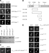
Ste20 localizes to the nucleus. (A) Sequence of the NLS and BR domain. The NLS is composed of two clusters of basic residues, whereas the BR domain consists of three clusters (BR-1, -2, and -3) (Takahashi and Pryciak, 2007). The second cluster of the NLS is identical to BR-1. (B) The Ste20 NLS is functional. Exponentially growing wild-type cells carrying the indicated Ste20 fragments fused to GST-GFP were incubated with galactose for 1 h. Cells were subsequently fixed with formaldehyde and stained with DAPI. One asterisk denotes the fusion with the peptide SGAGAGAGAGAIL. Two asterisks indicate addition of the sequence LGYFLFFEGGPGTQFAL. (C) Ste20 lacking the NLS is excluded from the nucleus. Logarithmically growing wild-type strains carrying the indicated GFP-STE20 alleles under control of a galactose-inducible promoter on a centromeric plasmid were cultivated in galactose-containing media for 1 h. Arrowheads indicate the nucleus. (D) Quantification of C. n > 100. (E) In few cells, Ste20 was enriched in the nucleus. Exponentially growing wild-type cells harboring GFP-Ste20 under control of a galactose-inducible promoter on a centromeric plasmid were incubated with galactose for 1 h.
Figure 4.
Figure 4.
Nuclear localization of Skm1 and Cla4. (A) Skm1 is strongly enriched in the nucleus. Exponentially growing cells carrying the indicated GFP-SKM1 alleles were induced for 1 h by the addition of galactose. Cells were fixed with formaldehyde and stained with DAPI. (B) Wild-type SKM1 and its derivatives are expressed at comparable levels. The indicated strains were incubated with galactose for 1 h. Cells were lysed and equal amounts of protein extract were analyzed by immunoblotting using antibodies against GFP and GAPDH (loading control). (C) Residues 201-230 are sufficient for nuclear targeting of Skm1. The Skm1 fragment was expressed as galactose-inducible GST-GFP fusion. (D) Fragments used to map the sequence that targets Cla4 to the nucleus. The kinase domain and regions that are required for membrane association are highlighted. The CRIB domain is the minimal Cdc42-binding motif (Cvrckova et al., 1995), a proline-rich region (Pro) mediates binding to the scaffold protein Bem1 (Winters and Pryciak, 2005), and the PH domain is required for the association with membrane phosphoinositides (Wild et al., 2004). (E) Cla4 fragments localize to the nucleus. Cla4 fragments were expressed as GST-GFP fusions from a galactose-inducible promoter in wild-type cells.
Figure 5.
Figure 5.
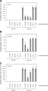
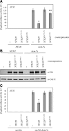
PAKs down-regulates the expression of DAN1, AUS1, and PDR11. (A) AUS1 expression is regulated by Ste20, Cla4, and Skm1. The indicated strains harboring a plasmid on which the lacZ was fused to the promoter region of AUS1 were grown in selective raffinose medium in the presence of ergosterol and Tween 80. The indicated genes were overexpressed for 18 h by the addition of galactose. Shown is the mean of β-galactosidase activity of at least six independent experiments with SD, *p < 0.0001, compared with hem1Δ cells without any overexpression. (B) Ste20 and Cla4 control the expression of DAN1. The indicated strains carrying a DAN1-lacZ plasmid were treated as described in A. Shown is the mean of β-galactosidase activity of at least six independent experiments. *p < 0.0001 and **p < 0.001, compared with hem1Δ cells without any overexpression. (C) PDR11 expression is negatively regulated by Cla4 and Skm1. β-Galactosidase activity of the indicated strains harboring a PDR11-lacZ plasmid was determined in at least 6 independent experiments. *p < 0.0001 and **p < 0.005, compared with hem1Δ cells without any overexpression.
Figure 6.
Figure 6.
Expression of AUS1, DAN1, and PDR11 in PAK deletion strains. (A) STE20 deletion results in higher AUS1 expression. The indicated strains carrying an AUS1-lacZ plasmid were grown in selective medium supplemented with ergosterol and Tween 80, and β-galactosidase activity was determined in at least six independent experiments. wt, wild type; *p < 0.001, compared with hem1Δ cells. (B) PAK deletion does not affect DAN1 levels. Shown is the mean β-galactosidase activity of at least six independent experiments with SD of the indicated strains harboring DAN1-lacZ on a plasmid. (C) Cells lacking SKM1 express higher PDR11 levels. β-Galactosidase activity of the indicated strains harboring a PDR11-lacZ plasmid was determined in at least 6 independent experiments. *p < 0.0005, compared with hem1Δ cells.
Figure 7.
Figure 7.
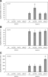
Down-regulation of AUS1 expression by PAKs depends on SUT1. (A) Effect of SUT1 deletion on AUS1 levels following STE20 overexpression. The β-galactosidase activity of the indicated strains was determined in six independent experiments as described in Figure 5A. *p < 0.0001 and **p < 0.05, compared with hem1Δ cells without STE20 overexpression. (B) Regulation of AUS1 expression by Cla4 depends on SUT1. Shown is the mean β-galactosidase activity of six independent experiments with SD of the indicated strains. *p < 0.0001 and **p < 0.05, compared with hem1Δ cells without CLA4 overexpression. (C) The down-regulation of AUS1 expression by Skm1 requires SUT1. β-Galactosidase activity of the indicated strains was determined in at least six independent experiments. *p < 0.0001, compared with hem1Δ cells without SKM1 overexpression. (D) Effect of SUT1 deletion on AUS1 expression in cells lacking STE20. The β-galactosidase activity of the indicated strains was determined in at least six independent experiments as described in Figure 6A. *p < 0.0005, compared with hem1Δ cells.
Figure 8.
Figure 8.
The regulation of AUS1 expression requires kinase activity and nuclear localization of Ste20. (A) Wild-type STE20 and mutant versions were overexpressed in the indicated strains as described in Figure 5A. Shown is the mean β-galactosidase activity of at least six independent experiments with SD, *p < 0.0001 and **p < 0.05, compared with hem1Δcells without STE20 overexpression. (B) Wild-type STE20 and its mutant alleles are expressed at comparable levels. The indicated strains were incubated with galactose for 18 h. Cells were lysed and equal amounts of protein extract were analyzed by immunoblotting. An N-terminal 3HA tag allowed the detection of Ste20. Cdc11 was used as loading control. (C) Wild-type and mutant alleles of STE20 expressed from the native STE20 promoter were integrated into ste20Δ and ste20Δ hem1Δ cells. AUS1 expression was determined in at least six independent experiments as described in Figure 6A. *p < 0.0005, compared with ste20Δ hem1Δ cells.
Figure 9.
Figure 9.
The down-regulation of PDR11 expression requires nuclear localization of Skm1. SKM1 and SKM1ΔNLS, respectively, were integrated into skm1Δ and skm1Δ hem1Δ cells. PDR11 expression was determined in at least six independent experiments as described in Figure 6C. *p < 0.0005, compared with skm1Δ hem1Δ cells.
Figure 10.
Figure 10.
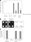
Ste20 regulates gene expression by two distinct mechanisms. (A) The down-regulation of AUS1 expression is independent of the MAPK kinase kinase Ste11. The β-galactosidase activity was determined in eight independent experiments as described in Figure 5A. *p < 0.0001, compared with hem1Δ cells without STE20 overexpression. (B) Haploid invasive growth does not require nuclear localization of Ste20. Cells (105) of the indicated strains were spotted on a YPD plate and incubated for 2 d at 30°C. Pictures were taken before and after gentle rinsing with water. (C) The NLS of Ste20 is not necessary for the formation of a mating projection. Exponentially growing cells of the indicated strains were incubated in selective medium with 1 μg/ml α-factor for 3 h. Cells were then fixed with formaldehyde and the percentage of cells with a mating projection was determined. Given is the mean of three independent experiments with SD bars (n > 100 for each experiment).
Figure 11.
Figure 11.
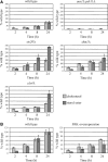
Ste20, Cla4, and Skm1 negatively regulate sterol uptake. (A) Ste20, Skm1, and Cla4 overaccumulate free sterols. Heme-deficient cells of the indicated genotype were cultured in media containing [14C]cholesterol for the indicated period of time. Cells were collected, and lipids were isolated, separated by TLC, and quantified by radioscanning. Data are normalized to free sterols and steryl ester levels in wild-type cells after 24 h of uptake. Values are means and SD of two independent experiments. The aus1Δ pdr11Δ mutant was used as negative control. (B) Overexpression of PAKs results in a decreased sterol import. For PAK overexpression, SKM1 and CLA4 were placed under control of the GAL1 promoter by genomic integration. These cells also harbored STE20 expressed from the GAL1 promoter on a plasmid. Wild-type cells carried the corresponding empty vector. Both strains were grown in SC-Ura medium supplemented with galactose and [14C]cholesterol. Sterols and steryl ester were analyzed as described in A.

Similar articles

Cited by

References

    1. Ahn S. H., Cheung W. L., Hsu J. Y., Diaz R. L., Smith M. M., Allis C. D. Sterile 20 kinase phosphorylates histone H2B at serine 10 during hydrogen peroxide-induced apoptosis in S. cerevisiae. Cell. 2005;120:25–36. - PubMed
    1. Alimardani P., Régnacq M., Moreau-Vauzelle C., Ferreira T., Rossignol T., Blondin B., Bergès T. SUT1-promoted sterol uptake involves the ABC transporter Aus1 and the mannoprotein Dan1 whose synergistic action is sufficient for this process. Biochem. J. 2004;381:195–202. - PMC - PubMed
    1. Alvarez F. J., Douglas L. M., Konopka J. B. Sterol-rich plasma membrane domains in fungi. Eukaryot. Cell. 2007;6:755–763. - PMC - PubMed
    1. Ash J., Wu C., Larocque R., Jamal M., Stevens W., Osborne M., Thomas D. Y., Whiteway M. Genetic analysis of the interface between Cdc42p and the CRIB domain of Ste20p in Saccharomyces cerevisiae. Genetics. 2003;63:9–20. - PMC - PubMed
    1. Bourot S., Karst F. Isolation and characterization of the Saccharomyces cerevisiae SUT1 gene involved in sterol uptake. Gene. 1995;165:97–102. - PubMed

Publication types

MeSH terms

Substances