. 2009 Nov;74(3):724-41.
doi: 10.1111/j.1365-2958.2009.06897.x. Epub 2009 Sep 28.

Genetic and biochemical analysis of the serine/threonine protein kinases PknA, PknB, PknG and PknL of Corynebacterium glutamicum: evidence for non-essentiality and for phosphorylation of OdhI and FtsZ by multiple kinases

Affiliations
Free PMC article

Genetic and biochemical analysis of the serine/threonine protein kinases PknA, PknB, PknG and PknL of Corynebacterium glutamicum: evidence for non-essentiality and for phosphorylation of OdhI and FtsZ by multiple kinases

Christian Schultz et al. Mol Microbiol. 2009 Nov.
Free PMC article

Abstract

We previously showed that the 2-oxoglutarate dehydrogenase inhibitor protein OdhI of Corynebacterium glutamicum is phosphorylated by PknG at Thr14, but that also additional serine/threonine protein kinases (STPKs) can phosphorylate OdhI. To identify these, a set of three single (DeltapknA, DeltapknB, DeltapknL), five double (DeltapknAG, DeltapknAL, DeltapknBG, DeltapknBL, DeltapknLG) and two triple deletion mutants (DeltapknALG, DeltapknBLG) were constructed. The existence of these mutants shows that PknA, PknB, PknG and PknL are not essential in C. glutamicum. Analysis of the OdhI phosphorylation status in the mutant strains revealed that all four STPKs can contribute to OdhI phosphorylation, with PknG being the most important one. Only mutants in which pknG was deleted showed a strong growth inhibition on agar plates containing glutamine as carbon and nitrogen source. Thr14 and Thr15 of OdhI were shown to be phosphorylated in vivo, either individually or simultaneously, and evidence for up to two additional phosphorylation sites was obtained. Dephosphorylation of OdhI was shown to be catalysed by the phospho-Ser/Thr protein phosphatase Ppp. Besides OdhI, the cell division protein FtsZ was identified as substrate of PknA, PknB and PknL and of the phosphatase Ppp, suggesting a role of these proteins in cell division.


Figures
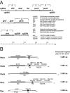
Fig. 1
Fig. 1
Genomic organization of pknA, pknB, pknG, pknL and ppp in C. glutamicum (A) and domain architecture of the corresponding proteins as predicted by PFAM, TMHMM or TMPred (B). KD, kinase domain; TM, transmembrane helix; PP2C, phosphatase domain; PASTA, penicillin-binding protein and serine/threonine kinase-associated domain.
Fig. 2
Fig. 2
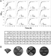
Growth of STPK and Ppp mutants of C. glutamicum. A. Growth of different C. glutamicum mutants (black symbols) in comparison with the wild type (white symbols) in BHI medium containing 4% (w/v) glucose. Mean values of triplicate experiments are shown. The final optical densities at 600 nm (OD600) with standard deviations and the maximal growth rates are listed below as well as the P-values from a t-test for pairwise comparisons of the mutants with the wild type. B. Growth on CGXII agar plates containing 100 mM l-glutamine as sole carbon and nitrogen source.
Fig. 3
Fig. 3
Cell morphology of STPK and Ppp mutants of C. glutamicum growing in BHI medium with 4% (w/v) glucose at 30°C (early exponential phase) was observed. Phase-contrast microscopy pictures are shown and 500 cells of each strain were counted to determine the frequency of cell length. The dotted lines represent the length range from 1.5 to 3 μm which covers 75% of the wild-type cells.
Fig. 4
Fig. 4
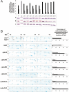
Analysis of the in vivo OdhI phosphorylation status in cells grown for 24 h in BHI medium with 4% (w/v) glucose. A. Western blot analysis of cell-free protein extracts (20 μg each) of the indicated C. glutamicum strains with OdhI antibodies. The experiment was performed in triplicate (a, b, c). The upper band in the Western blot represents singly or doubly phosphorylated OdhI, the lower band unphosphorylated OdhI (Schultz et al., 2007). The percentages (mean values with standard deviation) of unphosphorylated OdhI (black bars) to phosphorylated OdhI (grey bars) were calculated by densitometry. B. Two-dimensional polyacrylamide gel electrophoresis of cell-free protein extracts (300 μg each) of the indicated C. glutamicum strains. In the left part of the figure, sections of three 2D gels prepared with extracts of three independent cultures of the respective strain are shown. The three spots representing unphosphorylated, monophosphorylated and presumably diphosphorylated OdhI are circled and labelled with 0, 1 and 2 respectively. The relative amounts of the three OdhI forms were calculated by densitometric analysis of the OdhI spots. Normalization was performed using the spot labelled X that represents adenylate kinase (cg0648), a protein whose intracellular level is apparently not influenced by the deletion of STPKs or Ppp. The percentage of unphosphorylated OdhI is shown with black bars, that of monophosphorylated OdhI with grey bars, and that of presumably diphosphorylated OdhI by white bars. The spot labelled Y represents MenG (S-adenosylmethionine:2-demethylmenaquinone methyltransferase, cg1055) and is shown because of the small distance to the OdhI spot labelled 2.
Fig. 5
Fig. 5
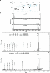
Identification of in vivo phosphorylation sites of OdhI. A. Analysis of the in vivo OdhI phosphorylation status by 2D gel electrophoresis of cell-free protein extract of C. glutamicumΔppp/pJC1-odhI. The strain was cultivated for 24 h in BHI medium with 4% (w/v) glucose and 300 μg of protein was used for separation by 2D-PAGE. The two spots representing native monophosphorylated and diphosphorylated OdhI are labelled as 1 and 2, respectively, and Strep-tagged monophosphorylated, diphosphorylated and presumably triphosphorylated OdhI are labelled with 1′, 2′ and 3′ respectively. B. MALDI-TOF-MS and MS/MS analysis of tryptically digested OdhI spots 1′ (panel a), 2′ (panel b) and 3′ (panel c) of strain C. glutamicumΔppp/pJC1-odhI. Peaks are labelled with their monoisotopic masses. The only phosphopeptide detected was the N-terminal one composed of amino acids 2–19. In the unphoshorylated state, the predicted mass (in the H+ form) is 1977.9 Da, in the monophosphorylated state 2057.9 Da, in the diphosphorylated state 2137.9 Da. In panels d and e, MALDI-TOF tandem MS of the 2057.9 Da peptide derived from spot 2′ and of the 2137.8 Da peptide derived from spot 3′ are shown respectively. β-Elimination of phosphoric acid (mass shifts of −18 Da or −36 Da compared to the unphosphorylated fragment after a single or a double β-elimination respectively) is indicated.
Fig. 6
Fig. 6
In vitro phosphorylation of OdhI by different STPKs. A. Kinase domains purified for in vitro analysis. PknA1−287 (34.0 kDa), PknB1−287 (33.4 kDa), PknG1−342 (40.1 kDa) and PknL1−287 (33.6 kDa), all containing an N-terminal decahistidine tag, were overproduced in E. coli and purified by Ni2+-chelate affinity chromatography. OdhIStrep (16.6 kDa) containing a C-terminal StrepTag-II was overproduced in E. coli and purified by StrepTactin affinity chromatography. Purified proteins were subjected to SDS-PAGE and stained with Coomassie brilliant blue. B. In vitro phosphorylation of OdhI by PknA1−287, PknB1−287, PknG1−342 and PknL1−287. The in vitro phosphorylation assays were performed as described in Experimental procedures either with non-radioactive ATP or with [γ-33P]-ATP. In the former case, the OdhI phosphorylation status was followed by Coomassie-stained SDS gels and by Western blot analysis with OdhI antibodies. In the latter case, the samples were subjected to autoradiography. Only the OdhI section is shown.
Fig. 7
Fig. 7
In vitro dephosphorylation of phosphorylated OdhI by Ppp. Ppp1−309 (34.7 kDa) containing an N-terminal decahistidine tag was overproduced in E. coli and purified by Ni2+-chelate affinity chromatography. The in vitro dephosphorylation assay was performed as described in Experimental procedures. Lane 1, unphosphorylated OdhIStrep (purified from E. coli BB1553/pAN3K-odhI); lane 2, phosphorylated OdhIStrep (purified from C. glutamicumΔppp/pJC1-odhI); lane 3–8, phosphorylated OdhIStrep 1, 5, 10, 15, 30 and 120 min after addition of the phosphatase Ppp1−309. The samples were subjected to SDS-PAGE and stained with Coomassie brilliant blue. The upper band represents singly or doubly phosphorylated OdhI, the lower band unphosphorylated OdhI (Schultz et al., 2007). The percentage of unphosphorylated (black bars) and phosphorylated OdhI (grey bars) was calculated by densitometric analysis.
Fig. 8
Fig. 8
Identification of FtsZ as an in vivo substrate of Ppp and as an in vitro substrate for PknA1−287, PknB1−287 and PknL1−287. A. Analysis of the FtsZ phosphorylation status in vivo by 2D gel electrophoresis of cell-free extracts of C. glutamicum wild type and C. glutamicumΔppp. Both strains were cultivated for 24 h in BHI medium with 4% (w/v) glucose and 300 μg of protein of cell-free extracts was used for separation by 2D-PAGE. For isoelectric focusing, IPG strips (GE Healthcare) with a pH gradient from 4.0 to 5.0 were used, for subsequent SDS-PAGE Excel SDS gradient gels 12–14% (GE Healthcare). In the left part of the figure, sections of three 2D gels prepared from cell extracts of three distinct cultures of the respective strain are shown. The four spots representing presumably unphosphorylated, monophosphorylated, diphosphorylated and triphosphorylated FtsZ are circled and labelled with 0, 1, 2 and 3 respectively. The relative amounts of the four detectable FtsZ isoforms were calculated by densitometric analysis of the FtsZ spots (spot 0, black bars; spot 1, dark grey bars; spot 2, light grey bars; spot 3, white bars). B. Full-length FtsZ (amino acid residues 1–442; 49.7 kDa) containing an N-terminal His tag was overproduced in E. coli and purified by Ni2+-chelate affinity chromatography. A sample was subjected to SDS-PAGE and stained with Coomassie brilliant blue. Purified FtsZ was incubated with PknA1−287, PknB1−287, PknG1−342 or PknL1−287 and [γ-33P]-ATP and analysed by autoradiography. C. FtsZ phosphopeptides and phosphorylation sites identified by MALDI-TOF-MS and ESI-TOF-MS of tryptic digests of spot 3 in the 2D gel of the Δppp mutant and of tryptic digests after in vitro phosphorylation of FtsZ with PknA1−287 and PknL1−287. The FtsZ sequence coverage was between 35% and 45% and did not include all serine and threonine residues present in FtsZ. Unambiguously identified phosphorylation sites are shown in bold and underlined.
Fig. 9
Fig. 9
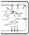
Model of STPK-dependent phosphorylation and Ppp-dependent dephosphorylation of OdhI and FtsZ in C. glutamicum.

Similar articles

Cited by

References

    1. Barthe P, Roumestand C, Canova MJ, Kremer L, Hurard C, Molle V, Cohen-Gonsaud M. Dynamic and structural characterization of a bacterial FHA protein reveals a new autoinhibition mechanism. Structure. 2009;17:568–578. - PubMed
    1. Bott M. Signal transduction by serine/threonine protein kinases in bacteria (Chapter 24) In: Krämer R, Jung K, editors. Bacterial Signaling. Weinheim: Wiley-VCH; 2010. pp. 427–447. in press.
    1. Brinkrolf K, Brune I, Tauch A. The transcriptional regulatory network of the amino acid producer Corynebacterium glutamicum. J Biotechnol. 2007;129:191–211. - PubMed
    1. Burkovski A, editor. Corynebacteria: Genomics and Molecular Biology. Norfolk, UK: Caister Academic Press; 2008.
    1. Eggeling L, Bott M, editors. Handbook of Corynebacterium glutamicum. Boca Raton, FL: CRC Press, Taylor & Francis Group.; 2005.

Publication types

MeSH terms

LinkOut - more resources