. 2009 Sep 24;35(6):830-40.
doi: 10.1016/j.molcel.2009.07.023.

Characterization of cytoplasmic caspase-2 activation by induced proximity

Affiliations

Characterization of cytoplasmic caspase-2 activation by induced proximity

Lisa Bouchier-Hayes et al. Mol Cell. .

Abstract

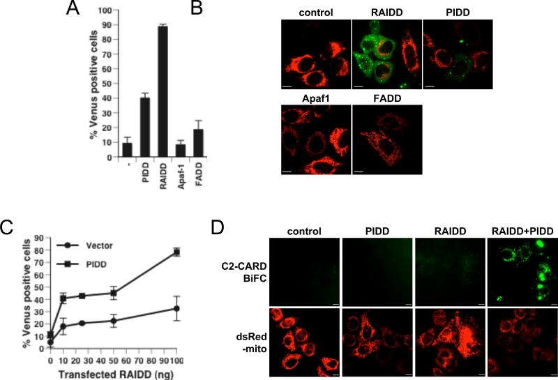
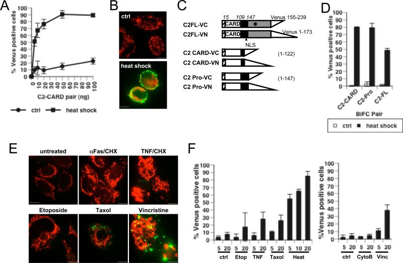
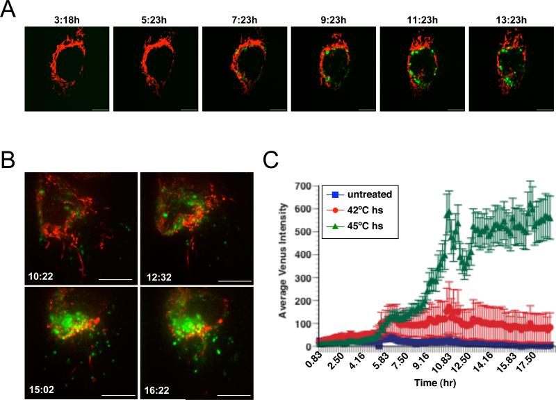
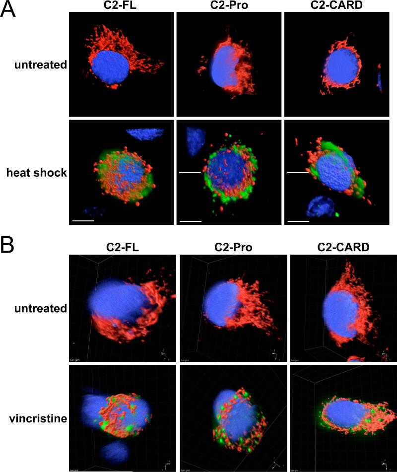
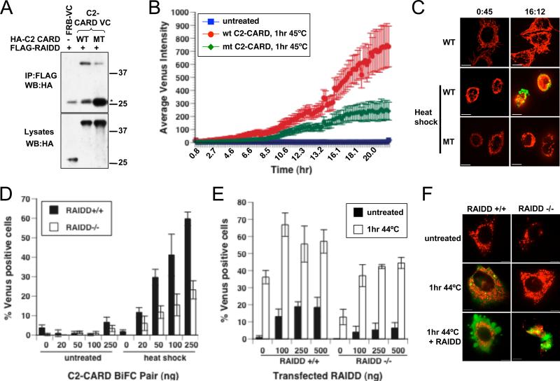
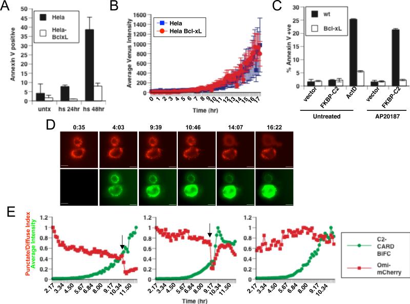
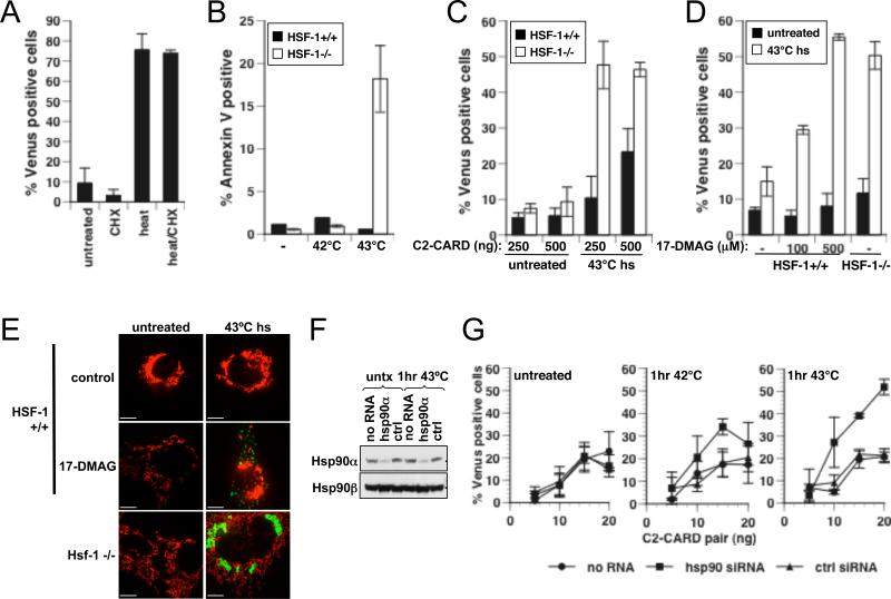
Caspase-2 is an initiator caspase activated in response to heat shock and other stressors that induce apoptosis. Activation of caspase-2 requires induced proximity resulting after recruitment to caspase-2 activation complexes such as the PIDDosome. We have adapted bimolecular fluorescence complementation (BiFC) to measure caspase-2 induced proximity in real time in single cells. Nonfluorescent fragments of the fluorescent protein Venus that can associate to reform the fluorescent complex were fused to caspase-2, allowing visualization and kinetic measurements of caspase-2 induced proximity after heat shock and other stresses. This revealed that the caspase-2 activation platform occurred in the cytosol and not in the nucleus in response to heat shock, DNA damage, cytoskeletal disruption, and other treatments. Activation, as measured by this approach, in response to heat shock was RAIDD dependent and upstream of mitochondrial outer-membrane permeabilization. Furthermore, we identify Hsp90alpha as a key negative regulator of heat shock-induced caspase-2 activation.


Figures

Figure 1
Figure 1
C2-CARD BiFC is induced by components of the PIDDosome. (A) Hela cells were transiently transfected with C2-CARD VN (20ng) and C2-CARD VC (20ng) along with 500ng of expression plasmids encoding PIDD, RAIDD, FADD or Apaf-1. All wells also received pshooter.dsRed-mito (10ng) as a reporter for transfection. 24 hr after transfection the percentage of dsRed-mito positive (red) cells that were Venus-positive (green) was determined from a minimum of 300 cells per well. Results represent triplicate counts with error bars representing standard deviation (B) Representative confocal images of cells from (A) are shown. (C) Hela cells were transiently transfected as in (A) with the indicated amounts of expression plasmid. 48 hr after transfection the percentage Venus-positive cells was determined as in (A). (D) Representative images of cells from (C) are shown. Scale bars represent 10μm.
Figure 2
Figure 2
Induced proximity of caspase-2 is induced by various stressors. (A) Hela cells were transiently transfected with the indicated amounts of each of C2-CARD VC, C2-CARD VN and pshooter.dsRed-mito (10ng). 24 hr later cells were either left untreated or heated for 1 hr at 45°C. All cells were treated with qVDOPH (20μM). Cells were assessed 24 hr after treatment for the percentage cells that were Venus-positive cells (green), determined from a minimum of 300 cells per well. Results represent triplicate counts with error bars representing standard deviation. (B) Representative images of cells from (A) are shown. (C) Schematic representation of the caspase-2 BiFC constructs. (*) indicates the active cysteine that was mutated to alanine in C2-FL. (D) Hela cells were transiently transfected with expression plasmids with C2-CARD, C2-Pro (20ng of each) or C2-FL BiFC plasmid pair (100ng of each) along with pshooter.dsRed-mito (10ng). Cells were treated and assessed as in (A). (E) Hela cells were transiently transfected with plasmids encoding C2-CARD VC (20ng), C2-CARD VN (20ng) and dsRed-mito (10ng) for 24 hr followed by treatment with anti-Fas (250ng/ml)/CHX (10μg/ml), TNF (10ng/ml)/CHX (10μg/ml), etoposide (5μM), taxol (10μg/ml) or vincristine (50μg/ml), in the presence of qVD-OPH (20μM). Representative confocal images were taken 24 hr after treatment. (F) Cells were transfected as in (E) for 24 hr followed by treatment with etoposide (Etop, 5μM), TNF (10ng/ml)/CHX (10μg/ml), taxol (Tax, 1μg/ml) or heat shock (45°C, 1 hr), cytochalasin B (CytoB, 20μg/ml) or vincristine (50μg/ml), with qVD-OPH (20μM). Cells were assessed as in (A). Scale bars represent 10μm
Figure 3
Figure 3
Kinetics of heat shock-induced C2-CARD BiFC. (A) Hela cells were transiently transfected with plasmids encoding C2-CARD VN (20ng), C2-CARD VC (20ng) and dsRed-mito (10ng). 24 hr later cells were incubated at 45°C for 1 hr plus qVD-OPH (20μM). The cells were returned to 37°C on a confocal microscope and images were taken every 6 min for 16 hr. Frames from the movie show BiFC (green) of a representative cell. (B) Hela cells were transfected and treated as in (A). Confocal images from a time-lapse with a 3 × 1.5μm z-stack are shown. (C) Hela cells were transfected as in (A). 24 hr later cells were either left untreated or heated at 42°C or 45°C for 1 hr with qVD-OPH (20μM). The cells were returned to 37°C on a confocal microscope and images were taken every 15 min for 16 hr. The average intensity of Venus was measured at each time point. Each line of the graph is an average of 21 (untreated), 35 (42°C), or 41 (45°C) individual cells and error bars represent SEM. Scale bars represent 10μm.
Figure 4
Figure 4
Heat shock-induced caspase-2 BiFC is cytoplasmic. (A) Hela cells were transiently transfected with expression plasmids encoding C2-FL VC (100ng), C2-FL VN (100ng) or with the indicated BiFC plasmid pairs (20ng of each) along with dsRed-mito (10ng). 24 hr after transfection cells were left untreated or heated at 45°C for 1 hr with qVD-OPH (20μM). Cells were assessed for BiFC (green) 24 hr after treatment. (B) Hela cells were transfected as in (A) and 24 hr after transfection cells were either left untreated or treated with vincristine (25μg/ml) with qVD-OPH (20μM). Cells were assessed 24 hr after treatment. Nuclei were stained with Draq5 (blue). Images are 3D isosurface rendering reconstructions composed from 0.1μm (A) or 0.2μm (B) serial confocal images through the z-plane of the cell. Scale bars represent 10μm.
Figure 5
Figure 5
Heat shock-induced C2-CARD BiFC requires RAIDD. (A) D83A/E87A C2-CARD mutant binds RAIDD with less efficiency than wild type. 293T cells were transiently transfected with plasmids encoding FLAG-RAIDD and HA-C2-CARD VC (WT), HA-C2-CARD VC D83A/E87A (MT) or a control protein (HAFRB-VC). FLAG-RAIDD was immunoprecipitated from cell lysates with anti-FLAG-agarose and blotted for the presence of C2-CARD with an anti-HA antibody. (*) indicates immunoglobulin light chain. (B) Hela cells were transiently transfected with plasmids encoding C2-CARD VN (20ng) and C2-CARD VC (20ng) or the D83A/E87A mutant C2-CARD BiFC pair (20ng of each) along with pshooter.dsRed-mito (10ng) and 24 hr later cells were incubated at 45°C for 1 hr with qVD-OPH (20μM). The cells were returned to 37°C on a confocal microscope and images were taken every 10 min for 20 hr. Each line of the graph represents the average intensity of Venus of 15 (untreated), 29 (WT C2-CARD, 1 hr 45°C), or 22 (MT C2-CARD, 1 hr 45°C) individual cells. Error bars represent SEM. (C) Representative images from the time-lapse are shown. (D) RAIDD−/− MEF and RAIDD+/+ MEF from littermate embryos were transiently transfected with the indicated amounts of expression plasmids encoding C2-CARD pair and dsRed-mito (10ng). 24 hr post-transfection cells were left untreated or heat shocked for 1 hr at 44°C with qVD-OPH (20μM). The percentage Venus-positive cells was determined at 24 hr from a minimum of 300 cells per well. Results represent triplicate counts with error bars representing standard deviation. (E) RAIDD−/− and RAIDD+/+ MEF were transiently transfected with the plasmids encoding the C2-CARD Venus pair (250ng of each) and dsRedmito (10ng) with the indicated amounts of an expression plasmid encoding RAIDD. 24 hr post-transfection cells were left untreated or heat shocked for 1 hr at 44°C with qVD-OPH (20μM). The percentage Venus-positive cells was determined as in (D). (F) Representative images of cells from (E) are shown. Scale bars represent 10μm.
Figure 6
Figure 6
Caspase-2 is activated by induced proximity upstream of mitochondria to induce apoptosis. (A) Hela cells or Hela cells stably expressing Bcl-xL were heated for 1 hr at 45°C. Apoptosis was assessed by flow cytometry for Annexin V binding at the indicated times. Error bars represent standard deviation. (B) Hela or Hela.Bcl-xL cells were transiently transfected with plasmids encoding C2-CARD pair (20ng of each) along with dsRed-mito (10ng) and 24 hr later were heated at 45°C for 1 hr with qVD-OPH (20μM). Cells were returned to 37°C on a confocal microscope and images were taken every 15 min for 16 hr. The average intensity of Venus was measured at each time point. Results are an average of 21 (Hela) and 21 (Hela-Bcl-xL) individual cells. Error bars represent SEM. (C) Hela or Hela.Bcl-xL cells that were stably expressing vector or caspase-2 fused to FKBP dimerization domain were left untreated, treated with ActD (1μM) or the dimerization drug (AP20187, 200nM) for 24 hr. Apoptosis was assessed as in (A). (D) Hela cells stably expressing Omi-mCherry were transiently transfected with expression plasmids encoding C2-CARD VN (20ng) and C2-CARD VC (20ng). 24 hr later cells were incubated at 45°C for 1 hr. The cells were returned to 37°C on a fluorescent microscope and images were taken every 6 min for 16 hr. Frames from the movie show representative cells undergoing BiFC (green) prior to release of Omi-mCherry from the mitochondria (red). Scale bars represent 10μm. (E) Graphs of three representative cells from a movie are shown. Each point on Omi-mCherry graph (red squares) represents the punctate/diffuse index and is scaled and aligned to each point on the caspase-2 BiFC graph (green circles) that represents the average intensity of Venus in the cell at 10 min intervals. Arrows show the point of onset of MOMP.
Figure 7
Figure 7
Hsp90α blocks caspase-2 activation. (A) Hela cells were transiently transfected with C2-CARD VC (20ng), C2-CARD VN (20ng) and dsRed-mito (10ng). 24 hr later cells were left untreated or heated for 1 hr at 45°C for 24 hr in the presence or absence of cycloheximide (CHX, 10μg/ml) with qVD-OPH (20μM). Cells were assessed 24 hr later for the percentage of Venus-positive cells, determined from a minimum of 300 cells per well. Results represent triplicate counts with error bars representing standard deviation. (B) HSF-1+/+ or HSF-1−/− MEF from littermate embryos were heat shocked at the indicated temperatures for 1 hr. 24 hr later apoptosis was assessed by flow cytometry for Annexin V binding. (C) HSF-1+/+ and HSF-1−/− MEF were transiently transfected with the indicated amounts of expression plasmids encoding the C2-CARD Venus pair and dsRed-mito (10ng). 24 hr post-transfection cells were left untreated or heat shocked for 1 hr at 43°C with qVD-OPH (20μM). The percentage of Venus-positive cells was determined at 24 hr as in (A). (D) HSF-1+/+ and HSF-1−/− MEF were transiently transfected with the plasmids encoding the C2-CARD Venus pair (250ng of each) and dsRed-mito (10ng). 24 hr post-transfection cells were left untreated or heat shocked for 1 hr at 43°C with qVDOPH (20μM), with or without 17-DMAG as indicated. The percentage Venus-positive cells was determined at 24 hr as in (A). (E) Representative images of cells from (D) are shown. Scale bars represent 10μm. (F) Hela cells were transfected with siRNA targeting human Hsp90α (100nM) or control siRNA (100nM). 24 hr post-transfection cells were left untreated or heat shocked for 1 hr at 43°C. Lysates were made at 72 hrs and assessed for Hsp90α and β expression. (G) Hela cells were transiently transfected with the indicated amounts of expression plasmids encoding the C2-CARD Venus pair and dsRedmito (10ng) along with the indicated siRNA (100nM). 24 hr post-transfection cells were left untreated or heat shocked for 1 hr at 42°C or 1hr at 43°C with qVD-OPH (20μM). The percentage Venus-positive cells was determined at 24 hr as in (A).

Comment in

  • A cut above the other caspases.
    Andersen JL, Kornbluth S. Andersen JL, et al. Mol Cell. 2009 Sep 24;35(6):733-4. doi: 10.1016/j.molcel.2009.09.001. Mol Cell. 2009. PMID: 19782021

Similar articles

Cited by

References

    1. Arlander S, Eapen A, Vroman B, McDonald R, Toft D, Karnitz L. Hsp90 inhibition depletes Chk1 and sensitizes tumor cells to replication stress. J Biol Chem. 2003;278:52572–52577. - PubMed
    1. Baliga BC, Colussi PA, Read SH, Dias MM, Jans DA, Kumar S. Role of prodomain in importin-mediated nuclear localization and activation of caspase-2. J Biol Chem. 2003;278:4899–4905. - PubMed
    1. Baliga BC, Read SH, Kumar S. The biochemical mechanism of caspase-2 activation. Cell Death Differ. 2004;11:1234–1241. - PubMed
    1. Baptiste-Okoh N, Barsotti A, Prives C. A role for caspase 2 and PIDD in the process of p53-mediated apoptosis. Proc Natl Acad Sci U S A. 2008;105:1937–1942. - PMC - PubMed
    1. Beere HM, Wolf BB, Cain K, Mosser DD, Mahboubi A, Kuwana T, Tailor P, Morimoto RI, Cohen GM, Green DR. Heat-shock protein 70 inhibits apoptosis by preventing recruitment of procaspase-9 to the Apaf-1 apoptosome. Nat Cell Biol. 2000;2:469–475. - PubMed

Publication types

MeSH terms