Evolutionary dynamics of GII.4 noroviruses over a 34-year period
- PMID: 19759138
- PMCID: PMC2772697
- DOI: 10.1128/JVI.00864-09
Evolutionary dynamics of GII.4 noroviruses over a 34-year period
Abstract
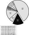
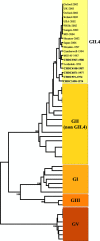
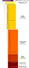
Noroviruses are a major cause of epidemic gastroenteritis in children and adults, and GII.4 has been the predominant genotype since its first documented occurrence in 1987. This study examined the evolutionary dynamics of GII.4 noroviruses over more than three decades to investigate possible mechanisms by which these viruses have emerged to become predominant. Stool samples (n = 5,424) from children hospitalized at the Children's Hospital in Washington, DC, between 1974 and 1991 were screened for the presence of noroviruses by a custom multiplex real-time reverse transcription-PCR. The complete genome sequences of five GII.4 noroviruses (three of which predate 1987 by more than a decade) in this archival collection were determined and compared to the sequences of contemporary strains. Evolutionary analysis determined that the GII.4 VP1 capsid gene evolved at a rate of 4.3 x 10(-3) nucleotide substitutions/site/year. Only six sites in the VP1 capsid protein were found to evolve under positive selection, most of them located in the shell domain. No unique mutations were observed in or around the two histoblood group antigen (HBGA) binding sites in the P region, indicating that this site has been conserved since the 1970s. The VP1 proteins from the 1974 to 1977 noroviruses contained a unique sequence of four consecutive amino acids in the P2 region, which formed an exposed protrusion on the modeled capsid structure. This protrusion and other observed sequence variations did not affect the HBGA binding profiles of recombinant virus-like particles derived from representative 1974 and 1977 noroviruses compared with more recent noroviruses. Our analysis of archival GII.4 norovirus strains suggests that this genotype has been circulating for more than three decades and provides new ancestral strain sequences for the analysis of GII.4 evolution.
Figures


Similar articles
-
Comparative evolution of GII.3 and GII.4 norovirus over a 31-year period.J Virol. 2011 Sep;85(17):8656-66. doi: 10.1128/JVI.00472-11. Epub 2011 Jun 29. J Virol. 2011. PMID: 21715504 Free PMC article.
-
Mechanisms of GII.4 norovirus persistence in human populations.PLoS Med. 2008 Feb;5(2):e31. doi: 10.1371/journal.pmed.0050031. PLoS Med. 2008. PMID: 18271619 Free PMC article.
-
Molecular epidemiology of noroviruses detected in Nepalese children with acute diarrhea between 2005 and 2011: increase and predominance of minor genotype GII.13.Infect Genet Evol. 2015 Mar;30:27-36. doi: 10.1016/j.meegid.2014.12.003. Epub 2014 Dec 9. Infect Genet Evol. 2015. PMID: 25497351
-
The resurgence of the norovirus GII.4 variant associated with sporadic gastroenteritis in the post-GII.17 period in South China, 2015 to 2017.BMC Infect Dis. 2019 Aug 6;19(1):696. doi: 10.1186/s12879-019-4331-6. BMC Infect Dis. 2019. PMID: 31387542 Free PMC article.
-
Molecular and Genetics-Based Systems for Tracing the Evolution and Exploring the Mechanisms of Human Norovirus Infections.Int J Mol Sci. 2023 May 22;24(10):9093. doi: 10.3390/ijms24109093. Int J Mol Sci. 2023. PMID: 37240438 Free PMC article. Review.
Cited by
-
Antigenic cartography reveals complexities of genetic determinants that lead to antigenic differences among pandemic GII.4 noroviruses.Proc Natl Acad Sci U S A. 2021 Mar 16;118(11):e2015874118. doi: 10.1073/pnas.2015874118. Proc Natl Acad Sci U S A. 2021. PMID: 33836574 Free PMC article.
-
Immune Imprinting Drives Human Norovirus Potential for Global Spread.mBio. 2022 Oct 26;13(5):e0186122. doi: 10.1128/mbio.01861-22. Epub 2022 Sep 14. mBio. 2022. PMID: 36102514 Free PMC article.
-
Diagnostic accuracy and analytical sensitivity of IDEIA Norovirus assay for routine screening of human norovirus.J Clin Microbiol. 2010 Aug;48(8):2770-8. doi: 10.1128/JCM.00654-10. Epub 2010 Jun 16. J Clin Microbiol. 2010. PMID: 20554813 Free PMC article.
-
Evidence for recombination between pandemic GII.4 norovirus strains New Orleans 2009 and Sydney 2012.J Clin Microbiol. 2013 Nov;51(11):3855-7. doi: 10.1128/JCM.01847-13. Epub 2013 Aug 21. J Clin Microbiol. 2013. PMID: 23966499 Free PMC article.
-
Seroprevalence of norovirus genogroup IV antibodies among humans, Italy, 2010-2011.Emerg Infect Dis. 2014 Nov;20(11):1828-32. doi: 10.3201/eid2011.131601. Emerg Infect Dis. 2014. PMID: 25340375 Free PMC article.
References
-
- Biebricher, C. K., and M. Eigen. 2006. What is a quasispecies? Curr. Top. Microbiol. Immunol. 299:1-31. - PubMed
Publication types
MeSH terms
Associated data
- Actions
- Actions
- Actions
- Actions
- Actions
Grants and funding
LinkOut - more resources
Full Text Sources
Other Literature Sources
Medical

