

A Wnt kinase network alters nuclear localization of TCF-1 in colon cancer
- PMID: 19749792
- PMCID: PMC2787979
- DOI: 10.1038/onc.2009.271
A Wnt kinase network alters nuclear localization of TCF-1 in colon cancer
Abstract
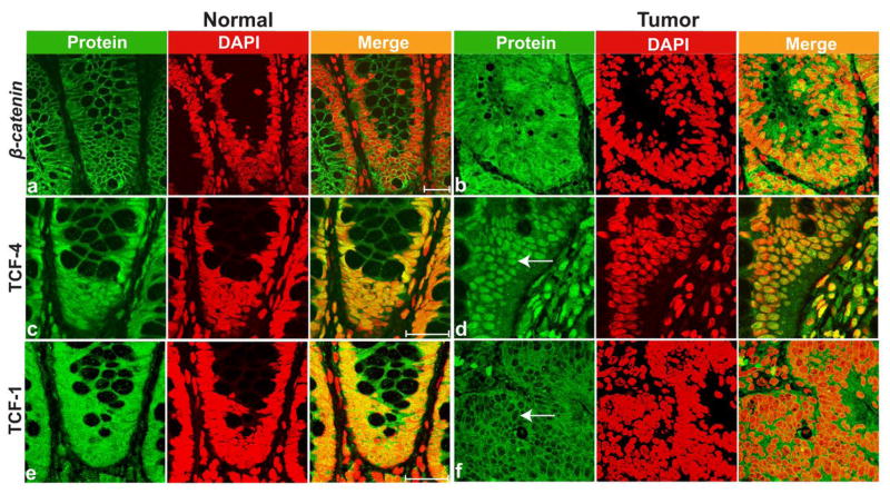
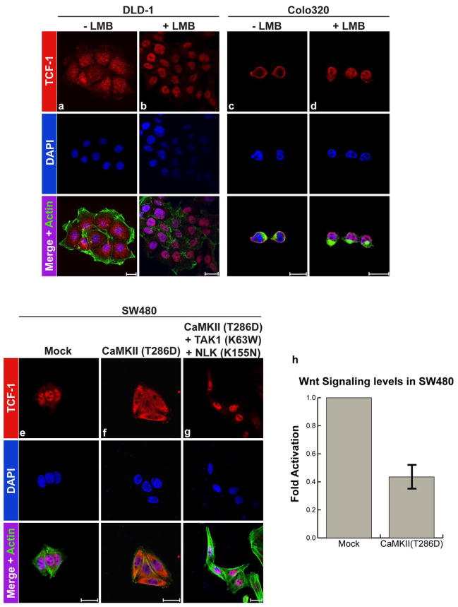
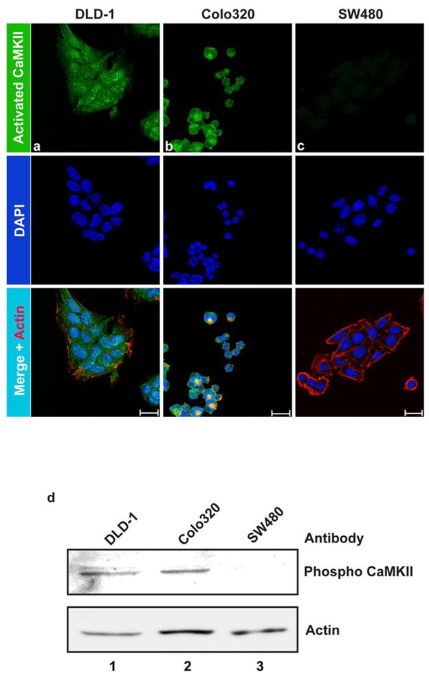
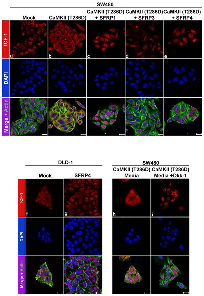
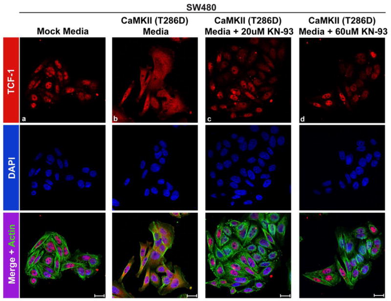
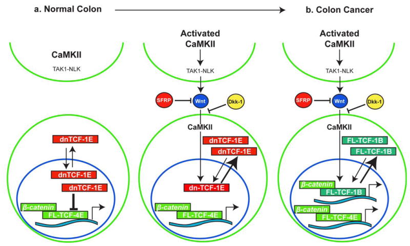
Constitutive activation of the Wnt/beta-catenin pathway has been implicated as the primary cause of colon cancer. However, the major transducers of Wnt signaling in the intestine, T-cell factor 1 (TCF-1) and TCF-4, have opposing functions. Knockout of TCF-4 suppresses growth and maintenance of crypt stem cells, whereas knockout of TCF-1 leads to adenomas. These phenotypes suggest that TCF-4 is Wnt-promoting, whereas TCF-1 acts like a tumor suppressor. Our study of TCF expression in human colon crypts reveals a mechanistic basis for this paradox. In normal colon cells, a dominant-negative isoform of TCF-1 (dnTCF-1) is expressed that is equally distributed between nuclear and cytoplasmic compartments. In colon cancer cells, TCF-1 is predominantly cytoplasmic. Localization is because of active nuclear export and is directed by an autocrine-acting Wnt ligand that requires Ca2+/calmodulin-dependent kinase II (CaMKII) activity for secretion and a downstream step in the export pathway. TCF-4 remains nuclear; its unopposed activity is accompanied by downregulation of dnTCF-1 and increased expression of full-length isoforms. Thus, the dnTCF-1 and TCF-4 balance is corrupted in cancer by two mechanisms, a Wnt/CaMKII kinase signal for nuclear export and decreased dnTCF-1 expression. We propose that dnTCF-1 provides homeostatic regulation of Wnt signaling and growth in normal colon, and the alterations in nuclear export and promoter usage contribute to aberrant Wnt activity in colon cancer.
Conflict of interest statement
The authors declare no conflict of interest.
Figures


Similar articles
-
Aberrant nuclear localization of EBP50 promotes colorectal carcinogenesis in xenotransplanted mice by modulating TCF-1 and β-catenin interactions.J Clin Invest. 2012 May;122(5):1881-94. doi: 10.1172/JCI45661. Epub 2012 Apr 2. J Clin Invest. 2012. PMID: 22466651 Free PMC article.
-
Phospholipase D1 drives a positive feedback loop to reinforce the Wnt/beta-catenin/TCF signaling axis.Cancer Res. 2010 May 15;70(10):4233-42. doi: 10.1158/0008-5472.CAN-09-3470. Epub 2010 May 4. Cancer Res. 2010. PMID: 20442281
-
NGX6 inhibits cell invasion and adhesion through suppression of Wnt/beta-catenin signal pathway in colon cancer.Acta Biochim Biophys Sin (Shanghai). 2010 Jul;42(7):450-6. doi: 10.1093/abbs/gmq049. Epub 2010 Jun 13. Acta Biochim Biophys Sin (Shanghai). 2010. PMID: 20705583 Free PMC article.
-
Lymphoid enhancer factor/T cell factor expression in colorectal cancer.Cancer Metastasis Rev. 2004 Jan-Jun;23(1-2):41-52. doi: 10.1023/a:1025858928620. Cancer Metastasis Rev. 2004. PMID: 15000148 Review.
-
The Yin-Yang of TCF/beta-catenin signaling.Adv Cancer Res. 2000;77:1-24. doi: 10.1016/s0065-230x(08)60783-6. Adv Cancer Res. 2000. PMID: 10549354 Review.
Cited by
-
Horizontal gene transfers with or without cell fusions in all categories of the living matter.Adv Exp Med Biol. 2011;714:5-89. doi: 10.1007/978-94-007-0782-5_2. Adv Exp Med Biol. 2011. PMID: 21506007 Free PMC article. Review.
-
SKLB-677, an FLT3 and Wnt/β-catenin signaling inhibitor, displays potent activity in models of FLT3-driven AML.Sci Rep. 2015 Oct 26;5:15646. doi: 10.1038/srep15646. Sci Rep. 2015. PMID: 26497577 Free PMC article.
-
A role for YY1 in repression of dominant negative LEF-1 expression in colon cancer.Nucleic Acids Res. 2010 Oct;38(19):6375-88. doi: 10.1093/nar/gkq492. Epub 2010 Jun 4. Nucleic Acids Res. 2010. PMID: 20525792 Free PMC article.
-
Dishevelled-1 DIX and PDZ domain lysine residues regulate oncogenic Wnt signaling.Oncotarget. 2021 Oct 26;12(22):2234-2251. doi: 10.18632/oncotarget.28089. eCollection 2021 Oct 26. Oncotarget. 2021. PMID: 34733415 Free PMC article.
-
The emerging role of CaMKII in cancer.Oncotarget. 2015 May 20;6(14):11725-34. doi: 10.18632/oncotarget.3955. Oncotarget. 2015. PMID: 25961153 Free PMC article. Review.
References
-
- Arce L, Yokoyama NN, Waterman ML. Diversity of LEF/TCF action in development and disease. Oncogene. 2006;25:7492–504. - PubMed
-
- Bienz M, Clevers H. Linking colorectal cancer to Wnt signaling. Cell. 2000;103:311–20. - PubMed
-
- Bovolenta P, Esteve P, Ruiz JM, Cisneros E, Lopez-Rios J. Beyond Wnt inhibition: new functions of secreted Frizzled-related proteins in development and disease. J Cell Sci. 2008;121:737–46. - PubMed
Publication types
MeSH terms
Substances
Grants and funding
- R01 HD36049/HD/NICHD NIH HHS/United States
- R01 HD36081/HD/NICHD NIH HHS/United States
- R01 CA096878-07/CA/NCI NIH HHS/United States
- R01 CA108697-01A1/CA/NCI NIH HHS/United States
- CA-82450/CA/NCI NIH HHS/United States
- R21DK071591/DK/NIDDK NIH HHS/United States
- K24 CA082450-08/CA/NCI NIH HHS/United States
- R21 DK071591-02/DK/NIDDK NIH HHS/United States
- R01 CA108697-04/CA/NCI NIH HHS/United States
- K24 CA082450/CA/NCI NIH HHS/United States
- R01 CA108697/CA/NCI NIH HHS/United States
- CA108697/CA/NCI NIH HHS/United States
- CA-62203/CA/NCI NIH HHS/United States
- R01 CA096878-04/CA/NCI NIH HHS/United States
- R01 CA108697-05/CA/NCI NIH HHS/United States
- R01 CA096878-05/CA/NCI NIH HHS/United States
- T32 CA113265/CA/NCI NIH HHS/United States
- R01 CA096878/CA/NCI NIH HHS/United States
- CA096878/CA/NCI NIH HHS/United States
- R01 CA108697-03/CA/NCI NIH HHS/United States
- K24 CA082450-07/CA/NCI NIH HHS/United States
- R01 HD036081/HD/NICHD NIH HHS/United States
- R01 CA096878-06/CA/NCI NIH HHS/United States
- R01 CA096878-01A1/CA/NCI NIH HHS/United States
- P30 CA062203/CA/NCI NIH HHS/United States
- R01 CA108697-02/CA/NCI NIH HHS/United States
- R21 DK071591/DK/NIDDK NIH HHS/United States
- R01 CA096878-03/CA/NCI NIH HHS/United States
- R01 CA096878-02/CA/NCI NIH HHS/United States
