. 2009 Nov;89(11):1275-90.
doi: 10.1038/labinvest.2009.93. Epub 2009 Sep 7.

Curcumin eliminates oxidized LDL roles in activating hepatic stellate cells by suppressing gene expression of lectin-like oxidized LDL receptor-1

Affiliations

Curcumin eliminates oxidized LDL roles in activating hepatic stellate cells by suppressing gene expression of lectin-like oxidized LDL receptor-1

Qiaohua Kang et al. Lab Invest. 2009 Nov.

Abstract

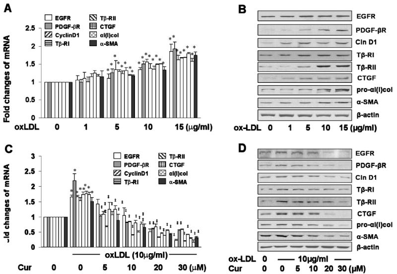
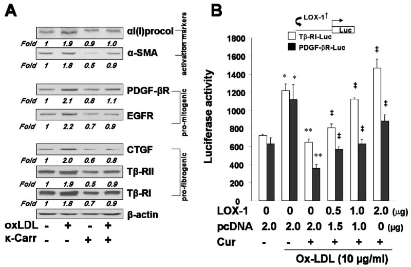
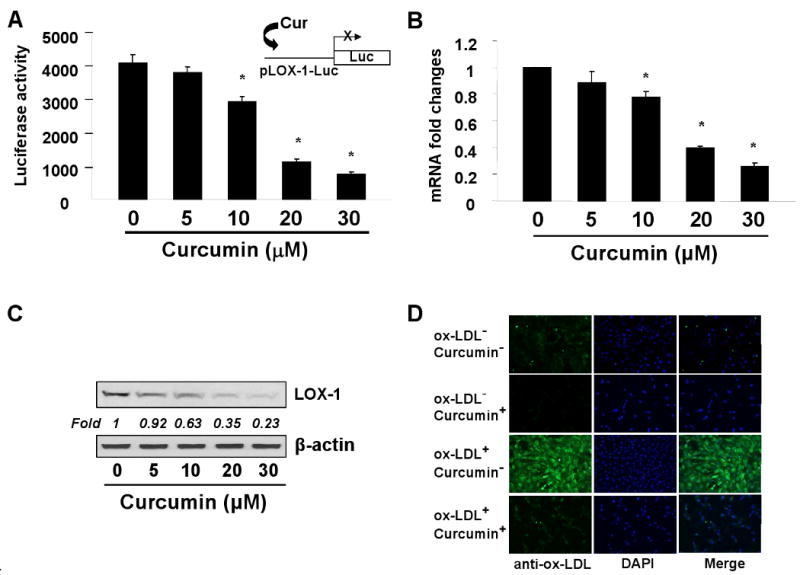
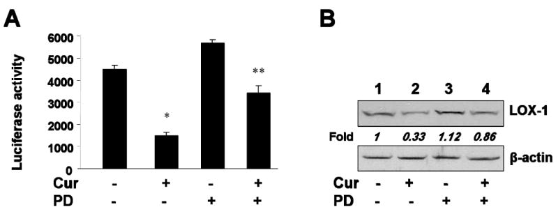
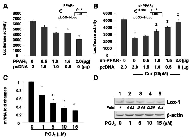
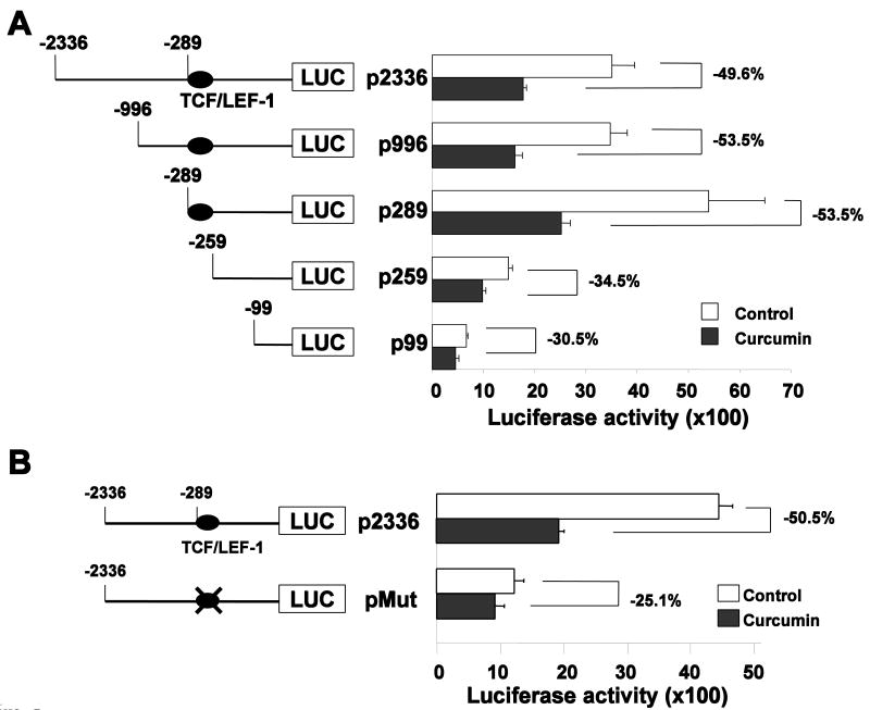
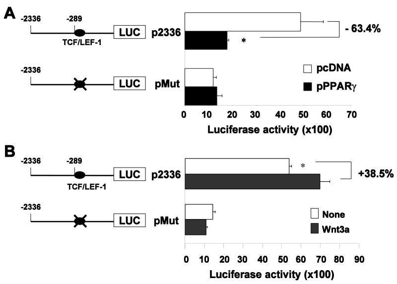
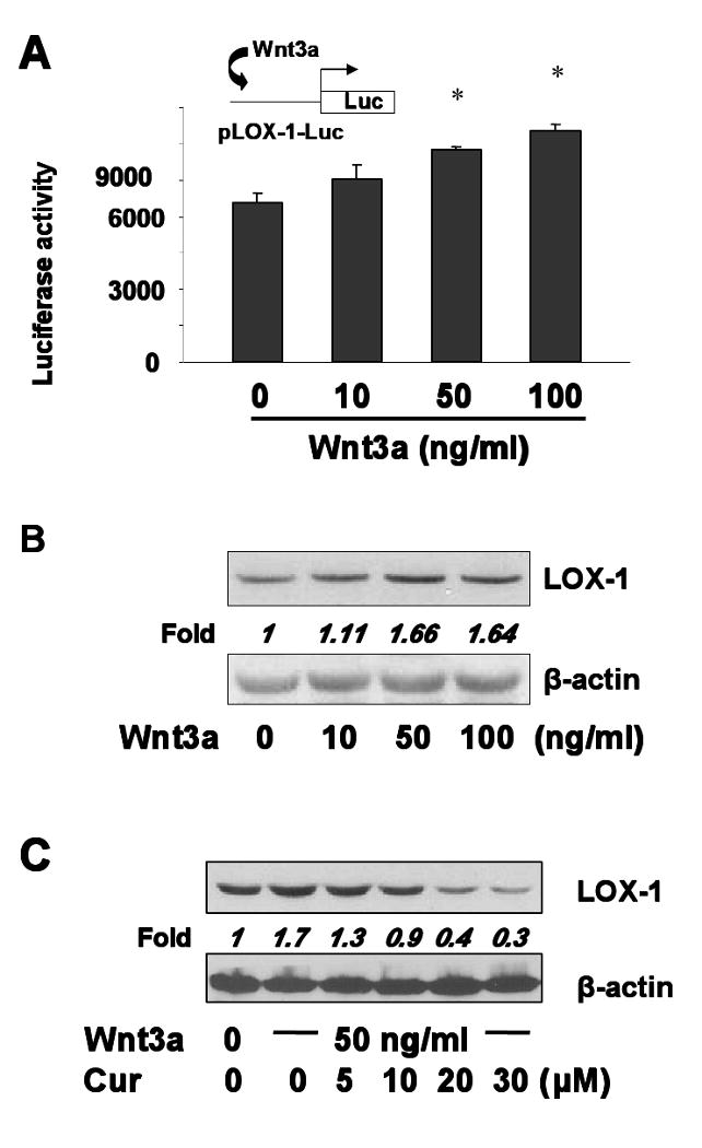
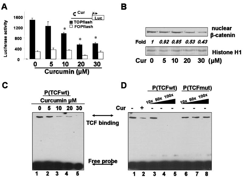
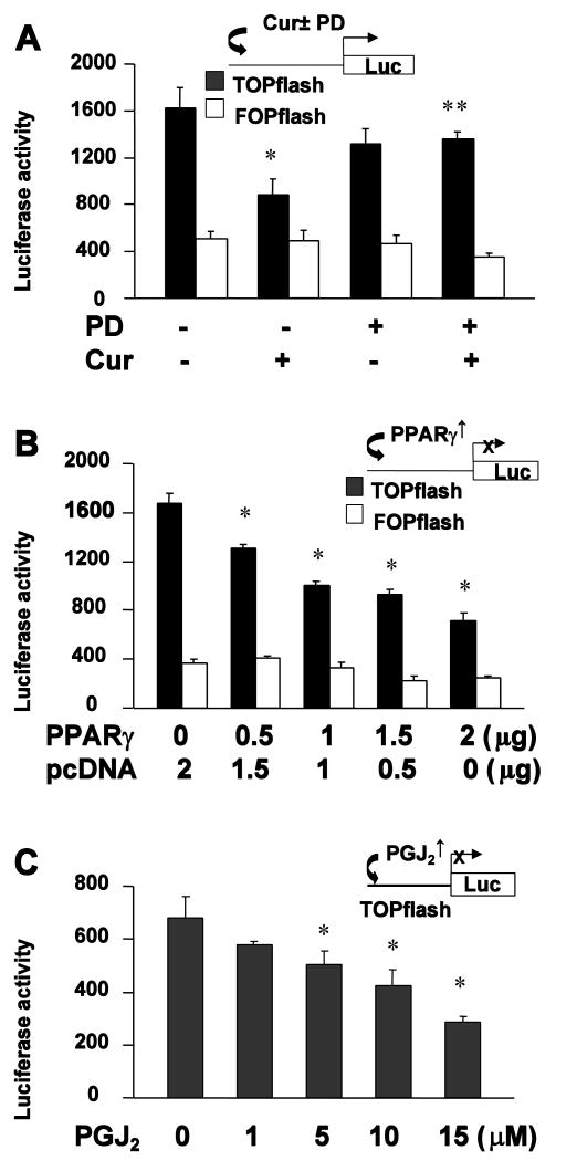
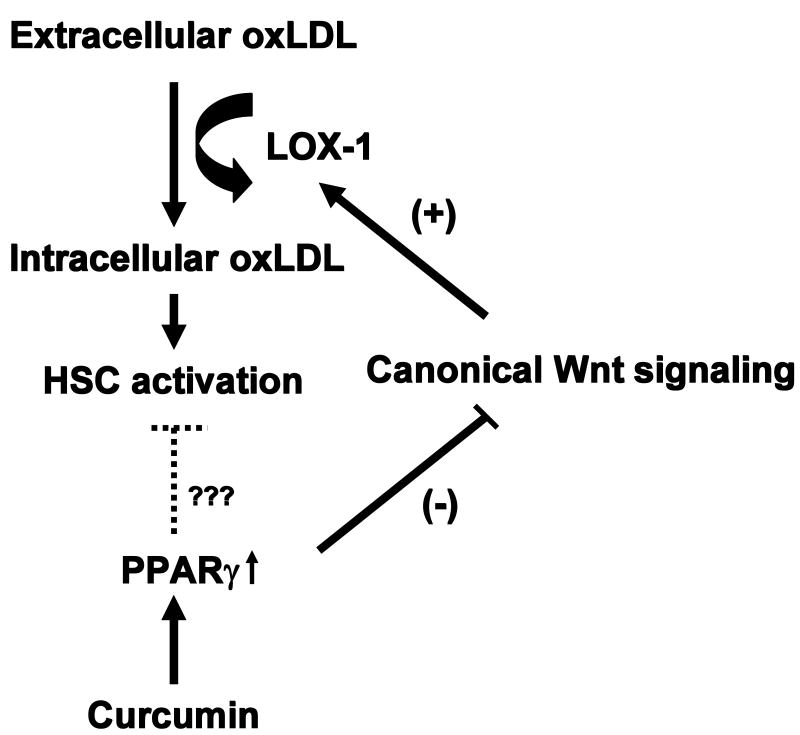
Type II diabetes mellitus (T2DM) is often accompanied by non-alcoholic steatohepatitis (NASH) and associated with hypercholesterolemia, that is, increased levels of plasma low-density lipoprotein (LDL) and oxidized LDL (ox-LDL). Approximately one-third of NASH develops hepatic fibrosis. The role of hypercholesterolemia in T2DM and NASH-associated hepatic fibrogenesis remains obscure. We previously reported that the phytochemical curcumin inhibited the activation of hepatic stellate cells (HSCs), the major effector cells during hepatic fibrogenesis, and protected the liver from fibrogenesis in vitro and in vivo. The aims of this study are to evaluate the role of ox-LDL in activation of HSCs, to assess curcumin effects on eliminating the role of ox-LDL, and to further explore the underlying mechanisms. In this report, we observe that ox-LDL alters the expression of genes closely relevant to HSC activation, which is eliminated by curcumin. Curcumin suppresses gene expression of lectin-like oxidized LDL receptor-1 (LOX-1), leading to the blockade of the transport of extracellular ox-LDL into cells. This suppressive effect of curcumin results from the interruption of Wnt signaling and the activation of peroxisome proliferator-activated receptor-gamma (PPARgamma). In conclusion, these results support our initial hypothesis and demonstrate that ox-LDL stimulates HSC activation, which is eliminated by curcumin by suppressing lox-1 expression by interrupting Wnt signaling and stimulating PPARgamma activity. These results provide novel insights into the role of ox-LDL in T2DM and NASH-associated hepatic fibrogenesis and mechanisms by which curcumin suppresses ox-LDL-induced HSC activation, as well as the implication of curcumin in the treatment of T2DM and NASH-associated hepatic fibrosis.


Conflict of interest statement

Disclosures Section: The authors have nothing to disclose.

Figures

Figure 1
Figure 1. ox-LDL stimulates expression of genes relevant to HSC activation, which is dose-dependently eliminated by curcumin
Serum-starved HSCs were stimulated with ox-LDL at indicated concentrations in serum-depleted media for 24 hr in the absence (A & B) or presence (C & D) of curcumin at 0-30 μM. Total RNA and whole cell protein extracts were respectively prepared from the cells for real-time PCR (A & C) and Western blotting analyses (B & D). *P<0.05 versus cells without treatment (n=3) (1st corresponding column on the left); ‡P<0.05 versus cells treated with ox-LDL only (the 2nd corresponding column). β-actin was used in Western blotting analyses as an internal control for equal protein loading. Representatives were from three independent experiments.
Figure 2
Figure 2. LOX-1 plays a mediating role in ox-LDL-induced HSC activation and in the curcumin elimination of the stimulatory effect of ox-LDL
(A). serum-starved HSCs were pre-treated with or without the LOX-1 antagonist κ-carrageenan at 250 μg/ml for 1 hr prior to the stimulation with or without ox-LDL (10 μg/ml) for additional 24 hr. Whole cell extracts were prepared for Western blotting analyses of genes relevant to HSC activation. β-actin was used in Western blotting analyses as an internal control for equal protein loading. Representatives were from three independent experiments. Italic numbers beneath blots were fold changes in the densities of the bands compared to the control without treatment in the blot (n=3), after normalization with the internal invariable control β-actin. Because of the limited space, standard deviations were not presented. (B). HSCs were co-transfected with LOX-1 cDNA expression plasmid pLOX-1 at indicated doses plus pPDGF-βR-Luc, or pTβ-RI-Luc. A total of 4.5 μg of plasmid DNA per well was used for co-transfection of HSCs in 6-well culture plates. It included 2 μg of pPDGF-βR-Luc or pTβ-RI-Luc, 0.5 μg of pSV-β-gal, and 2.0 μg of pLOX-1 at indicated doses plus the empty vector pcDNA. The latter was used to ensure an equal amount of total DNA in transfection assays. After recovery, cells were stimulated with or without ox-LDL (10 μg/ml) in the presence or absence of curcumin (20 μM) in serum-depleted media for 24 hr. Luciferase activities were expressed as relative units after β-galactosidase normalization (means ± s.d.; n≥6). *P<0.05 versus cells without treatment (the corresponding 1st column); **P<0.05 versus cells treated only with ox-LDL (the corresponding 2nd column); ‡P<0.05 versus cells treated with ox-LDL plus curcumin (the corresponding 3rd column). The floating schema denoted the luciferase reporter construct pPDGF-βR-Luc or pTβ-RI-Luc in use and forced expression of LOX-1 cDNA in the system.
Figure 3
Figure 3. Curcumin suppresses lox-1 expression in activated HSCs in vitro
(A). Luciferase activity assays of cells transiently transfected with the lox-1 promoter luciferase reporter pLOX-1-Luc, and treated with curcumin after transfection. Luciferase activities were expressed as relative units after β-galactosidase normalization (means ± s.d.; n≥6). *P<0.05 versus cells without treatment (the first column on the left). The floating schema denoted the luciferase reporter construct pLOX-1-Luc in use and the application of curcumin to the system. (B). Real-time PCR analyses of the steady-state levels of LOX-1 mRNA in cells treated with curcumin. mRNA fold changes were calculated as stated in Materials and Methods. Values were expressed as means ± s.d. (n≥3). *P<0.05 versus the untreated control (the first column on the left); (C). Western blotting analyses of LOX-1 in cells treated with curcumin at indicated concentrations. β-actin was used as an internal control for equal protein loading. Representative was from three independent experiments. Italic numbers beneath the blot were fold changes in the densities of the bands compared to the control without treatment in the blot (n=3), after normalization with the internal invariable control β-actin. (D). Immuno-staining of cultured HSCs treated with or without ox-LDL (10 μg/ml) plus or minus curcumin (10 μM) for evaluating the impact of curcumin on the abundance of intracellular ox-LDL. DAPI in mounting solution was used for staining nuclei. Representative views were presented.
Figure 4
Figure 4. The activation of PPARγ is required for curcumin to inhibit lox-1 expression in activated HSCs in vitro
Semi-confluent HSCs were pretreated with or without the PPARγ antagonist PD68235 (20μM) for 30 min prior to the addition of curcumin at 20μM for additional 24 hr. (A). Luciferase activity assays of HSCs transfected with the plasmid pLOX-1-Luc, followed by the above treatment. Luciferase activities were expressed as relative units after β-galactosidase normalization (means ± s.d.; n≥6). *P<0.05 versus cells with no treatment (the first column on the left). **P<0.05 versus cells treated with curcumin only (the second column). (B). Western blotting analyses of LOX-1 in cultured HSCs with the above treatment. β-actin was used as an internal control for equal protein loading. Representative was from three independent experiments. Italic numbers beneath the blot were fold changes in the densities of the bands compared to the control without treatment in the blot (n=3), after normalization with β-actin. Because of the limited space, standard deviations were not presented.
Figure 5
Figure 5. The activation of PPARγ results in the suppression of lox-1 expression in activated HSCs in vitro
(A) and (B): Semi-confluent HSCs were co-transfected with the plasmid pLOX-1-Luc and the plasmid pPPARγ (A), or its mutant counterpart pdn-PPARγ (B), at indicated doses. After recovery, cells were treated with or without curcumin (20μM) for 24 hr. Luciferase activities were expressed as relative units after β-galactosidase normalization (means ± s.d.; n≥6). *P<0.05 versus cells without pPPARγ, or pdn-PPARγ, (the first column on the left). ‡P<0.05 versus cells transfected with no pdn-PPARγ and treated with curcumin only (the second column). The floating schema denoted pLOX-1-Luc in use and co-transfected plasmid pPPARγ or pdn-PPARγ ± curcumin in the system. (C) and (D): Serum-starved HSCs were treated with the natural PPARγ agonist PGJ2 at 0-15μM for 24 hr. (C). Real-time PCR assays of LOX-1 mRNA. mRNA fold changes were expressed as means ± s.d. (n≥3). *P<0.05 versus the untreated control (the first column on the left); (D). Western blotting analyses of LOX-1. β-actin was used as an internal control for equal protein loading. Representative was from three independent experiments. Italic numbers beneath the blot were fold changes in the densities of the bands compared to the control without treatment in the blot (n=3), after normalization with β-actin. Because of the limited space, standard deviations were not presented.
Figure 6
Figure 6. Identification of putative curcumin response elements in the lox-1 promoter in HSCs
Semi-confluent HSCs were transiently transfected with a group of LOX-1 promoter luciferase reporter plasmids. A total of 3.5μg of plasmid DNA per well was added to HSCs in 6-well culture plates. It included 3μg of a LOX-1 promoter reporter plasmid and 0.5μg of pSV-β-gal. After recovery, cells were treated with or without curcumin at 20μM for 24 hr. Luciferase activities were expressed as relative units after β-galactosidase normalization (means ± s.d.; n≥6). The percentages indicated the reduction in luciferase activities in cells treated with curcumin, compared to those in corresponding cells without curcumin treatment. (A). Luciferase activity assays of cells transfected with a group of plasmids with various lengths of the lox-1 5′-flanking promoter region. (B) Luciferase activity assays of HSCs transfected with the wild-type pLOX-1-Luc, or its mutant counterpart pLOX-1(mut)-Luc with site-directed mutations in the putative TCF/LEF-1 binding site.
Figure 7
Figure 7. The TCF/LEF-1 binding site in the lox-1 promoter mediates Wnt signaling and PPARγ in the regulation of the lox-1 promoter activity in HSCs
(A). Luciferase activity assays of HSCs co-transfected with pLOX-1-Luc, or pLOX-1(mut)-Luc, plus pPPARγ, or the empty control vector pcDNA. A total of 4.5μg of plasmid DNA per well was used for co-transfection of HSCs in 6-well culture plates. It included 2μg of pLOX-1-Luc, or pLOX-1(mut)-Luc, 0.5μg of pSV-β-gal and 2.0μg of pPPARγ or pcDNA. After recovery, cells were cultured in DMEM with FBS (10%) for 24 hr. (B). Luciferase activity assays of HSCs transfected with pLOX-1-Luc, or pLOX-1(mut)-Luc. Cells were treated with or without Wnt3a (50ng/ml) in serum-free medium for 24 hr. A total of 3.5μg of plasmid DNA per well was added to HSCs in 6-well culture plates. It included 3μg of pLOX-1-Luc, or pLOX-1(mut)-Luc, and 0.5μg of pSV-β-gal. Luciferase activities were expressed as relative units after β-galactosidase normalization (means ± s.d.; n≥6). The percentages indicated the changes in luciferase activities, compared to corresponding control cells (the white column).
Figure 8
Figure 8. Activation of canonical Wnt signaling induces lox-1 expression in activated HSCs, which is dose-dependently eliminated by curcumin
(A). HSCs were transiently transfected with pLOX-1-Luc and subsequently treated with Wnt3a at indicated concentrations in serum-free medium for 24 hr. *P< 0.05 vs. cells with no treatment (the first column). Luciferase activities were expressed as relative units after β-galactosidase normalization (means ± s.d.) (n=6). The floating schema denoted pLOX-1-Luc in use and the application of Wnt3a to the system. (B) and (C). Western blotting analyses of LOX-1 in HSCs treated with Wnt3a at indicated concentrations in the absence (B) or presence (C) of curcumin at various concentrations in serum-depleted media for 24 hr. β-actin was used as an internal control for equal protein loading. Representative was from three independent experiments. Italic numbers beneath the blot were fold changes in the densities of the bands compared to the control without treatment in the blot (n=3), after normalization with β-actin. Because of the limited space, standard deviations were not presented.
Figure 9
Figure 9. Curcumin interrupts canonical Wnt signaling in activated HSCs in vitro
(A). Luciferase activity assays of HSCs transiently transfected with the plasmid TOPflash or FOPflash, followed by the treatment with curcumin at indicated concentrations for 24 hr. (n=6). *P< 0.05 vs. cells with no treatment (the first column). The floating schema denoted the canonical Wnt signaling luciferase reporter construct TOPflash or its mutant counterpart FOPflash in use and the application of curcumin to the system. (B). Semi-confluent HSCs were treated with curcumin at indicated concentrations for 24 hr. Total nuclear extracts were prepared for Western blotting analyses of β-catenin. Histone H1 was used as an invariant control for equal nuclear protein loading. Representative was from three independent experiments. Italic numbers beneath the blot were fold changes in the densities of the bands compared to the control without treatment in the blot (n=3), after normalization with Histone H1. Because of the limited space, standard deviations were not presented. (C). EMSA of nuclear protein extracts from HSCs treated with various concentrations of curcumin using the biotin-labeled probe P(TCFwt), which contained the consensus TCF/LEF binding site found in the lox-1 promoter. (D). EMSA competition assays of nuclear protein extracts from HSCs treated with or without curcumin (Cur) at 20 μM using the biotin-labeled probe P(TCFwt) and a 10-, 50-, or 100-fold excess of the unlabeled P(TCFwt) (lanes 3-5), or an unlabeled probe P(TCFmut) (lanes 6-8). The latter probe contained the consensus TCF/LEF binding site found in the lox-1 promoter with site-directed mutations. Representatives of EMSA were shown from 3 independent experiments.
Figure 10
Figure 10. The activation of PPARγ by curcumin interrupts canonical Wnt signaling in activated HSCs in vitro
(A) and (B): HSCs were transiently transfected with the plasmid TOPflash or FOPflash. After recovery, cells were incubated in DMEM with FBS (10%) with treatment for 24 hr. *P< 0.05 vs. cells with no treatment (the first column). Luciferase activities were expressed as relative units after β-galactosidase normalization (means ± s.d.) (n=6). The floating schemas denote the plasmid TOPflash or its mutant FOPflash in use and the application of treatments to the system. (A). Luciferase activity assays of cells pretreated with PD68235 (20μM) for 30min prior to the addition of curcumin (20μM) for additional 24 hr. (B). Luciferase activity assays of cells co-transfected with pPPARγ at indicated doses. A total of 4.5μg of plasmid DNA per well was used for co-transfection of HSCs in 6-well culture plates. It included 2μg of TOPflash, or FOPflash, 0.5μg of pSV-β-gal and 2.0μg of pPPARγ plus pcDNA. The latter was used to ensure an equal amount of total DNA in transfection assays. (C). HSCs were transfected with TOPflash and treated with PGJ2 at indicated doses in serum-depleted medium for 24 hr. Luciferase activities were expressed as relative units after β-galactosidase normalization (means ± s.d.) (n=6). *P< 0.05 vs. cells with no treatment (the first column). The floating schema denoted the plasmid TOPflash in use and the application of PGJ2 to the system.
Figure 11
Figure 11. A simplified action model for curcumin to inhibit ox-LDL-induced HSC activation
Extracellular ox-LDL is transported into HSCs, mediated by LOX-1, leading to the stimulation of HSC activation. This process is blocked by the curcumin-caused suppression of LOX-1 gene expression by activating PPARγ and interrupting Wnt signaling.

Comment in

  • Inside lab invest.
    [No authors listed] [No authors listed] Lab Invest. 2009 Nov;89(11):1190-1. doi: 10.1038/labinvest.2009.114. Lab Invest. 2009. PMID: 19861966 No abstract available.

Similar articles

Cited by

References

    1. Maiese K, Morhan SD, Chong ZZ. Oxidative stress biology and cell injury during type 1 and type 2 diabetes mellitus. Curr Neurovasc Res. 2007;4:63–71. - PMC - PubMed
    1. Tsochatzis E, Papatheodoridis GV, Manesis EK, Kafiri G, Tiniakos DG, Archimandritis AJ. Metabolic syndrome is associated with severe fibrosis in chronic viral hepatitis and non-alcoholic steatohepatitis. Aliment Pharmacol Ther. 2008;27:80–89. - PubMed
    1. Jacqueminet S, Lebray P, Morra R, Munteanu M, Devers L, Messous D, Bernard M, Hartemann-Heurtier A, Imbert-Bismut F, Ratziu V, Grimaldi A, Poynard T. Screening for liver fibrosis by using a noninvasive biomarker in patients with diabetes. Clin Gastroenterol Hepatol. 2008;6:828–831. - PubMed
    1. Clark JM. The epidemiology of nonalcoholic fatty liver disease in adults. J Clin Gastroenterol. 2006;40(1):S5–10. - PubMed
    1. Holvoet P, Lee DH, Steffes M, Gross M, Jacobs DR., Jr Association between circulating oxidized low-density lipoprotein and incidence of the metabolic syndrome. Jama. 2008;299:2287–2293. - PMC - PubMed

Publication types

MeSH terms