. 2009 Aug 31;4(8):e6868.
doi: 10.1371/journal.pone.0006868.

Regulation of PERK signaling and leukemic cell survival by a novel cytosolic isoform of the UPR regulator GRP78/BiP

Affiliations

Regulation of PERK signaling and leukemic cell survival by a novel cytosolic isoform of the UPR regulator GRP78/BiP

Min Ni et al. PLoS One. .

Abstract

The unfolded protein response (UPR) is an evolutionarily conserved mechanism to allow cells to adapt to stress targeting the endoplasmic reticulum (ER). Induction of ER chaperone GRP78/BiP increases protein folding capacity; as such it represents a major survival arm of UPR. Considering the central importance of the UPR in regulating cell survival and death, evidence is emerging that cells evolve feedback regulatory pathways to modulate the key UPR executors, however, the precise mechanisms remain to be elucidated. Here, we report the fortuitous discovery of GRP78va, a novel isoform of GRP78 generated by alternative splicing (retention of intron 1) and alternative translation initiation. Bioinformatic and biochemical analyses revealed that expression of GRP78va is enhanced by ER stress and is notably elevated in human leukemic cells and leukemia patients. In contrast to the canonical GRP78 which is primarily an ER lumenal protein, GRP78va is devoid of the ER signaling peptide and is cytosolic. Through specific knockdown of endogenous GRP78va by siRNA without affecting canonical GRP78, we showed that GRP78va promotes cell survival under ER stress. We further demonstrated that GRP78va has the ability to regulate PERK signaling and that GRP78va is able to interact with and antagonize PERK inhibitor P58(IPK). Our study describes the discovery of GRP78va, a novel cytosolic isoform of GRP78/BiP, and the first characterization of the modulation of UPR signaling via alternative splicing of nuclear pre-mRNA. Our study further reveals a novel survival mechanism in leukemic cells and other cell types where GRP78va is expressed.


Conflict of interest statement

Competing Interests: The authors have declared that no competing interests exist.

Figures

Figure 1
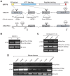
Figure 1. Detection of the alternative splicing transcript Grp78va of mouse Grp78.
A. Schematic representation of GRP78 protein domains and canonical mGrp78 and alternative transcripts. KDEL refers to the ER retention signal. Arrows labeled as p1 to p5 indicate positions of the primers used in RT-PCR. The sequence of 5′ splice site (5′ss) and 3′ splice site (3′ss) of intron 1 are aligned with the optimal splice site sequences. The translational initiation codon (AUG) and stop codon (UAG) of Grp78 are located in exon 1 and exon 8 respectively. Intron 1 contains a stop codon UAA. The underlined AUG in exon 3 is a putative initiation site of Grp78va embedded in a consensus Kozak sequence. B. Detection of Grp78va transcript in mouse NIH3T3 cells by RT-PCR. NIH3T3 cells were either non-treated (−) or treated with 300 nM thapsigargin (Tg) for 16 hours and the total RNA were subjected to RT-PCR with the indicated primer sets. β-actin levels served as control. C. RT-PCR identification of Grp78va transcript in E11.5 mouse embryos and indicated organs. D. Detection of Grp78va transcript in adult mouse tissues. Total RNA extracted from the spleen, kidney, liver, heart, intestine, lung, cerebral cortex and cerebellum of a 5 wk old mouse were subjected to RT-PCR with the indicated primer sets.
Figure 2
Figure 2. Identification of the Grp78va transcript in human cell lines and leukemia patient samples.
A. Expression of the Grp78va transcript was determined by RT-PCR with the total RNA from the indicated human cell lines, which were either non-treated (−) or treated (+) with Tg (300 nM) for 16 hours. PCR was performed with specific primers for the human homologue of mGrp78va shown in Figure 1A. B,C. Quantitative real-time PCR to compare relative expression levels of Grp78va and total Grp78 transcripts in different human cell lines respectively. D. Summary of Grp78va transcript expression in leukemia patients detected by RT-PCR. RNA samples from the indicated types of leukemia patients were used. β-actin levels served as control. The patient samples are either from peripheral blood or bone marrow (asterisk). The Grp78 transcript levels after normalization against β-actin in normal individuals and leukemia patients are shown.
Figure 3
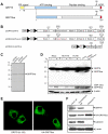
Figure 3. Characterization of human GRP78va protein.
A. Schematic diagram comparing the protein domains of canonical GRP78 and GRP78va. The carboxy-terminal region recognized by the anti-GRP78 monoclonal antibody (Ab) is indicated. B. Schematic diagram of the indicated GRP78va expression plasmids. The base mutations in the 5′ splice site of intron 1 in pcDNA/Grp78va-sm are indicated in red. C. Coupled in vitro transcription and translation assay to detect the protein products from pcDNA/Grp78va and pcDNA/Grp78va-sm plasmids. D. Detection of endogenous GRP78va protein in human cell lines. The post-nuclear (PN) fractions from non-treated or Tg-treated HeLa, HCT116 and HL-60 cells were subjected to Western blot with the anti-GRP78 monoclonal antibody and β-actin served as loading control. GRP78va expression in 293T cells transfected with pcDNA/Grp78va-sm was used as positive control. A darker exposure of the GRP78va bands is shown below the entire blot. E. Immunofluorescence staining of GRP78 and GRP78va. HeLa cells were transfected with pcDNA-Grp78 (left panel) or pcDNA/HA-Grp78va (right panel). The cells were stained with anti-GRP78 antibody (H-129) or anti-HA antibody as indicated, followed by FITC-conjugated secondary antibodies. The confocal immunofluorescence images are shown. F. Cytosolic localization of GRP78va determined by the cell permeabilization assay. HeLa cells transiently transfected with pcDNA/HA-GRP78va were treated with 0.01% digitonin for 5 minutes. Intact cells, digitonin-permeabilized cells, and the released fraction were analyzed by Western blots for the proteins as indicated.
Figure 4
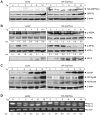
Figure 4. GRP78va enhances PERK signaling.
A. Western blots to confirm the ectopical expression of GRP78va. HeLa cells stably transfected with vector or pcDNA/HA-Grp78va were treated with Tg (300 nM) for the indicated time. HA-GRP78va and endogenous GRP78 were detected by anti-HA and anti-GRP78 antibody respectively. β-actin levels served as loading control. B. Western blots were performed to detect the kinetics and magnitude of UPR signaling markers identified on the right following Tg treatment for the indicated time points. The experiments were repeated twice. The representative results are shown with the ratio (p/t) of p-PERK/PERK or p-eIF2α/eIF2α indicated under each panel with the non-treated control cells set as 1.0. C. Detection of the CHOP activation and ATF6 cleavage following Tg treatment for the indicated time points. The uncleaved (p90) and cleaved (p50) forms of ATF6 are indicated. β-actin levels served as loading control. D. RT-PCR to detect Xbp1 splicing following Tg treatment for the indicated time. The positions of the Xbp1 unspliced (u) and spliced (s) forms were indicated and the ratio of spliced to unspliced form (s/u) is indicated below. The experiments were repeated three times and the representative results are shown.
Figure 5
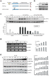
Figure 5. Mutation in ATP binding domain of GRP78va attenuates eIF2α phosphorylation.
A. Schematic representation of human GRP78, GRP78va, and the ATP binding mutant GRP78va-G80D. The position of mutation site is indicated. B. Western blots to detect ectopic expression of GRP78va and the mutant in stably transfected HeLa cell lines. The GRP78va proteins and endogenous canonical GRP78 were detected by the anti-HA and anti-GRP78 antibody respectively, with β-actin as loading control. C. Time course analysis of eIF2α phosphorylation in Tg-treated HeLa cells stably expressing HA-GRP78va or the mutant. Western blots were performed to determine total and phospho-eIF2α levels (upper panel). The ratio of phospho-eIF2α to total eIF2α from two independent experiments are summarized and expressed as the mean with the indicated standard deviation (SD) (lower panel). P values for all time points are indicated. D. Time course analyses of Tg induction of Grp78va and canonical Grp78 transcripts by RT-PCR. HL-60 cells were treated with Tg (300 nM) for the indicated time and the total RNA was subjected to RT-PCR (left panel). The experiments were repeated three times. The results are summarized and expressed as the mean of the normalized Grp78va or canonical Grp78 levels with the indicated SD (right panel). E. Time course analyses of Tg induction of canonical GRP78, GRP78va, P58IPK and eIF2α phosphorylation (p-eIF2α). HL-60 cells treated with Tg (300 nM) for the indicated time were used for Western blot for detection of the indicated proteins with β-actin as loading control (left panel). The experiments were repeated twice. The levels of canonical GRP78, GRP78va and P58IPK normalized to β-actin and the ratio of phospho-eIF2α to total eIF2α are summarized and plotted respectively (right panel).
Figure 6
Figure 6. Physical and functional interactions between GRP78va and P58IPK.
A. Co-immunoprecipitation of GRP78va with P58IPK. 293T cells co-transfected with pcDNA/P58IPK-FLAG (schematic drawing shown above) and pcDNA/HA-Grp78va were either non-treated (−) or treated with Tg (300 nM) for 16 hours. Immunoprecipitations were performed with either anti-FLAG antibody or control mouse IgG, followed by Western blot with the respective antibodies. The input levels are shown. B. Reduced binding between FLAG-P58IPK and ATP binding mutant of GRP78va. 293T cells were co-transfected with pcDNA/FLAG-P58IPK and pcDNA/HA-Grp78va or the mutant Grp78va-G80D. Immunoprecipitations were performed with anti-FLAG antibody or control IgG, followed by Western blot. C. Cytosolic localization of P58IPK determined by the cell permeabilization assay. HeLa cells transiently transfected with pcDNA/P58IPK-FLAG were permeabilized by 0.01% digitonin for 5 minutes. The various fractions were subjected to Western blots for the proteins as indicated. P58IPK-FLAG(SS) designates P58IPK with a slower electrophoretic mobility being released from the permeabilized cells, consistent with retention of signal sequence. D. Overexpression of FLAG-P58IPK suppressed eIF2α phosphorylation mediated by GRP78va. Following transient transfection of the P58IPK expression plasmid or vector into the indicated stably transfected HeLa cell lines, Western blots were performed to probe for the indicated proteins. The experiments were repeated twice. The representative Western blots are shown with the ratio (p/t) of phospho-eIF2α to total eIF2α indicated. E. Overexpression of GRP78va reduced endogenous P58IPK levels. Western blots were performed in HeLa cells stably transfected with vector or HA-GRP78va expression plasmid. The experiments were repeated three times. The representative Western blots are shown and the P58IPK levels normalized to β-actin are indicated below. F. Knockdown of GRP78va increased endogenous P58IPK level. Western blots were performed with HeLa cells transfected with control siRNA (siCtrl) or Grp78va siRNA (siGrp78va) to detect endogenous P58IPK and β-actin level. The cells were either non-treated (−) or treated (+) with Tg (300 nM) for 16 hours. The P58IPK levels normalized to β-actin are shown below the immunoblots. Statistical comparisons were made between Tg-treated cells transfected with siCtrl or siGrp78va, p = 0.03.
Figure 7
Figure 7. GRP78va protects HL-60 cells from ER stress-induced cell death.
A, B. Knockdown of GRP78va in HL-60 cells. RT-PCR (A, left panel) or quantitative real-time PCR (A, right panel) was used to determine the level of Grp78va transcript in HL-60 cells transfected with the indicated siRNA. Western blot. B was performed to determine the levels of the proteins indicated. C. Inhibition of Tg-induced eIF2α phosphorylation by knockdown of GRP78va. HL-60 cells were transfected with either siCtrl or siGrp78va. After treatment with Tg (300 nM) for the indicated time, phospho-eIF2α and total eIF2α were detected by Western blot (upper panel). The experiments were repeated three times. The results are summarized and the ratio of p-eIF2α/eIF2α is plotted with SD (lower panel). The ratio of the non-treated, siCtrl transfected cells was set as 1. P values are indicated. D. Western blot of caspase-7, cleaved caspase-3 and β-actin in siRNA-transfected HL-60 cells treated with Tg for 24 hours. E. Soft-agar clonogenic survival assay for HL-60 cells after transfection with the indicated siRNA for 72 hours and then treated with Tg (300 nM) for the indicated time.

Similar articles

Cited by

References

    1. Rutkowski DT, Kaufman RJ. A trip to the ER: coping with stress. Trends Cell Biol. 2004;14:20–28. - PubMed
    1. Ron D, Walter P. Signal integration in the endoplasmic reticulum unfolded protein response. Nat Rev Mol Cell Biol. 2007;8:519–529. - PubMed
    1. Lee AS. The glucose-regulated proteins: stress induction and clinical applications. Trends Biochem Sci. 2001;26:504–510. - PubMed
    1. Hendershot LM. The ER function BiP is a master regulator of ER function. Mt Sinai J Med. 2004;71:289–297. - PubMed
    1. Li J, Lee AS. Stress induction of GRP78/BiP and its role in cancer. Curr Mol Med. 2006;6:45–54. - PubMed

Publication types