. 2009 Dec;20(12):1665-78.
doi: 10.1089/hum.2009.123.

Pharmacological activation of guanine nucleotide exchange factors for the small GTPase Rap1 recruits high-affinity beta1 integrins as coreceptors for parvovirus B19: improved ex vivo gene transfer to human erythroid progenitor cells

Affiliations

Pharmacological activation of guanine nucleotide exchange factors for the small GTPase Rap1 recruits high-affinity beta1 integrins as coreceptors for parvovirus B19: improved ex vivo gene transfer to human erythroid progenitor cells

Kirsten A K Weigel-Van Aken. Hum Gene Ther. 2009 Dec.

Abstract

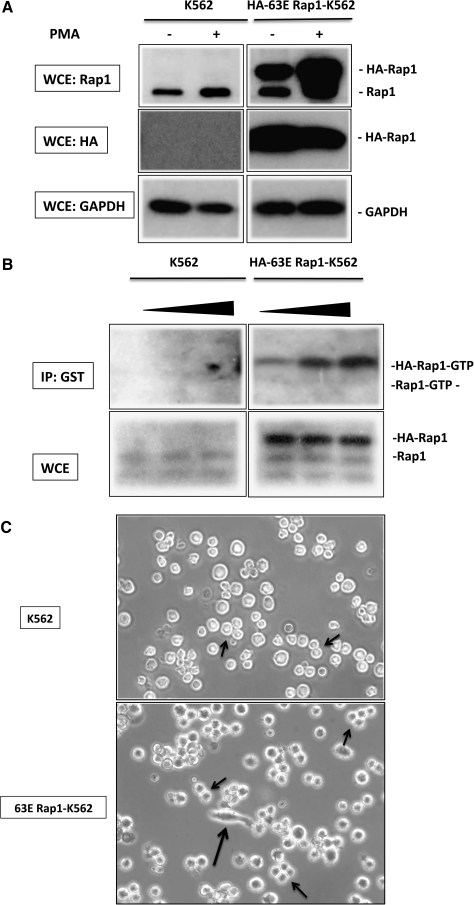
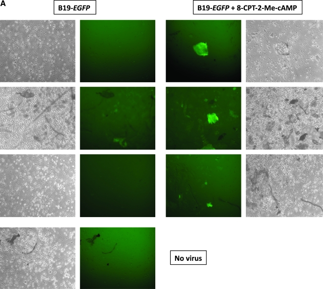
Parvovirus B19 has potential as a gene therapy vector because of its restricted tropism for human erythroid progenitor cells in the bone marrow. B19 binds to the cell surface through P antigen and we identified activated beta(1) integrins as coreceptors for internalization. Because differentiation with phorbol ester induces beta(1) integrin coreceptor activity, but cell differentiation is not desirable in gene transfer to human progenitor cells and one of the downstream effectors of phorbol esters is the small GTPase Rap1, the role of Rap1 in the recruitment of beta(1) integrins on hematopoietic cells was examined. Expression of a constitutively active Rap1 (63E) was sufficient to recruit beta(1) integrin coreceptors in erythroleukemic K562 cells by inducing high-affinity integrin conformation. A crucial role of actin polymerization in Rap1-mediated beta(1) integrin recruitment was documented by complete inhibition of the 63E Rap1 effect with low-dose cytochalasin D and by the ability of a constitutively active mutant of the actin cytoskeleton regulator Rac1 to sensitize K562 cells to the pharmacological activation of endogenous Rap1, using the Rap1 exchange factor-specific 8-pCPT-2'-O-Me-cAMP [8-(4-chlorophenylthio)-2'-O-methyladenosine-3',5'-cyclic monophosphate]. Interestingly, in primary human erythroid progenitor cells, 8-pCPT-2'-O-Me-cAMP was sufficient to significantly increase B19-mediated gene transfer, suggesting that these cells possess the cytoskeleton organization capacity required for efficient recruitment of beta(1) integrins by brief pharmacological stimulation of Rap1 GTP loading. Because 8-pCPT-2'-O-Me-cAMP has been implicated in enhanced homing of progenitor cells, these results identify a novel tool with which to optimize ex vivo B19-mediated gene transfer and potentially improve homing of transduced cells by Rap1-beta(1) integrin activation with 8-pCPT-2'-O-Me-cAMP.


Figures

FIG. 1.
FIG. 1.
Expression of constitutively active Rap1 (63E Rap1) in K562 cells. (A) K562 cells were stably transfected with a plasmid encoding a hemagglutinin (HA)-tagged 63E Rap1 protein. Total Rap1 levels were detected by immunoblot with either a Rap1 antibody (top) or an HA antibody (middle). Phorbol ester (PMA) differentiation of K562 cells slightly increased endogenous Rap1 levels (top left) but had no major effect on the expression of transfected HA-63E Rap1 (middle right). (B) Levels of active, GTP-loaded Rap1 were determined in K562 and 63E Rap1-expressing K562 cells, using a glutathione S-transferase (GST)-fused Rap binding domain (RBD) from the downstream effector RalGDS that preferentially binds the GTP-bound form of Rap1. GTP-loaded Rap1 was precipitated with increasing amounts of GST–RalGDS RBD in 63E Rap1-transfected K562 cells (top right), but not untransfected control K562 cells (top left). Total levels of endogenous and HA-tagged 63E Rap1 in whole cell extracts are shown in the bottom panels. (C) Phase-contrast microscopy of untransfected and 63E Rap1-transfected K562 cells demonstrates the typical rounded cell morphology and loose cell–cell interactions (small arrows) in untransfected cells (top panel) and elongated cell shapes (long arrow) and tight cell–cell adhesions (short arrows) in 63E Rap1-transfected cells (bottom panel). GAPDH, glyceraldehyde-3-phosphate dehydrogenase; IP, immunoprecipitation; WCE, whole cell extract.
FIG. 2.
FIG. 2.
Constitutively active Rap1 is sufficient to convert parvovirus B19-resistant undifferentiated K562 cells into parvovirus B19-permissive cells. (A) Untreated K562 cells, K562 cells treated for 15 min with the Rap1 GEF-specific cAMP analog 8-pCPT-2′-O-Me-cAMP, and 63E Rap1-expressing K562 cells were infected with recombinant parvovirus B19-Luc vector and luciferase activity was determined in cell extracts 24 hr postinfection. Experiments were performed three times and all measurements shown represent means and SEM. (B) Control and 63E Rap1-expressing K562 cells were incubated with high-affinity-stabilizing (N29) or low-affinity-stabilizing β1 (P4C10 and JB1A), β2, and β3 integrin antibodies before infection with recombinant parvovirus B19-Luc vector. For β1 integrin cross-linking, incubation with low-affinity-stabilizing antibody (JB1A) was followed by incubation with cross-linking anti-mouse IgG antibodies (JB+sec.) before infection with recombinant parvovirus B19-Luc vector. Luciferase activity was determined in cell extracts 24 hr postinfection. Experiments were performed three times and all measurements shown represent means and SEM. (C) Expression of constitutively active Rap1 does not affect surface expression of β1, β2, or β3 integrin. Cells were washed with cold PBS–1% BSA, incubated with primary anti-integrin antibodies and fluorescein isothiocyanate (FITC)-conjugated secondary anti-mouse IgG antibodies, and analyzed by flow cytometry. (D) Expression of constitutively active Rap1 induces high-affinity conformation in β1 integrins. Untreated K562 cells, K562 cells treated for 15 min with the Rap1 GEF-specific cAMP analog 8-pCPT-2′-O-Me-cAMP, and 63E Rap1-expressing K562 cells were stained with JB1A anti-β1 integrin antibodies (to determine the total level of β1 integrins) and high-affinity conformation-specific HUTS-21 β1 integrin antibodies and FITC- and phycoerythrin (PE)-labeled secondary antibodies, respectively, and analyzed by flow cytometry. The percentage of HUTS-21-positive β1 integrins present on the cell surface is shown. Experiments were performed three times and all measurements shown represent means and SEM.
FIG. 3.
FIG. 3.
Functional recruitment of β1 integrin coreceptors by constitutively active Rap1 is dependent on de novo actin polymerization. (A) Control and 63E Rap1-expressing K562 cells were preincubated with low (10 nM) and high (500 nM) concentrations of the actin filament elongation blocker cytochalasin D (CD) and subsequently infected with recombinant parvovirus B19-Luc vector. Luciferase activity was determined in cell extracts 24 hr postinfection. (B) Control, 63E Rap1-expressing, and Q61L Rac1-expressing K562 cells were either left untreated or treated with 8-pCPT-2′-O-Me-cAMP for 15 min, stained with JB1A anti-β1 integrin antibody and high-affinity-specific HUTS-21 β1 integrin antibody and with FITC- and PE-labeled secondary antibodies and analyzed by flow cytometry. The percentage of HUTS-21-positive β1 integrin present on the cell surface is shown. (C) Control and Q61L Rac1-expressing K562 cells were either left untreated or treated with 8-pCPT-2′-O-Me-cAMP for 15 min and infected with recombinant parvovirus B19-Luc vector. Luciferase activity was determined in cell extracts 24 hr postinfection. RLU, relative light units.
FIG. 4.
FIG. 4.
Murine NIH3T3 fibroblasts are transduced by parvovirus B19 vectors and transduction efficiency can be increased by pharmacological Rap1 activation. (A) Expression of β1 integrins was determined in cell extracts of β1−/− murine fibroblasts (left lane) and NIH3T3 fibroblasts (right lane) by immunoblot. (B) Wild-type (wt) parvovirus B19 enters and traffics to the nucleus efficiently in NIH3T3 cells. NIH3T3 cells were incubated with wild-type AAV2 (left two lanes) and wild-type parvovirus B19 particles (right two lanes) for 2 hr at 37°C to allow viral binding and entry. Uninternalized virions were removed by extensive trypsin treatment, cells were lysed, and cytoplasmic (C) and nuclear (N) fractions were isolated. Viral genomes were detected in cytoplasmic and nuclear fractions by Southern blot hybridization with 32P-labeled wild-type AAV2- and wild-type B19-specific DNA probes. (C) NIH3T3 cells were either left untreated or treated with 8-pCPT-2′-O-Me-cAMP for 15 min, and subsequently infected with recombinant parvovirus B19-EGFP vector. The percentage of EGFP-positive cells was determined by flow cytometry 24 hr postinfection.
FIG. 4.
FIG. 4.
Murine NIH3T3 fibroblasts are transduced by parvovirus B19 vectors and transduction efficiency can be increased by pharmacological Rap1 activation. (A) Expression of β1 integrins was determined in cell extracts of β1−/− murine fibroblasts (left lane) and NIH3T3 fibroblasts (right lane) by immunoblot. (B) Wild-type (wt) parvovirus B19 enters and traffics to the nucleus efficiently in NIH3T3 cells. NIH3T3 cells were incubated with wild-type AAV2 (left two lanes) and wild-type parvovirus B19 particles (right two lanes) for 2 hr at 37°C to allow viral binding and entry. Uninternalized virions were removed by extensive trypsin treatment, cells were lysed, and cytoplasmic (C) and nuclear (N) fractions were isolated. Viral genomes were detected in cytoplasmic and nuclear fractions by Southern blot hybridization with 32P-labeled wild-type AAV2- and wild-type B19-specific DNA probes. (C) NIH3T3 cells were either left untreated or treated with 8-pCPT-2′-O-Me-cAMP for 15 min, and subsequently infected with recombinant parvovirus B19-EGFP vector. The percentage of EGFP-positive cells was determined by flow cytometry 24 hr postinfection.
FIG. 5.
FIG. 5.
Pharmacological induction of endogenous Rap1 GTP loading significantly increases parvovirus B19 transduction of primary human erythroid progenitor cells. (A) Human erythroid progenitor cells were either left untreated (left) or treated with 8-pCPT-2′-O-Me-cAMP for 15 min (right), and subsequently infected with recombinant parvovirus B19-EGFP vector. Fluorescence microscopy and phase-contrast micrographs were taken 36 hr postinfection. (B) Human erythroid progenitor cells were either left untreated or treated with 8-pCPT-2′-O-Me-cAMP for 15 min, and infected with recombinant parvovirus B19-EGFP vector. The percentage of EGFP-positive cells was determined 36 hr postinfection by flow cytometry. (C) Human erythroid progenitor cells from two different donors were either left in suspension cultures (−FN) or cultured on fibronectin-coated (+FN) plates overnight. Cells were either left untreated or treated with 8-pCPT-2′-O-Me-cAMP for 15 min, and infected with recombinant parvovirus B19-EGFP vector. The percentage of EGFP-positive cells was determined 36 hr postinfection by flow cytometry. Experiments were performed three times and all measurements shown represent means and SEM. P values are calculated by Student t test.
FIG. 5.
FIG. 5.
Pharmacological induction of endogenous Rap1 GTP loading significantly increases parvovirus B19 transduction of primary human erythroid progenitor cells. (A) Human erythroid progenitor cells were either left untreated (left) or treated with 8-pCPT-2′-O-Me-cAMP for 15 min (right), and subsequently infected with recombinant parvovirus B19-EGFP vector. Fluorescence microscopy and phase-contrast micrographs were taken 36 hr postinfection. (B) Human erythroid progenitor cells were either left untreated or treated with 8-pCPT-2′-O-Me-cAMP for 15 min, and infected with recombinant parvovirus B19-EGFP vector. The percentage of EGFP-positive cells was determined 36 hr postinfection by flow cytometry. (C) Human erythroid progenitor cells from two different donors were either left in suspension cultures (−FN) or cultured on fibronectin-coated (+FN) plates overnight. Cells were either left untreated or treated with 8-pCPT-2′-O-Me-cAMP for 15 min, and infected with recombinant parvovirus B19-EGFP vector. The percentage of EGFP-positive cells was determined 36 hr postinfection by flow cytometry. Experiments were performed three times and all measurements shown represent means and SEM. P values are calculated by Student t test.

Similar articles

Cited by

References

    1. Abou-Kheir W. Isaac B. Yamaguchi H. Cox D. Membrane targeting of WAVE2 is not sufficient for WAVE2-dependent actin polymerization: A role for IRSp53 in mediating the interaction between Rac and WAVE2. J. Cell Sci. 2008;121:379–390. - PMC - PubMed
    1. Arai A. Nosaka Y. Kanda E. Yamamoto K. Miyasaka N. Miura O. Rap1 is activated by erythropoietin or interleukin-3 and is involved in regulation of β1 integrin-mediated hematopoietic cell adhesion. J. Biol. Chem. 2001;276:10453–10462. - PubMed
    1. Arthur W.T. Quilliam L.A. Cooper J.A. Rap1 promotes cell spreading by localizing Rac guanine nucleotide exchange factors. J. Cell Biol. 2004;167:111–122. - PMC - PubMed
    1. Berger G. Quarck R. Tenza D. Levy-Toledano S. De Gunzburg J. Cramer E.M. Ultrastructural localization of the small GTP-binding protein Rap1 in human platelets and megakaryocytes. Br. J. Haematol. 1994;88:372–382. - PubMed
    1. Bertoni A. Tadokoro S. Eto K. Pampori N. Parise L.V. White G.C. Shattil S.J. Relationships between Rap1b, affinity modulation of integrin αIIbβ3, and the actin cytoskeleton. J. Biol. Chem. 2002;277:25715–25721. - PubMed

Publication types

MeSH terms