. 2009 Aug;5(8):e1000554.
doi: 10.1371/journal.ppat.1000554. Epub 2009 Aug 21.

CpG methylation controls reactivation of HIV from latency

Affiliations

CpG methylation controls reactivation of HIV from latency

Jana Blazkova et al. PLoS Pathog. 2009 Aug.

Abstract

DNA methylation of retroviral promoters and enhancers localized in the provirus 5' long terminal repeat (LTR) is considered to be a mechanism of transcriptional suppression that allows retroviruses to evade host immune responses and antiretroviral drugs. However, the role of DNA methylation in the control of HIV-1 latency has never been unambiguously demonstrated, in contrast to the apparent importance of transcriptional interference and chromatin structure, and has never been studied in HIV-1-infected patients. Here, we show in an in vitro model of reactivable latency and in a latent reservoir of HIV-1-infected patients that CpG methylation of the HIV-1 5' LTR is an additional epigenetic restriction mechanism, which controls resistance of latent HIV-1 to reactivation signals and thus determines the stability of the HIV-1 latency. CpG methylation acts as a late event during establishment of HIV-1 latency and is not required for the initial provirus silencing. Indeed, the latent reservoir of some aviremic patients contained high proportions of the non-methylated 5' LTR. The latency controlled solely by transcriptional interference and by chromatin-dependent mechanisms in the absence of significant promoter DNA methylation tends to be leaky and easily reactivable. In the latent reservoir of HIV-1-infected individuals without detectable plasma viremia, we found HIV-1 promoters and enhancers to be hypermethylated and resistant to reactivation, as opposed to the hypomethylated 5' LTR in viremic patients. However, even dense methylation of the HIV-1 5'LTR did not confer complete resistance to reactivation of latent HIV-1 with some histone deacetylase inhibitors, protein kinase C agonists, TNF-alpha, and their combinations with 5-aza-2deoxycytidine: the densely methylated HIV-1 promoter was most efficiently reactivated in virtual absence of T cell activation by suberoylanilide hydroxamic acid. Tight but incomplete control of HIV-1 latency by CpG methylation might have important implications for strategies aimed at eradicating HIV-1 infection.


Conflict of interest statement

The authors have declared that no competing interests exist.

Figures
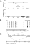
Figure 1
Figure 1. Methylation profiles of latent promoters of HIV-1 LTR-driven vectors integrated in Jurkat cells.
(A) CpG methylation levels of the 5′ LTR of latent HIV-1 LTR-driven retroviral vector pEV731 coding the HIV-1 protein Tat and EGFP in Jurkat clonal cell lines A2, A8, G10, and H12, and of the latent complete EGFP-coding HIV-1 in the JNLGFP cell line. Methylation levels are presented as a mean±standard error of percentages of methylated CpGs in cloned HIV-1 promoters for each cell line. Only DNA sequences with at least 95% conversion of cytosines outside CpGs were taken into account. Levels of EGFP expression (mean±standard error) in all five cell lines are shown bellow. Significance of the difference in methylation levels between cell lines was calculated by non-parametric, two-side Mann-Whitney test. (B) Methylation pattern of the 5′ LTR in the H12 cell line. Analysis of ten promoter molecules is shown as a linear array of open circles representing nonmethylated CpG residues and closed circles representing methylated CpG residues. Shown in the rectangle representing the LTR regions U3, R, and U5 of the pEV731 vector is the distribution of CpG dinucleotides. (C) Integration site of the latent HIV-1 “mini-virus” in the H12 cell line. Gray rectangles represent the exons of ubiquilin. See Table S2 for the flanking sequences. mCpG, methylated CpG residues.
Figure 2
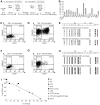
Figure 2. Negative correlation of CpG density in the 5′ LTR and reactivation of HIV-1 provirus.
(A) Flow chart protocol showing two consecutive cycles of 24-h stimulation of H12 cells with TNF-α and PMA, followed by the first and second cell sorting of EGFP-negative cells. Negative sortings are separated by 24-day and followed by 60-day cultivation periods in the absence of any exogeneous stimulator to reach steady-state culture conditions. In the frames are shown the percentages of EGFP-negative cells determined at the moment of cell sorting and the percentages of methylated CpGs in HIV-1 promoter determined in the EGFP-negative cell population just after cell separation. (B) Reactivation levels of HIV-1 promoters in clones harboring latent HIV-1. The percentages of EGFP+ cells in cell clones exposed to TNF-α (10 ng/ml) and PMA (10 nM) for 24 h are shown. The cell clones were prepared by sublimit dilution of the population of H12 cells 60 days after the second sorting of EGFP-negative cells (as shown in flow chart protocol, panel A, and in Figure S1, panel I) and analyzed after the outgrowth of cell clones 20 days later. The cell culture from which the clones were derived was analyzed under the same conditions, 20 days after cell cloning (H12, 2×EGFP). (C,D) Immunofluorescence of EGFP and CD69 in 1B6 clone stimulated with TNF-α (10 ng/ml) and PMA (10 nM) for 24 h (D) and in non-stimulated control (C). (E) CpG methylation profile of the 5′ LTR in non-stimulated 1B6 clone and in 1B6 clone stimulated for 24 h. (F,G) Immunofluorescence of EGFP and CD69 in 2D12 clone stimulated with TNF-α (10 ng/ml) and PMA (10 nM) for 24 h (G) and in non-stimulated control (F). (H) CpG methylation profile of the 5′ LTR in non-stimulated 2D12 clone and in 2D12 clone stimulated for 24 h. (I) Negative correlation of CpG density in the 5′ LTR of HIV-1 provirus with EGFP expression in latently infected Jurkat clonal cell lines exposed to TNF-α and PMA. Open circles, nonmethylated CpG residues; closed circles, methylated CpG residues (mCpG).
Figure 3
Figure 3. DNA methylation levels of the 5′ LTR after reactivation of HIV-1 promoter from latency.
H12 and 2D12 cells activated with TNF-α and PMA for 24 h (86% EGFP+ H12 cells and 28% of EGFP+ 2D12 cells) were separated by FACS according to the expression of EGFP and analyzed for CpG methylation. Percentages of methylated CpGs are presented as a mean±SEM.
Figure 4
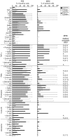
Figure 4. Reactivation of HIV-1 latency in clones harboring proviruses with densely (2D12) and weakly (H12) methylated 5′ LTR.
Percentages of EGFP+, CD69+, and EGFP+/CD69+ cells in the population of H12 and 2D12 clonal cell lines stimulated with the indicated substances were determined by means of flow cytometry. All data were obtained by gating for live cells after vital staining with Hoechst 33258. Results that represent duplicates or several independent experiments are presented as means±SD. Additive (A) and synergistic (S) effects between two inducers of HIV-1 reactivation are assigned for 2D12 cell line. The index of synergism (in brackets) was determined from the following formula: the percentage of EGFP+ cells after stimulation with the combination of stimulators divided by the sum of percentages of EGFP+ cells after stimulation with the stimulators separately. As synergistic were considered the combinations resulting in the index of synergism >1.5. As additive were considered the combinations resulting in the index of synergism ≤1.5 and in >30% increase of stimulation in comparison to stimulation with one out of two stimulators.
Figure 5
Figure 5. Chromatin structures associated with active and latent states of HIV-1 promoter.
Histone 3 (H3) modifications of HIV-1 promoter in the H12 and 2D12 cell lines were analyzed by means of the quantitative ChIP assay using the indicated antibodies before (NI) and after stimulation with TNF-α (10 ng/ml) and PMA (10 nM) (H3CT, anti-H3 C-terminus antibody; diMeH3K4, anti-H3 dimethylated on lysine 4 antibody; AcH3, anti-acetyl-H3 antibody; triMeH3K9, anti-H3 trimethylated on lysine 9 antibody, triMeH3K27, anti-H3 trimethylated on lysine 27 antibody). The equal quantities of HIV promoter immunoprecipitated with anti-H3 C-terminal domain antibody in both cell lines before and after stimulation shows that the total quantity of H3 remained constant. As a control, the cell extracts were immunoprecipitated with normal rabbit IgG. The amount of immunoprecipitated material was normalized to the input DNA. Note that quantities of immunoprecipitated DNA cannot be compared between different H3 modifications, as quantitation is not absolute and depends on antibody affinity. The experiments were performed in triplicates.
Figure 6
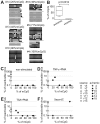
Figure 6. Methylation analysis of HIV-1 promoter in memory CD4+ T cells purified from HIV-1-infected individuals.
Hypermethylation of the HIV-1 5′ LTR in HIV-1-infected long-term aviremic individuals contrasts with hypomethylation of the HIV-1 5′ LTR in viremic patients and negatively correlates with reactivation of the HIV-1 provirus. (A) CpG methylation patterns in the bisulfite-treated HIV-1 5′ LTR sequences of aviremic patients were clustered using neighbor-joining method into several groups within each patient (separated by horizontal bars) with a strong bootstrap support (>990/1000). The sequences within each group differed in addition in about 1 to 10 point-mutations. Note that the variability of bisulfite-treated sequences is underestimated due to the conversion of the majority of cytosine residues to thymine (except for cytosine in 5-methylcytosine residues). Open circles, nonmethylated CpG residues; closed circles, methylated CpG residues. (B) Percentage of methylated CpGs (mCpG) in the 5′ LTR of HIV-1 in long-term aviremic (n = 6) and viremic (n = 7) patients. p, non-parametric, two side Mann-Whitney test. (C–F) Reactivation of HIV-1 provirus in memory CD4+ T cells obtained from patients with different levels of CpG methylation. Memory CD4+ T cells cultured for three days in the presence of reactivating agents were 10-fold serially diluted in duplicate and co-cultured with PHA-activated CD4+ T cells from an allogeneic healthy donor. HIV-1 replication was followed by determination of p24 in the cell-free supernatant. Percentage of reactivable provirus is presented as a ratio of end-point dilutions of patients' CD4+ T cells producing HIV-1 virus to the number of DNA proviral copies in the cell quantity equivalent to end-point dilution and determined by quantitative PCR, as indicated in Table 1. (C) Non-stimulated CD4+ T cells. (D) CD4+ T cells stimulated with TNF-α at 10 ng/ml and 10-nM PMA. (E) CD4+ T cells stimulated with 500-nM TSA and 10-nM PMA. (F) CD4+ T cells stimulated with 5-µM 5-aza-dC. (C–F) Open symbols, viremic patients; closed symbols, aviremic patients.
Figure 7
Figure 7. Two-step model of epigenetic control of HIV-1 latency.
In the state of reactivable latency exemplified by the H12 cell line, the HIV-1 promoter is hypomethylated, histone 3 (H3) is methylated on lysine 4 (MeK4), but deacetylated by histone deacetylases (HDAC), nuc-0 and nuc-1 are in proximal position; chromatin is condensed. After reactivation with TNF-α and PMA, the hypomethylated HIV-1 promoter-associated H3 is acetylated by means of histone acetyltransferases (HAT). Nucleosomes nuc-0 and nuc-1 are in distal position. In the “locked” silent state exemplified by the 2D12 cell line, the HIV-1 promoter is methylated by DNMT, and H3 is demethylated on K4 and methylated on K9 (MeK9) and K27 (MeK27). After reactivation of the hypermethylated HIV-1 promoter the HIV-1 LTR-associated H3 remains methylated on K27 but is demethylated on K9; CpG methylation is not changed and the repression is overcome by excess of transcription factors. The percentage of cells harboring reactivated HIV-1 is markedly lower in the clone with the “locked” highly methylated promoter (2D12) than in the clone with hypomethylated promoter (H12). Open circles, nonmethylated CpG residues; closed circles, methylated CpG residues, TFs, transcription factors.

Similar articles

Cited by

References

    1. Chun TW, Engel D, Mizell SB, Hallahan CW, Fischette M, et al. Effect of interleukin-2 on the pool of latently infected, resting CD4+ T cells in HIV-1-infected patients receiving highly active anti-retroviral therapy. Nat Med. 1999;5:651–655. - PubMed
    1. Davey RT, Jr, Bhat N, Yoder C, Chun TW, Metcalf JA, et al. HIV-1 and T cell dynamics after interruption of highly active antiretroviral therapy (HAART) in patients with a history of sustained viral suppression. Proc Natl Acad Sci U S A. 1999;96:15109–15114. - PMC - PubMed
    1. Strain MC, Gunthard HF, Havlir DV, Ignacio CC, Smith DM, et al. Heterogeneous clearance rates of long-lived lymphocytes infected with HIV: intrinsic stability predicts lifelong persistence. Proc Natl Acad Sci U S A. 2003;100:4819–4824. - PMC - PubMed
    1. Ramratnam B, Mittler JE, Zhang L, Boden D, Hurley A, et al. The decay of the latent reservoir of replication-competent HIV-1 is inversely correlated with the extent of residual viral replication during prolonged anti-retroviral therapy. Nat Med. 2000;6:82–85. - PubMed
    1. Finzi D, Blankson J, Siliciano JD, Margolick JB, Chadwick K, et al. Latent infection of CD4+ T cells provides a mechanism for lifelong persistence of HIV-1, even in patients on effective combination therapy. Nat Med. 1999;5:512–517. - PubMed

Publication types

MeSH terms