. 2009 Oct;20(20):4324-34.
doi: 10.1091/mbc.e08-11-1158. Epub 2009 Aug 19.

Protein tyrosine phosphatase epsilon regulates integrin-mediated podosome stability in osteoclasts by activating Src

Affiliations

Protein tyrosine phosphatase epsilon regulates integrin-mediated podosome stability in osteoclasts by activating Src

Shira Granot-Attas et al. Mol Biol Cell. 2009 Oct.

Abstract

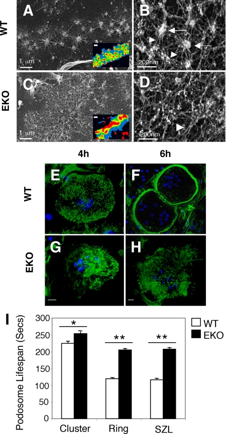
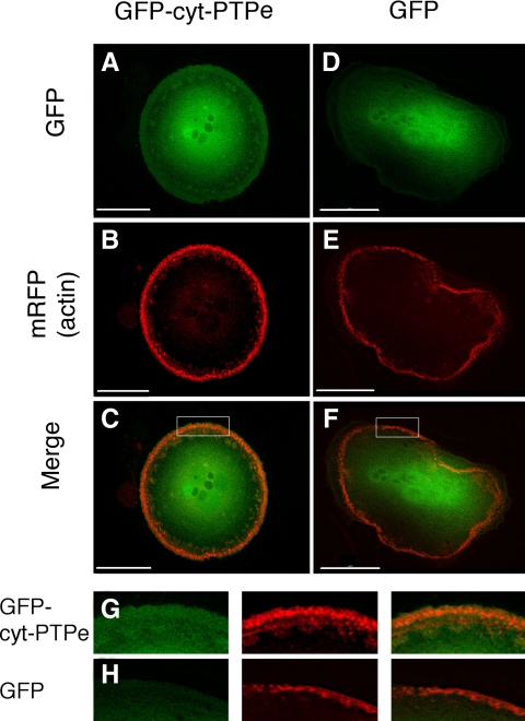
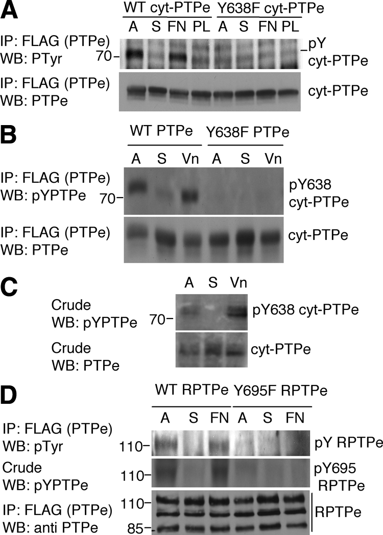
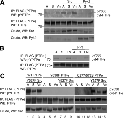
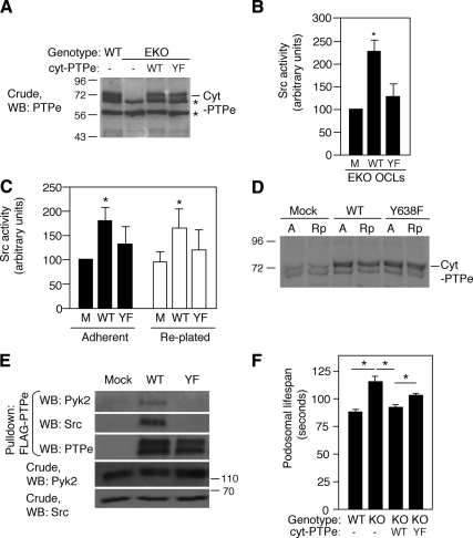
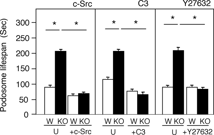
The nonreceptor isoform of tyrosine phosphatase epsilon (cyt-PTPe) supports osteoclast adhesion and activity in vivo, leading to increased bone mass in female mice lacking PTPe (EKO mice). The structure and organization of the podosomal adhesion structures of EKO osteoclasts are abnormal; the molecular mechanism behind this is unknown. We show here that EKO podosomes are disorganized, unusually stable, and reorganize poorly in response to physical contact. Phosphorylation and activities of Src, Pyk2, and Rac are decreased and Rho activity is increased in EKO osteoclasts, suggesting that integrin signaling is defective in these cells. Integrin activation regulates cyt-PTPe by inducing Src-dependent phosphorylation of cyt-PTPe at Y638. This phosphorylation event is crucial because wild-type-but not Y638F-cyt-PTPe binds and further activates Src and restores normal stability to podosomes in EKO osteoclasts. Increasing Src activity or inhibiting Rho or its downstream effector Rho kinase in EKO osteoclasts rescues their podosomal stability phenotype, indicating that cyt-PTPe affects podosome stability by functioning upstream of these molecules. We conclude that cyt-PTPe participates in a feedback loop that ensures proper Src activation downstream of integrins, thus linking integrin signaling with Src activation and accurate organization and stability of podosomes in osteoclasts.


Figures

Figure 1.
Figure 1.
Lack of PTPe disrupts podosomal structure and dynamics. (A–D) Lack of PTPe disrupts podosomal structure. Ventral membranes from wild-type (WT; A and B) or PTPe-deficient (EKO; C and D) primary OCLs were examined by high-resolution scanning electron microscopy. The SZL of WT cells contains punctate podosomal cores (arrows; B) interconnected by actin fibers (arrowheads; B) (Luxenburg et al., 2007); EKO cells do not contain visible cores (D). Insets in A and C show the SZL of intact OCLs stained for actin (red) and vinculin (blue), similar to (Chiusaroli et al., 2004). Bars, 1 μm (A and C) and 0.2 μm (B and D) or 5 μm (insets in A and C). (E–H) EKO OCLs do not organize their podosomes well after replating. OCLs were differentiated from bone marrow of WT or EKO mice in collagen gels, lifted, replated onto glass slides, stained for actin (green) and DNA (blue) at the indicated time points after plating, and analyzed by confocal microscopy. Pictures shown are representative of the WT (E and F) versus EKO (G and H) cultures. Bars, 10 μm. (I) Lack of PTPe increases podosomal stability. WT and EKO OCLs prepared from actin-GFP mice were grown on glass and examined by live-cell imaging. Shown is the average time between appearance and disappearance of individual GFP-labeled actin podosomal cores. N = 203–317 individual podosomes from seven to eight different cells per bar. *p = 0.00095, **p < 1 × 10⁻⁶. See numerical data in Supplemental Table 1.
Figure 2.
Figure 2.
Altered activities of Src, Pyk2, Rho, and Rac in EKO OCLs. (A) Left, Src was precipitated from primary WT or EKO OCLs and its activity was assayed in vitro; Src activity from EKO OCLs was 58.9 ± 12.7% of WT, N = 4, p = 0.018 by paired Student's t test. Right, autophosphorylation of Src at Y416 is reduced in lysates of primary EKO OCLs. NS, nonspecific band. (B) Phosphorylation of Pyk2 is reduced at Y402 in EKO OCLs. (C). Lysates of primary OCLs prepared from bone marrow of WT or EKO mice were subjected to a pull-down assay using the Rho binding domain of Rhotekin fused to GST. Left, amounts of active (GTP-bound) Rho are increased by 107 ± 37% in EKO cells relative to WT (mean ± SE, normalized to Rho expression levels in each sample; *p = 0.045, by paired two-tailed Student's t test; N = 4). Right, representative protein blot depicting levels of Rho-GTP (top), total Rho (middle), and cyt-PTPe (bottom). (D) Similar to C, using a GST fusion of the p21 activated kinase 1 (PAK1) to pull down GTP-bound Rac. GTP-Rac is decreased by 39 ± 8% in EKO relative to WT cells (*p = 0.041, by paired two-tailed Student's t test; N = 3). NS, nonspecific band.
Figure 3.
Figure 3.
cyt-PTPe is found near podosomes. RAW264.7 cells were cotransfected with actin-monomeric RFP and either cyt-PTPe-GFP (A–C) or GFP alone (D–F), allowed to differentiate on glass slides into OCLs, and examined with a Delta Vision deconvolution fluorescence microscope. G and H depict higher magnification of the regions indicated in C and F, respectively. Note the significantly weaker GFP staining at the cell periphery, but not center, in D versus A. Bars, 30 μm.
Figure 4.
Figure 4.
Integrin activation induces C-terminal phosphorylation of PTPe. (A) WT or Y638F cyt-PTPe were expressed in CHOβ3 cells. Cells were grown on plastic plates (A), serum-starved, lifted, and maintained in suspension for 60 min (S), and then plated on plates coated with fibronectin (FN) or poly-l-lysine (PL) for 10 min. After lysis, PTPe was precipitated and analyzed for phospho-tyrosine content by protein blotting with a general anti-p-tyrosine antibody. (B) Similar to A except that PTPe was expressed in RAW264.7 OCLs and phosphorylation of PTPe was examined using an anti C-terminal phospho-PTPe antibody (Berman-Golan and Elson, 2007). Cells were plated on vitronectin (Vn)-coated plates for 30 min. (C) Similar to B, using primary mouse OCLs differentiated from bone marrow of WT mice. (D) Similar to A, using CHOβ3 cells expressing the receptor-type form of PTPe, RPTPe, and its C-terminal Y-to-F mutant Y695 RPTPe. Y695 in RPTPe is identical to Y638 in cyt-PTPe. Mass markers are in kilodaltons.
Figure 5.
Figure 5.
Src induces phosphorylation of cyt-PTPe after integrin activation. (A) WT FLAG-tagged cyt-PTPe was expressed in RAW264.7 OCLs along with Src or Pyk2. Lifting and replating of the cells and analysis of cyt-PTPe phosphorylation were performed as in Figure 4B. (B) WT cyt-PTPe was expressed in CHOβ3 cells. Cells were treated with 10 μM PP1 for 30 min as indicated and analyzed as in Figure 4A. (C) WT, Y638F, or C277/572S cyt-PTPe were expressed in RAW264.7 OCLs with constitutively active Y527F Src as indicated. Cells were treated and analyzed as in Figure 4B. In all panels, A, adherent cells; S, suspended cells; Vn, replated on vitronectin; and FN, replated on fibronectin. Mass markers are in kilodaltons.
Figure 6.
Figure 6.
cyt-PTPe activates Src, associates with Src and Pyk2, and shortens podosomal life span in a phosphorylation-dependent manner. (A) WT or Y638F (YF) cyt-PTPe were expressed by adenoviral infection in OCLs differentiated from bone marrow of EKO mice. Blot shows expression of endogenous cyt-PTPe in WT OCLs and expression of exogenous cyt-PTPe in EKO cells. *, nonspecific bands. (B). Activity of endogenous Src was analyzed in EKO OCLs expressing WT or Y638F cyt-PTPe. Care was taken to analyze cultures in which expression of WT and of Y638F cyt-PTPe was similar, as in A. M, mock-infected control cells. Activity of Src was increased in the presence of WT or Y638F cyt-PTPe to 230 ± 35% or to 130 ± 28%, respectively, relative to mock-infected cells (mean ± SE; normalized to Src expression in each sample. *p = 0.026 by paired, two-tailed Student's t test. N = 5). (C) Expression of cyt-PTPe in RAW264.7 cells activates Src. WT or Y638F (YF) cyt-PTPe was expressed at similar levels in RAW264.7 cells, which were then differentiated with M-CSF and RANKL. Cells were analyzed either adherent or 10 min after replating. Activity of Src was increased in the presence of WT cyt-PTPe to 178 ± 24% relative to mock-transfected cells (M). *p ≤ 0.04, by paired two-tailed Student's t test; N = 7. (D) Expression of endogenous cyt-PTPe (Mock) or of exogenous (+ endogenous) cyt-PTPe (WT, Y638F lanes) in adherent (A) or replated (Rp) RAW264.7 cells. *, nonspecific bands. (E) WT, but not Y638F, cyt-PTPe associates with Src and Pyk2 in OCLs. Crude lysates of primary OCLs from WT mice were subjected to an in vitro pull-down assay using purified FLAG-tagged WT or Y638F cyt-PTPe. Mass markers are in kilodaltons. (F). WT, but not Y638F, cyt-PTPe shortens the life span of SZL podosomes from EKO OCLs. WT and EKO OCLs prepared from actin-GFP mice were grown on glass, infected with adenoviral vectors expressing WT or Y638F cyt-PTPe, and examined by live-cell imaging. All four cultures were also infected with adenovirus-RFP to identify infected cells. Shown is the average time between appearance and disappearance of individual GFP-labeled actin podosomal cores. *, p ≤ 3.4 × 10⁻⁶, N = 79–346 podosomes from two to four distinct cells per bar. See numerical data in Supplemental Table 2.
Figure 7.
Figure 7.
The abnormally long life spans of SZL podosomes in EKO OCLs are shortened by correcting abnormalities in Src or in downstream signaling elements. Left, WT (W) and EKO (KO) primary OCLs expressing an actin-GFP transgene were infected (+c-Src) or not (U) with adenovirus containing Src. Cells were coinfected with adeno-RFP to allow identification of infected cells. The life spans of the GFP-labeled podosome cores of these cells were measured by live-cell imaging. Middle, similar to left, except that cells were treated (+C3) or not (U) with 2 μg/ml cell-permeable C3 exoenzyme for 4 h and were not infected with adeno-RFP. Right, similar to middle, except that cells were treated or not with 10 μM Y27632 for 2 h. In all panels, asterisk (*) indicates p < 10−6, N = 64–416 podosomes from four to five individual cells per bar. See numerical data in Supplemental Table 3.
Figure 8.
Figure 8.
Schematic model suggesting how cyt-PTPe participates in activation of Src downstream of activated integrins in osteoclasts. Activation of integrins results in partial activation of Src by mechanisms that do not include cyt-PTPe as discussed. Src then participates in phosphorylation of cyt-PTPe at Y638; pY638 cyt-PTPe activates Src further, ensuring Src is sufficiently active to perform downstream roles, leading ultimately to normal stability of podosomes.

Similar articles

Cited by

References

    1. Abreu M. T., Penton P. C., Kwok V., Vachon E., Shalloway D., Vidali L., Lee W., McCulloch C. A., Downey G. P. Tyrosine phosphatase PTPalpha regulates focal adhesion remodeling through Rac1 activation. Am. J. Physiol. Cell Physiol. 2008;294:C931–C944. - PMC - PubMed
    1. Amoui M., Sheng M. H., Chen S. T., Baylink D. J., Lau K. H. A transmembrane osteoclastic protein-tyrosine phosphatase regulates osteoclast activity in part by promoting osteoclast survival through c-Src-dependent activation of NFkappaB and JNK2. Arch. Biochem. Biophys. 2007;463:47–59. - PubMed
    1. Aoki K., Didomenico E., Sims N. A., Mukhopadhyay K., Neff L., Houghton A., Amling M., Levy J. B., Horne W. C., Baron R. The tyrosine phosphatase SHP-1 is a negative regulator of osteoclastogenesis and osteoclast resorbing activity: increased resorption and osteopenia in me(v)/me(v) mutant mice. Bone. 1999;25:261–267. - PubMed
    1. Arias-Salgado E. G., Lizano S., Sarkar S., Brugge J. S., Ginsberg M. H., Shattil S. J. Src kinase activation by direct interaction with the integrin beta cytoplasmic domain. Proc. Natl. Acad. Sci. USA. 2003;100:13298–13302. - PMC - PubMed
    1. Arthur W. T., Petch L. A., Burridge K. Integrin engagement suppresses RhoA activity via a c-Src-dependent mechanism. Curr. Biol. 2000;10:719–722. - PubMed

Publication types

MeSH terms

LinkOut - more resources