

doi: 10.1002/ejlt.200800117.
A review of lipidomic technologies applicable to sphingolipidomics and their relevant applications
Affiliations
- PMID: 19690629
- PMCID: PMC2727941
- DOI: 10.1002/ejlt.200800117
Item in Clipboard
A review of lipidomic technologies applicable to sphingolipidomics and their relevant applications
Eur J Lipid Sci Technol.
2009.
Abstract
Sphingolipidomics, a branch of lipidomics, focuses on the large-scale study of the cellular sphingolipidomes. In the current review, two main approaches for the analysis of cellular sphingolipidomes (i.e. LC-MS- or LC-MS/MS-based approach and shotgun lipidomics-based approach) are briefly discussed. Their advantages, some considerations of these methods, and recent applications of these approaches are summarized. It is the authors' sincere hope that this review article will add to the readers understanding of the advantages and limitations of each developed method for the analysis of a cellular sphingolipidome.
Conflict of interest statement
The authors have declared no conflict of interest.
Figures
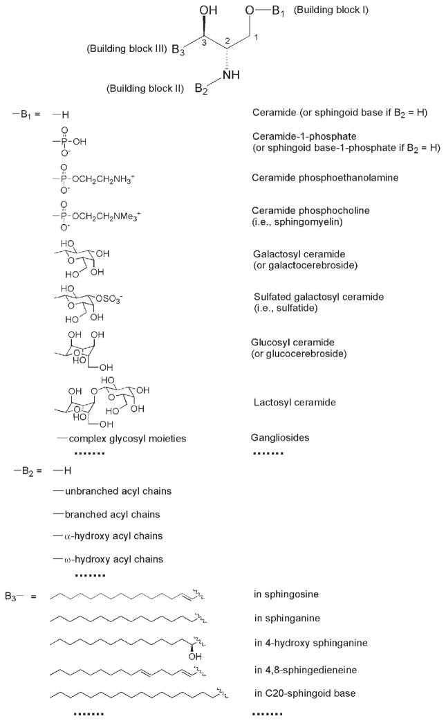
General structure of sphingoid-based lipids. The building block B1 represents a different polar moiety (linked to the oxygen at the C1 position of the sphingoid base). The building block B2 represents fatty acyl chains (acylated to the primary amine at the C2 position of the sphingoid base) with or without the presence of a hydroxyl group which is usually located at the α- or ω-position. The building block B3 represents the aliphatic chains in all of the possible sphingoid bases, which are carbon-carbon linked to the C3 position of sphingoid bases and vary with the aliphatic chain length, degree of unsaturation, the presence of branches, and the presence of an additional hydroxyl group. This illustration has been modified from ref. [11] with permission.
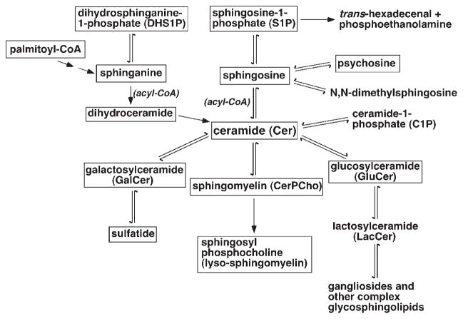
A simplified network of the common sphingolipid classes and other related lipids in the mammalian sphingolipidome. The sphingolipid classes with frames are those that can be quantitatively analyzed by shotgun (sphingo)lipidomics at its current stage.
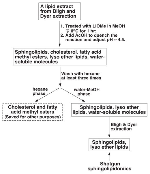
A schematic illustration of sample preparation for shotgun sphingolipidomics (adapted from ref. [26] with permission).
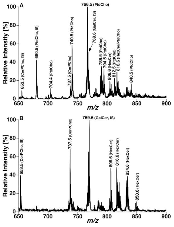
Shotgun lipidomics analyses of sphingolipid molecular species before and after treatment of a mouse cortex lipid extract with lithium methoxide in the positive-ion mode in the presence of a small amount of LiOH. The mass spectra (A) and (B) were acquired directly from a lipid extract of mouse cortex before and after treatment with lithium methoxide, respectively, as illustrated in Fig. 3. IS denotes internal standard. Both spectra are displayed after being normalized to the base peak in each spectrum.
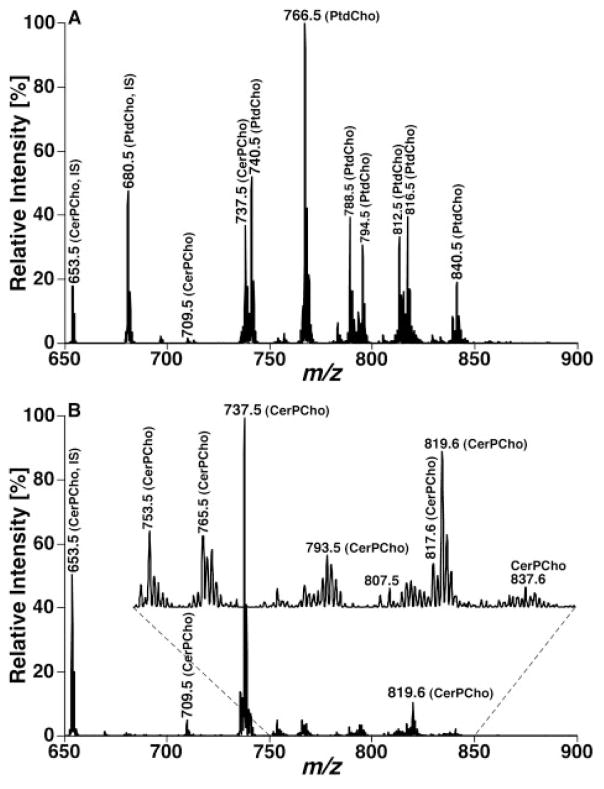
Shotgun lipidomics analyses of sphingolipid molecular species before and after treatment of a mouse cortex lipid extract with lithium methoxide in the NL mode in the presence of a small amount of LiOH. The mass spectra in (A) and (B) were acquired by using the NL scanning of 183.1 u (i.e. phosphocholine) from the lipid extract of mouse cortex before and after treatment with lithium methoxide, respectively, as illustrated in Fig. 3. IS denotes internal standard. The ion peaks in (B) represent lithiated CerPCho molecular species. Both spectra are displayed after being normalized to the base peak in each spectrum.
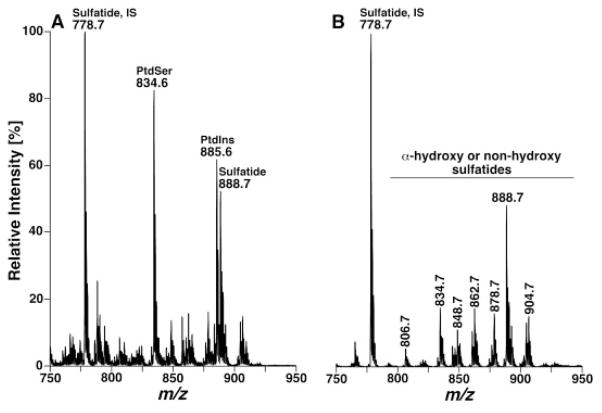
Shotgun lipidomics analyses of sulfatide molecular species before and after treatment of a mouse cortex lipid extract with lithium methoxide in the negative-ion mode. The mass spectra in (A) and (B) were acquired directly from a lipid extract of mouse cortex before and after treatment with lithium methoxide, respectively, as illustrated in Fig. 3, by using a nanomate device. IS denotes internal standard. Both mass spectra are displayed after being normalized to the base peak in each spectrum.
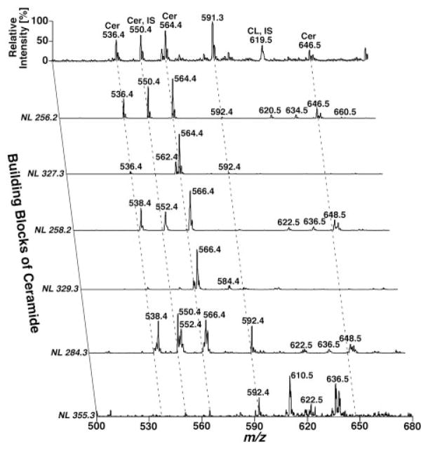
Two-dimensional mass spectrometric analyses of ceramide molecular species from a lipid extract of human brain temporal cerebellar white matter in a shotgun sphingolipidomics approach. The lipid sample from human temporal white matter for shotgun sphingolipidomics was prepared as illustrated in Fig. 3 in the presence of 1 nmol C17:1 ceramide/mg protein. A conventional ESI mass spectrum in the negative-ion mode was acquired prior to analysis of the building blocks of ceramide molecular species by NL scanning. These building blocks of ceramide molecular species include sphingoid bases of sphingosine (NL 256.2 and NL 327.3), sphinganine (NL 258.2 and NL 329.3), and C20-sphingoid base (NL 284.3 and NL 355.3) with or without the presence of a hydroxyl group in the fatty amide chains as previously described [74]. IS denotes internal standard. All mass spectra are displayed after normalization to the base peak in each individual spectrum.
Similar articles
-
Shotgun lipidomics: electrospray ionization mass spectrometric analysis and quantitation of cellular lipidomes directly from crude extracts of biological samples.Mass Spectrom Rev. 2005 May-Jun;24(3):367-412. doi: 10.1002/mas.20023. Mass Spectrom Rev. 2005. PMID: 15389848 Review.
-
Mass Spectrometry-based Lipidomics and Its Application to Biomedical Research.J Lifestyle Med. 2014 Mar;4(1):17-33. doi: 10.15280/jlm.2014.4.1.17. Epub 2014 Mar 31. J Lifestyle Med. 2014. PMID: 26064851 Free PMC article. Review.
-
Structure-specific, quantitative methods for analysis of sphingolipids by liquid chromatography-tandem mass spectrometry: "inside-out" sphingolipidomics.Methods Enzymol. 2007;432:83-115. doi: 10.1016/S0076-6879(07)32004-1. Methods Enzymol. 2007. PMID: 17954214
-
Analytical methods in sphingolipidomics: Quantitative and profiling approaches in food analysis.J Chromatogr A. 2016 Jan 8;1428:16-38. doi: 10.1016/j.chroma.2015.07.110. Epub 2015 Aug 1. J Chromatogr A. 2016. PMID: 26275862 Review.
-
Sphingolipidomics analysis of large clinical cohorts. Part 2: Potential impact and applications.Biochem Biophys Res Commun. 2018 Oct 7;504(3):602-607. doi: 10.1016/j.bbrc.2018.04.075. Epub 2018 Apr 19. Biochem Biophys Res Commun. 2018. PMID: 29654757 Review.
Cited by
-
Distribution and functions of sterols and sphingolipids.Cold Spring Harb Perspect Biol. 2011 May 1;3(5):a004762. doi: 10.1101/cshperspect.a004762. Cold Spring Harb Perspect Biol. 2011. PMID: 21454248 Free PMC article. Review.
-
Shotgun lipidomics in substantiating lipid peroxidation in redox biology: Methods and applications.Redox Biol. 2017 Aug;12:946-955. doi: 10.1016/j.redox.2017.04.030. Epub 2017 Apr 24. Redox Biol. 2017. PMID: 28494428 Free PMC article. Review.
-
Multi-Omics Approaches and Radiation on Lipid Metabolism in Toothed Whales.Life (Basel). 2021 Apr 20;11(4):364. doi: 10.3390/life11040364. Life (Basel). 2021. PMID: 33923876 Free PMC article. Review.
-
Sphingolipidomics in Translational Sepsis Research-Biomedical Considerations and Perspectives.Front Med (Lausanne). 2021 Jan 20;7:616578. doi: 10.3389/fmed.2020.616578. eCollection 2020. Front Med (Lausanne). 2021. PMID: 33553212 Free PMC article. Review.
-
Recent advances in the mass spectrometric analysis of glycosphingolipidome - A review.Anal Chim Acta. 2020 Oct 2;1132:134-155. doi: 10.1016/j.aca.2020.05.051. Epub 2020 May 24. Anal Chim Acta. 2020. PMID: 32980104 Free PMC article. Review.
References
-
- Kiechle FL, Zhang X, Holland-Staley CA. The -omics era and its impact. Arch Pathol Lab Med. 2004;128:1337–1345. - PubMed
-
- Altman RB, Rubin DL, Klein TE. An “omics” view of drug development. Drug Dev Res. 2004;62:81–85.
-
- Duncan MW. Omics and its 15 minutes. Exp Biol Med. 2007;232:471–472. - PubMed
-
- Han X, Gross RW. Global analyses of cellular lipidomes directly from crude extracts of biological samples by ESI mass spectrometry: A bridge to lipidomics. J Lipid Res. 2003;44:1071–1079. - PubMed
Grants and funding
LinkOut - more resources
Full Text Sources