. 2009 Sep;128(1):16-24.
doi: 10.1111/j.1365-2567.2009.03042.x.

Domain analyses of the Runx1 transcription factor responsible for modulating T-cell receptor-beta/CD4 and interleukin-4/interferon-gamma expression in CD4(+) peripheral T lymphocytes

Affiliations

Domain analyses of the Runx1 transcription factor responsible for modulating T-cell receptor-beta/CD4 and interleukin-4/interferon-gamma expression in CD4(+) peripheral T lymphocytes

Ryuji Uchino. Immunology. 2009 Sep.

Abstract

The Runx1 transcription factor is one of the master regulators of T-lymphocyte differentiation. There have been several reports trying to assign a domain within the Runx1 protein that is responsible for gene expression in thymocytes. The Runx1 domains involved in regulating the expression of several genes in peripheral CD4(+) T cells were analysed. It was observed that Runx1 over-expression enhanced the surface expression of CD4 and CD69 molecules via its activation domain and VWRPY domain, and decreased that of T-cell receptor-beta via its activation domain. Runx1 over-expression enhanced interferon-gamma expression via its activation and VWRPY domains, and abolished interleukin-4 expression through its activation domain. Transduction of Runx1 did not down-regulate CD4 expression until 72 hr of culture, but the repression of CD4 expression became evident after 96 hr. The main region responsible for repressing CD4 expression was the inhibitory domain of Runx1. Taken together, these results lead to a proposal that the regions in Runx1 responsible for modulating gene expression are distinct in thymocytes and in peripheral CD4(+) T cells.


Figures

Figure 1
Figure 1
Schematic illustration and expression of Runx1-mutated proteins. (a) Runx1 and its deletion mutants. Numbers indicate the positions of the amino acid residues from the N-terminus of the murine Runx1 protein. RD, Runt domain; AD, transcription activation domain; ID, transcription inhibition domain. Each protein is tagged with an haemagglutinin (HA) epitope at its N-terminal end. (b) Expression of Runx1-mutated proteins. CD4+ T cells were isolated from the spleens of BALB/c mice, cultured in the presence of anti-CD3/anti-CD28 antibodies and interleukin-2 (IL-2) for 24 hr, and infected by retroviruses carrying pMX–nerve growth factor receptor (NGFR), pMX–Runx1, pMX–Runx1ΔGr, pMX–Runx1ΔCS, pMX–Runx1ΔC, or pMX–Runx1ΔCL. After 72 hr incubation, the NGFR-positive cells were sorted. Protein extracts were processed for immunoblot analyses using an anti-HA antibody. Results represent the representative result from two independent experiments that yielded very similar results.
Figure 2
Figure 2
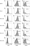
Expression profiles of CD4, CD69 and T-cell receptor-β (TCR-β) in Runx1-transduced CD4+ T cells. CD4+ T cells were cultured in the presence of anti-CD3/anti-CD28 antibodies and interleukin-2 (IL-2) for 24 hr, infected by retroviruses carrying pMX–nerve growth factor receptor (NGFR), pMX–Runx1, pMX–Runx1ΔGr, pMX–Runx1ΔCS, pMX–Runx1ΔC, or pMX–Runx1ΔCL, and incubated further for 72 hr. Cells were restimulated with phorbol 12-myristate 13-acetate and ionomycin for 5 hr, and then processed for flow cytometry. Three independent experiments were performed, with essentially the same results obtained for each experiment. Representative profiles of CD4, CD69 and TCR-β expression are shown for the NGFRhi gated population. The vertical lines in the CD4 panels represent the mean fluorescence intensity in pMX–NGFR-infected cells, which should serve as a standard to compare the effects of the various Runx1 mutants. The mean CD4 intensities were 2·6, 5·2, 3·3, 2·3, 2·5 and 1·8 for the pMX–NGFR-, pMX–Runx1-, pMX–Runx1ΔGr-, pMX–Runx1ΔCS-, pMX–Runx1ΔC-, and pMX–Runx1ΔCL-infected cells, respectively. Note that the fluorescence intensities are on a logarithmic scale. The numbers in the middle and right panels represent the percentages of CD69hi and TCR-βhi populations, respectively.
Figure 3
Figure 3
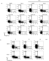
Expression profiles of intracellular interleukin-4 (IL-4) and interferon-γ (IFN-γ) in Runx1-transduced CD4+ T cells. (a) CD4+ T cells were cultured in the presence of anti-CD3/anti-CD28 antibodies and IL-2 for 48 hr, infected by retroviruses carrying pMX–nerve growth factor receptor (NGFR), pMX–Runx1, pMX–Runx1ΔGr, pMX–Runx1ΔCS, pMX–Runx1ΔC, or pMX–Runx1ΔCL, and incubated further for 7 days. In the right panels, excess IL-4 was also added throughout the culture. The cells were restimulated with phorbol 12-myristate 13-acetate (PMA) and ionomycin, and processed for flow cytometry analysis. Three independent experiments were performed, with essentially the same results obtained each time. Representative profiles of intracellular IL-4 and IFN-γ expression are shown for the NGFRhi gated population. The numbers represent the percentages of cells in each quadrant. (b) Effect of a TGF-β receptor inhibitor. CD4+ T cells were cultured in the presence of anti-CD3/anti-CD28 antibodies and IL-2 for 48 hr, infected by retroviruses carrying pMX–NGFR or pMX–Runx1, and incubated further for 7 days. In the lower panels, SB431542 was added throughout the culture at the indicated concentrations (μm). The cells were restimulated with PMA and ionomycin, and were processed for flow cytometry analysis of intracellular IL-4 and IFN-γ. Three independent experiments were performed, with essentially the same results obtained each time. Representative profiles of intracellular IL-4 and IFN-γ expression are shown for the NGFRhi gated population. The numbers represent the percentages of cells in each quadrant.
Figure 4
Figure 4
Long-term effects of Runx1 transduction on CD4 expression. (a) CD4+ T cells were cultured in the presence of anti-CD3/anti-CD28 antibodies and interleukin-2 (IL-2) for 24 hr, infected by retroviruses carrying pMX–nerve growth factor receptor (NGFR), pMX–Runx1, pMX–Runx1ΔGr, pMX–Runx1ΔCS, pMX–Runx1ΔC, or pMX–Runx1ΔCL, incubated further for 24, 48, 72, 96, 120, or 144 hr, and processed for flow cytometry. Three independent experiments were performed, with essentially the same results obtained each time. Representative profiles of CD4 expression at 72 and 144 hr postinfection are shown for the NGFRhi gated population. The numbers represent the percentages of the CD4low population. Note that the percentages of the CD4low fractions did not increase in the NGFR-negative populations of pMX–Runx1-infected cells, even at 144 hr (data not shown). (b) The percentages of CD4low populations are depicted at 24, 48, 72, 96, 120, and 144 hr postinfection.

Similar articles

Cited by

References

    1. Egawa T, Tillman RE, Naoe Y, Taniuchi I, Littman DR. The role of the Runx transcription factors in thymocyte differentiation and in homeostasis of naive T cells. J Exp Med. 2007;204:1945–57. - PMC - PubMed
    1. Okuda T, Takeda K, Fujita Y, et al. Biological characteristics of the leukemia-associated transcriptional factor AML1 disclosed by hematopoietic rescue of AML1-deficient embryonic stem cells by using a knock-in strategy. Mol Cell Biol. 2000;20:319–28. - PMC - PubMed
    1. Kagoshima H, Shigesada K, Satake M, Ito Y, Miyoshi H, Ohki M, Pepling M, Gergen P. The Runt domain identifies a new family of heteromeric transcriptional regulators. Trends Genet. 1993;9:338–41. - PubMed
    1. Meyers S, Downing JR, Hiebert SW. Identification of AML-1 and the (8;21) translocation protein (AML-1/ETO) as sequence-specific DNA-binding proteins: the runt homology domain is required for DNA binding and protein–protein interactions. Mol Cell Biol. 1993;13:6336–45. - PMC - PubMed
    1. Kitabayashi I, Yokoyama A, Shimizu K, Ohki M. Interaction and functional cooperation of the leukemia-associated factors AML1 and p300 in myeloid cell differentiation. EMBO J. 1998;17:2994–3004. - PMC - PubMed

MeSH terms