. 2009 Aug 14;35(3):280-90.
doi: 10.1016/j.molcel.2009.06.010.

Structure of the s5a:k48-linked diubiquitin complex and its interactions with rpn13

Affiliations

Structure of the s5a:k48-linked diubiquitin complex and its interactions with rpn13

Naixia Zhang et al. Mol Cell. .

Abstract

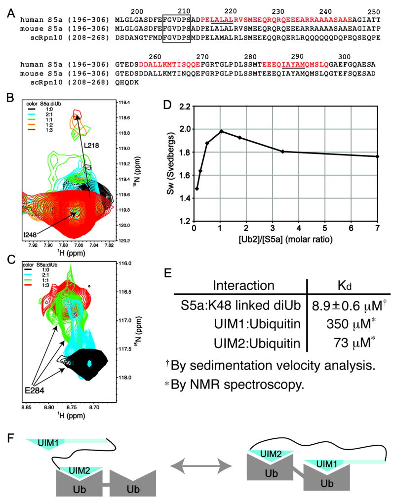
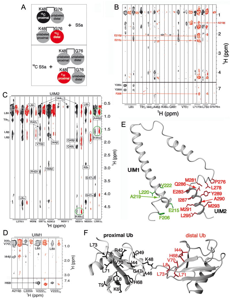
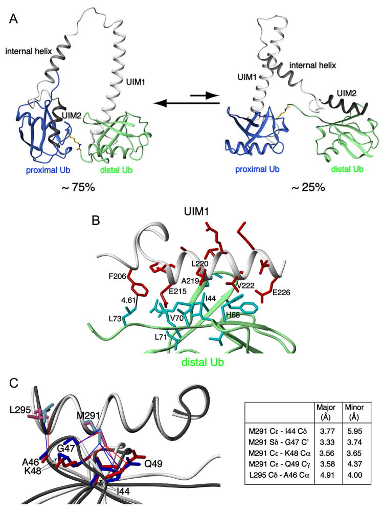
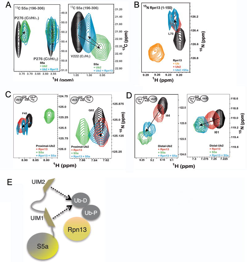
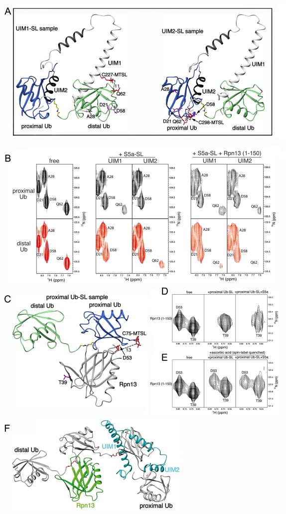
Degradation by the proteasome typically requires substrate ubiquitination. Two ubiquitin receptors exist in the proteasome, S5a/Rpn10 and Rpn13. Whereas Rpn13 has only one ubiquitin-binding surface, S5a binds ubiquitin with two independent ubiquitin-interacting motifs (UIMs). Here, we use nuclear magnetic resonance (NMR) and analytical ultracentrifugation to define at atomic level resolution how S5a binds K48-linked diubiquitin, in which K48 of one ubiquitin subunit (the "proximal" one) is covalently bonded to G76 of the other (the "distal" subunit). We demonstrate that S5a's UIMs bind the two subunits simultaneously with a preference for UIM2 binding to the proximal subunit while UIM1 binds to the distal one. In addition, NMR experiments reveal that Rpn13 and S5a bind K48-linked diubiquitin simultaneously with subunit specificity, and a model structure of S5a and Rpn13 bound to K48-linked polyubiquitin is provided. Altogether, our data demonstrate that S5a is highly adaptive and cooperative toward binding ubiquitin chains.


Figures

Figure 1
Figure 1. S5a’s affinity for diubiquitin is enhanced by its two UIMs binding to ubiquitin subunits simultaneously
(A) Sequence alignment of the ubiquitin-binding region of human and mouse S5a with their Saccharomyces cerevisiae homologue Rpn10. The helices of S5a are highlighted in red and a strictly conserved region N-terminal to UIM1 is boxed. The UIM’s hallmark LALAL/IAYAM motif is underlined. (B) UIM1 resonances shift and broaden upon binding to K48-linked diubiquitin. 1H,15N HSQC spectra expanded to view L218 are displayed for 15N labeled S5a (196–306) alone (black) and at 2:1 (blue), 1:1 (green), 1:2 (orange), and 1:3 (red) S5a:K48-linked diubiquitin molar ratio. The amide resonance of I248, which is not involved in diubiquitin-binding and does not shift with increasing quantities of diubiquitin, overlaps with the L218’s unbound resonance. (C) UIM2 resonances exhibit slow exchange dynamics upon binding to K48-linked diubiquitin. 1H, 15N HSQC spectra expanded to view E284 are displayed for S5a alone (black) and at 2:1 (blue), 1:1 (green), and 1:3 (red) S5a:K48-linked diubiquitin molar ratio. (D) Sedimentation velocity results reveal S5a (196–306) to bind K48-linked diubiquitin with 1:1 stoichiometry and with an affinity of 8.9±0.6 μM. Experimentally determined averaged sedimentation coefficients (diamonds) are plotted. (E) Binding affinities are displayed for S5a (196–306) binding to diubiquitin, as determined with the data of (D), and for each UIM binding to monoubiquitin (Ryu et al., 2003; Wang et al., 2005). (F) Model depicting the two S5a:diubiquitin states present at 1:1 molar ratio. The weaker binding UIM1 exchanges between an unbound and bound state. Ub, ubiquitin; UIM1, S5a’s ubiquitin interacting motif 1; UIM2, S5a’s ubiquitin interacting motif 2.
Figure 2
Figure 2. S5a prefers to bind K48-linked diubiquitin with its UIM1 bound to the distal subunit while its UIM2 binds the proximal one
(A) Schematic illustrating the NMR samples used for the 13C half-filtered NOESY spectra displayed in (B; top panel) and (C and D; bottom panel). Coloring matches the associated spectrum, such that the labeled black and red subunits produced the spectra displayed in black and red, respectively. (B) 13C half-filtered NOESY experiments recorded on K48-linked diubiquitin with either its proximal (black) or distal (red) subunit 13C labeled and mixed with unlabeled S5a (196–306). Abundant NOE interactions are observed between UIM2 and the proximal subunit as well as between UIM1 and the distal subunit. Selected interactions are labeled and greater detail is provided in Supplementary Figure 3A. (C, D) 13C half-filtered NOESY spectra recorded with 13C-labeled S5a (196–306) and 3-fold molar excess unlabeled K48-linked diubiquitin (black) or K48-linked diubiquitin with its proximal subunit deuterated (red). Most interactions with UIM2 residues are mitigated by 2H labeling the proximal subunit (C), whereas those involving UIM1 resonances are not (D). Breakthrough diagonal peaks, which should be treated as noise, are circled in grey. NOE crosspeaks involving UIM2 that are only slightly affected by deuterating the proximal subunit are circled in green; these indicate the presence of a minor species in which UIM2 binds the distal subunit. The sidechain atoms of (E) S5a and (F) diubiquitin residues that form intermolecular NOE contacts in the major binding species are highlighted with UIM1 and UIM2 in green and red, respectively, and diubiquitin’s proximal and distal subunits in black and red, respectively.
Figure 3
Figure 3. Structure of S5a:K48-linked diubiquitin complexes
(A) Ribbon representation of the major and minor binding modes for S5a:K48-linked diubiquitin with S5a’s UIM1 and UIM2 motifs in white and black, respectively, and diubiquitin’s proximal and distal subunits in blue and green, respectively. Diubiquitin’s K48-G76 linkage is highlighted in yellow. (B) Expanded view of the UIM1:distal ubiquitin complex reveals critical interactions. Residues of UIM1 and distal ubiquitin are highlighted in red and cyan, respectively, whereas their ribbon structures are grey and green respectively. (C) Multiple contacts become more compact when UIM2 binds to the proximal subunit of K48-linked diubiquitin. UIM2’s M291 and L295 are displayed bound to proximal (pink and red) or distal (blue) ubiquitin. Selected distances are listed between M291 and L295 of UIM2 and proximal or distal ubiquitin atoms.
Figure 4
Figure 4. Ubiquitin receptors Rpn13 and S5a bind K48-linked diubiquitin simultaneously
A) 1H, 13C HSQC experiments recorded on 13C labeled S5a (196–306) alone (black) or with equimolar K48-linked diubiquitin (green) or K48-linked diubiquitin and Rpn13 (1–150) (blue) reveal P276 (left panel) and V222 (right panel) to interact with diubiquitin when Rpn13 is present. B) 1H, 15N HSQC experiments on 15N labeled Rpn13 (1–150) alone (black) and with monoubiquitin (orange), K48-linked diubiquitin (red) or K48-linked diubiquitin and S5a (196–306) (blue) reveals L73 to shift to its diubiquitin-bound state when S5a is present. K48-linked diubiquitin with its (C) proximal or (D) distal subunit 15N labeled alone (black) or with Rpn13 (1–150) (red), S5a (196–306) (green), or both of these receptors (blue) indicates shifting that mimics Rpn13 binding to the proximal subunit, but S5a binding to the distal one, as shown for F45 and Q62 of the proximal subunit and I44 and I61 of the distal subunit. Additional data is provided in Supplementary Figure 6. (E) Model illustrating Rpn13 bound to K48-linked diubiquitin’s proximal subunit and S5a’s UIMs interacting dynamically with the distal one.
Figure 5
Figure 5. MTSL-labeling demonstrates hRpn13’s preference for K48-linked diubiquitin’s proximal subunit as S5a’s two UIMs bind the distal subunit
(A) Ribbon representation of the major S5a (196–306):K48-linked diubiquitin structure displaying MTSL covalently attached to C227 (left) or C298 (right). Each of these cysteines was introduced for UIM1 or UIM2 spin labeling by MTSL. Q62 is also displayed as well as approximate distances in Å between its backbone amide proton atom and MTSL’s single-electron center (oxygen atom). It is worth noting that MTSL and the cysteine sidechain atoms are flexible such that the distances shown are only approximations. D21, A28 and D58 are also displayed and highlighted in purple, as they are used for comparison. (B) Expanded view of 1H, 15N HSQC spectra for the proximal (top panels) or distal (bottom panels) subunit of K48-linked diubiquitin alone (left panel) with added MTSL labeled C227 (UIM1) or C298 (UIM2) only (middle panel), or with hRpn13 (1–150) in addition (right panel), as labeled. S5a-SL refers to MTSL labeled S5a (196–306). (C) Ribbon representation of Rpn13:K48-linked diubiquitin structure displaying MTSL covalently attached to C75 of the proximal ubiquitin. D53 of hRpn13 is displayed as well as the approximate distance in Å between its backbone amide proton atom and MTSL’s single-electron center (oxygen atom). T39 is displayed and highlighted in purple, as it is used for comparison. (D) Expanded view of 1H, 15N HSQC spectra for the hRpn13 (1–150) alone (left panel) with MTSL labeled K48-linked diubiquitin only (middle panel), or with S5a (196–306) in addition (right panel), as labeled. Proximal Ub-SL refers to K48 diubiquitin with its proximal subunit MTSL labeled. (E) Expanded view of 1H, 15N HSQC spectra for the hRpn13 (1–150) alone (left panel) and with either spin label quenched K48-linked diubiquitin G75C only (middle panel) or with unlabeled S5a (196–306) in addition (right panel). All spectra of protein mixtures were acquired with all components at 0.3 mM. (F) Model of one possible binding configuration for Rpn13 (1–150) (green) and S5a (196–306) (blue) simultaneously bound to K48-linked tetraubiquitin (grey with K48-G76 linker region in red). In a tetraubiquitin chain, a ubiquitin subunit is available to each ubiquitin-binding module and K48 and G76 of the interior subunits are both engaged in isopeptide bonds with neighboring units. It is worth noting that the highly dynamic nature of S5a binding to polyubiquitin ensures that other configurations are possible.

Similar articles

Cited by

References

    1. Bertolaet BL, Clarke DJ, Wolff M, Watson MH, Henze M, Divita G, Reed SI. UBA domains of DNA damage-inducible proteins interact with ubiquitin. Nat Struct Biol. 2001;8:417–422. - PubMed
    1. Ciechanover A. The ubiquitin-proteasome proteolytic pathway. Cell. 1994;79:13–21. - PubMed
    1. Ciechanover A, Finley D, Varshavsky A. Ubiquitin dependence of selective protein degradation demonstrated in the mammalian cell cycle mutant ts85. Cell. 1984;37:57–66. - PubMed
    1. Deveraux Q, Ustrell V, Pickart C, Rechsteiner M. A 26 S protease subunit that binds ubiquitin conjugates. J Biol Chem. 1994;269:7059–7061. - PubMed
    1. Elsasser S, Gali RR, Schwickart M, Larsen CN, Leggett DS, Muller B, Feng MT, Tubing F, Dittmar GA, Finley D. Proteasome subunit Rpn1 binds ubiquitin-like protein domains. Nat Cell Biol. 2002;4:725–730. - PubMed

Publication types

MeSH terms

LinkOut - more resources