

Polysaccharides derived from Yamoa (Funtumia elastica) prime gammadelta T cells in vitro and enhance innate immune responses in vivo
- PMID: 19671448
- PMCID: PMC2749908
- DOI: 10.1016/j.intimp.2009.07.015
Polysaccharides derived from Yamoa (Funtumia elastica) prime gammadelta T cells in vitro and enhance innate immune responses in vivo
Abstract
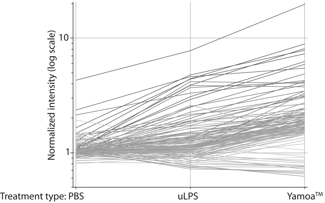
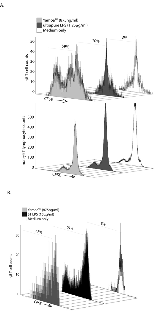
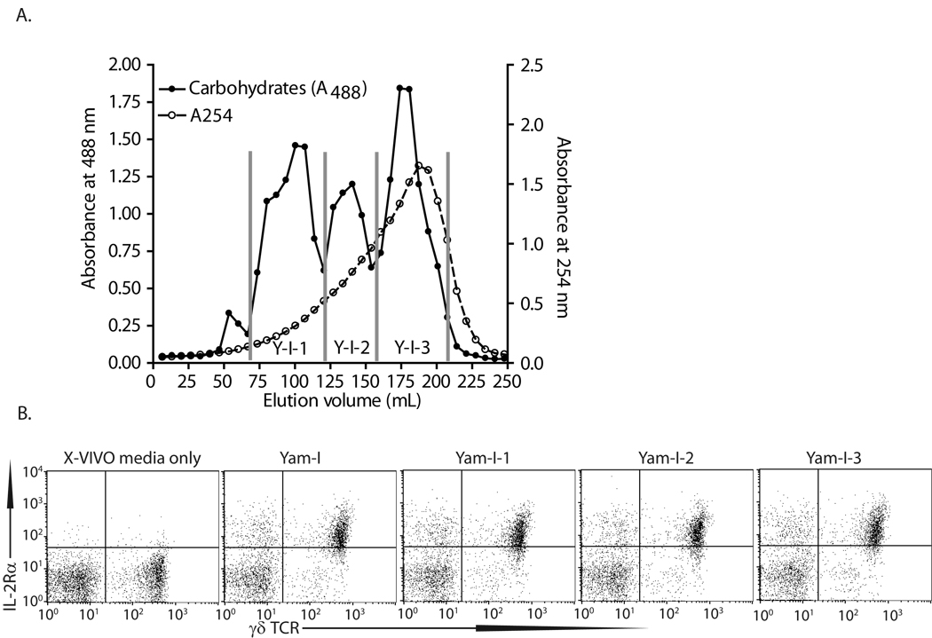
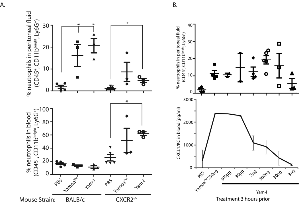
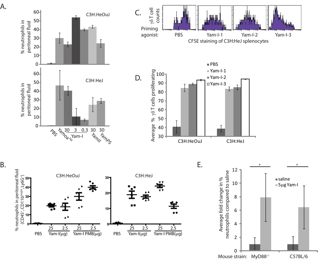
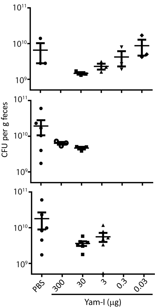
Yamoa (ground bark of Funtumia elastica tree) is marketed and sold as a dietary supplement with anecdotal therapeutic effects in the treatment of asthma and hay fever. We determined that Yamoa and Yamoa-derived polysaccharides affected innate immunity, in part, by priming gammadelta T cells. Gene expression patterns in purified bovine gammadelta T cells and monocytes induced by Yamoa were similar to those induced by ultrapure lipopolysaccharide (uLPS). In the presence of accessory cells, Yamoa had priming effects that were similar to those of LPS on bovine and murine gammadelta T cells, but much more potent than LPS on human gammadelta T cells. The bioactive component of Yamoa was delineated to a complex polysaccharide fraction (Yam-I). Intraperitoneal injection of Yamoa and Yam-I in mice induced rapid increases in peritoneal neutrophils directed by changes in chemokine expression. In support of a unique agonist found in Yam-I, similar peritonitis responses were also observed in TLR4- and MyD88-deficient mice. Therapeutic treatment with Yam-I resulted in decreased bacterial counts in feces from mice with Salmonella enterica serotype typhimurium (ST)-induced enterocolitis. This characterization of the immune stimulatory properties of polysaccharides derived from Yamoa suggests mechanisms for the anecdotal positive effects of its ingestion and that these polysaccharides show potential for application in innate protection from disease.
Figures
Similar articles
-
Innate immunity mediated by MyD88 signal is not essential for induction of lipopolysaccharide-specific B cell responses but is indispensable for protection against Salmonella enterica serovar Typhimurium infection.J Immunol. 2009 Feb 15;182(4):2305-12. doi: 10.4049/jimmunol.0801980. J Immunol. 2009. PMID: 19201885 Free PMC article.
-
Polysaccharides isolated from Açaí fruit induce innate immune responses.PLoS One. 2011 Feb 28;6(2):e17301. doi: 10.1371/journal.pone.0017301. PLoS One. 2011. PMID: 21386979 Free PMC article.
-
Mucosal lymphatic-derived gammadelta T cells respond early to experimental Salmonella enterocolitis by increasing expression of IL-2R alpha.Cell Immunol. 2007 Mar;246(1):8-16. doi: 10.1016/j.cellimm.2007.04.006. Epub 2007 Jun 15. Cell Immunol. 2007. PMID: 17574223 Free PMC article.
-
Adjuvant materials that enhance bovine γδ T cell responses.Vet Immunol Immunopathol. 2016 Nov 15;181:30-38. doi: 10.1016/j.vetimm.2016.03.010. Epub 2016 Mar 17. Vet Immunol Immunopathol. 2016. PMID: 27021513 Review.
-
Neutrophil influx during non-typhoidal salmonellosis: who is in the driver's seat?FEMS Immunol Med Microbiol. 2006 Apr;46(3):320-9. doi: 10.1111/j.1574-695X.2006.00051.x. FEMS Immunol Med Microbiol. 2006. PMID: 16553804 Review.
Cited by
-
Defining the nature of human γδ T cells: a biographical sketch of the highly empathetic.Cell Mol Immunol. 2013 Jan;10(1):21-9. doi: 10.1038/cmi.2012.44. Epub 2012 Oct 22. Cell Mol Immunol. 2013. PMID: 23085947 Free PMC article. Review.
-
Bupleurum polysaccharides attenuates lipopolysaccharide-induced inflammation via modulating Toll-like receptor 4 signaling.PLoS One. 2013 Oct 22;8(10):e78051. doi: 10.1371/journal.pone.0078051. eCollection 2013. PLoS One. 2013. PMID: 24167596 Free PMC article.
-
Oenothein B, a cyclic dimeric ellagitannin isolated from Epilobium angustifolium, enhances IFNγ production by lymphocytes.PLoS One. 2012;7(11):e50546. doi: 10.1371/journal.pone.0050546. Epub 2012 Nov 30. PLoS One. 2012. PMID: 23226309 Free PMC article.
-
Natural Polysaccharides and Their Derivates: A Promising Natural Adjuvant for Tumor Immunotherapy.Front Pharmacol. 2021 Apr 14;12:621813. doi: 10.3389/fphar.2021.621813. eCollection 2021. Front Pharmacol. 2021. PMID: 33935714 Free PMC article. Review.
-
An open-label dosing study to evaluate the safety and effects of a dietary plant-derived polysaccharide supplement on the N-glycosylation status of serum glycoproteins in healthy subjects.Eur J Clin Nutr. 2011 May;65(5):648-56. doi: 10.1038/ejcn.2010.263. Epub 2011 Jan 12. Eur J Clin Nutr. 2011. PMID: 21224866 Free PMC article.
References
-
- Holderness J, Jackiw L, Kimmel E, Kerns HMM, Radke M, Hedges JF, Petrie C, McCurley P, Glee PM, Palecanda A, Jutila MA. Select plant tannins induce IL-2Rα up-regulation and augment cell division in γδ T cells. J Immunol. 2007;179:6468–6478. - PubMed
-
- Hayday AC. [gamma][delta] cells: a right time and a right place for a conserved third way of protection. Ann Rev Immunol. 2000;18:975–1026. - PubMed
-
- Rivas A, Koide J, Cleary ML, Engleman EG. Evidence for involvement of the gamma, delta T cell antigen receptor in cytotoxicity mediated by human alloantigen-specific T cell clones. J Immunol. 1989;142:1840–1846. - PubMed
Publication types
MeSH terms
Substances
Grants and funding
LinkOut - more resources
Full Text Sources
Molecular Biology Databases
