. 2009 Aug 25;106(34):14466-71.
doi: 10.1073/pnas.0900190106. Epub 2009 Aug 7.

Essential global role of CDC14 in DNA synthesis revealed by chromosome underreplication unrecognized by checkpoints in cdc14 mutants

Affiliations

Essential global role of CDC14 in DNA synthesis revealed by chromosome underreplication unrecognized by checkpoints in cdc14 mutants

Stanimir Dulev et al. Proc Natl Acad Sci U S A. .

Abstract

The CDC14 family of multifunctional evolutionarily conserved phosphatases includes major regulators of mitosis in eukaryotes and of DNA damage response in humans. The CDC14 function is also crucial for accurate chromosome segregation, which is exemplified by its absolute requirement in yeast for the anaphase segregation of nucleolar organizers; however the nature of this essential pathway is not understood. Upon investigation of the rDNA nondisjunction phenomenon, it was found that cdc14 mutants fail to complete replication of this locus. Moreover, other late-replicating genomic regions (10% of the genome) are also underreplicated in cdc14 mutants undergoing anaphase. This selective genome-wide replication defect is due to dosage insufficiency of replication factors in the nucleus, which stems from two defects, both contingent on the reduced CDC14 function in the preceding mitosis. First, a constitutive nuclear import defect results in a drastic dosage decrease for those replication proteins that are regulated by nuclear transport. Particularly, essential RPA subunits display both lower mRNA and protein levels, as well as abnormal cytoplasmic localization. Second, the reduced transcription of MBF and SBF-controlled genes in G1 leads to the reduction in protein levels of many proteins involved in DNA replication. The failure to complete replication of late replicons is the primary reason for chromosome nondisjunction upon CDC14 dysfunction. As the genome-wide slow-down of DNA replication does not trigger checkpoints [Lengronne A, Schwob E (2002) Mol Cell 9:1067-1078], CDC14 mutations pose an overwhelming challenge to genome stability, both generating chromosome damage and undermining the checkpoint control mechanisms.


Conflict of interest statement

The authors declare no conflict of interest.

Figures
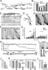
Fig. 1.
Fig. 1.
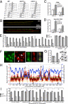
Nucleolar missegregation in cdc14 is consistent with rDNA under-replication. (A) Putative linking of rDNA sister chromatids due to under-replication, leading to the segregation failure. Only three repeats are shown for simplicity. Dotted lines - paths of replication forks. (B) rARS cannot maintain minichromosomes in cdc14 cells. The minichromosome stability was tested in isogenic strains under selective conditions at 23 °C. TRP1/CEN3 minichromosomes had various replication origins: full-length rDNA, short rARS-containing fragments, full-length ARS1 and a truncated (weakened) ARS1 (ars1′). Minichromosomes containing rARS (pAS1071) or full-length rDNA repeat (pAS1072) as a single replicator failed to propagate in cdc14 cells: all surviving Trp+ clones contained TRP1 integration. The rARS is not completely inactive in cdc14-1 cells, as indicated by the synergistic restoration of minichromosome stability when ars1′ and rARS are combined (last column). (C) Schematic of a single rDNA repeat progressing through replication stages. Southern hybridization probes and restriction sites used for 2D gel analyses (SfoI or StuI) are shown. (D) Replication intermediates distribution on a neutral-neutral two-dimensional gel. (E) Replication intermediates in asynchronous wild type and cdc14 mutant at permissive temperature. Wild type and cdc14-1 cells were grown at 23 °C. Purified DNA was digested with SfoI, enriched by ssDNA affinity chromatography, separated by 2D electrophoresis and analyzed by Southern blotting. The arrow: relative enrichment of the bubble arc. (F) cdc14 mutants arrested at 37 °C still contain rDNA replication intermediates. Cultures were arrested in G1 for 2 h at 23 °C, 1 h at 37 °C, and released at 37 °C for 3 h. Anaphase-arrested cells were analyzed as in E, except that rDNA was digested with StuI. To exclude the possibility that the observed retarded replication is a property of a hidden (non-CDC14) mutation, the cdc14-1 allele was cloned by PCR and reintroduced it into a wild-type strain by gene replacement (cdc14 integrated). (G) The cdc14 mutation delays progression of rDNA replication. Cultures were synchronously released from G1 arrest as in F, time-course cell samples were collected every 15 min and analyzed as in E. RFB and Y-arc signals were normalized to the 30 min and 15 min points, respectively, as signals at these time points were similar for wild type and cdc14. (H) BrdU incorporation in rDNA is progressively inhibited in cdc14 cells in the RNA PolI-transcribed region. Both strains were arrested as in G and BrdU was added to the media 30 min before the release from G1 into fresh YPD containing BrdU. BrdU incorporation was analyzed 2 h after release by ChIP/qPCR using rDNA probes as in (14). Shaded area is duplicated to show RFB surroundings. (I) Inhibition of transcription reduces underreplication in cdc14. The cdc14-1 mutant was arrested and released as in F in the absence or presence of thiolutin (87 μM), which was added in the mid-S-phase. The RFB and Y-arc signals were quantified and normalized to the corresponding values without thiolutin. (J) Curing cdc14 strains of 2-μm episomes significantly rescues nucleolar missegregation. Four independent cir0 clones were analyzed after arresting cells as in F. Minimum 200 nuclei were scored. (K) Metaphase delay suppresses both the rDNA replication delay and nucleolar segregation defects in cdc14 mutants. pGal:CDC20 and control cells were arrested in YPRG media in G1 and then arrested in metaphase (the Cdc20p depletion after addition of dextrose) with inactivated Cdc14p (37 °C). Cultures were grown in YPRG and split in half after the G1 arrest, one-half was released to YPRG at 37 °C for 5 h (Gal), while another half was released to YPD at 37 °C (to arrest cells in metaphase). Cells were harvested after 3 h, re-suspended in prewarmed YPRG media and incubated at 37 °C for 2 h (Gal Dex Gal). Continuous CDC20 expression (Gal) was used to control for possible overexpression effects unrelated to the mitotic arrest itself. Replication intermediates were analyzed as in E; the nucleolus segregation was scored by Sik1p-RFP, in 200 anaphase cells. (L) ARS1 integration in the rDNA locus partially rescues nucleolar nondisjunction in cdc14. Three control cdc14 cultures and three independent clones with ARS1 integration into rDNA were grown at 23 °C and arrested at 37 °C for 3 h. Cells were fixed and stained with DAPI, nucleolar segregation was monitored by Sik1p-RFP, with two sets of 100 cells scored per each strain.
Fig. 2.
Fig. 2.
cdc14-specific DNA under-replication is not limited to rDNA. (A) DNA content (FACS) in a time-course experiment. Cultures were grown at 23 °C, arrested in G1 with α-factor, shifted to 37 °C for 30 min in YPD + BrdU and then released at 37 °C in YPD + BrdU for combing analyses in (B–D). (B) Representative image of combed DNA fibers showing fully replicated molecules and unreplicated molecules (red only) detected both with anti-ssDNA (red) and anti-BrdU (green) antibodies. Analysis was performed both for total genomic DNA and specifically for rDNA after cutting genomic DNA with restriction enzymes (BamHI and XhoI) that cleave every 5–10 kb, but leave the 1.5-Mb rDNA array intact. (C and D) Percent of DNA fibers (≥100 kb) largely unreplicated after DNA combing. DNA was analyzed at the indicated time for both total genomic DNA and rDNA. Plotting the percentage of unreplicated DNA fibers (lacking BrdU signal for longer than 100 kb) with respect to time after release shows the progression and completion of S phase. (E) BrdU incorporation is reduced at telomeres in cdc14. Strains, arrest and labeling procedures are as in A. BrdU incorporation into DNA was analyzed (2 h after release into 37 °C/nocodazole from G1) by ChIP/qPCR using the probes covering the telomeric regions of two chromosomes (15R and 9L). Neither probe set is unique for these locations, usually corresponding to 2–6 sites at other telomeres. The early-replicating regions of chromosome III were analyzed similarly, as a control. (F) Telomere segregation is impaired in cdc14. Strains carrying SIR3:mRFP gene replacement were arrested as in Fig. 1F, stained with DAPI (green), and scored for Sir3p-mRFP signal position (red). Unlike wild type, cdc14 had blocked telomere segregation (arrows and higher magnification), and prominent additional signs of asymmetric segregation (asterisk). (G) Telomere missegregation in cdc14-3 is suppressed by the loss of two-μm episomes. Four independent cir0 clones were analyzed after arresting cells as in Fig. 1F. Minimum 200 nuclei were scored. TEL5R-adjacent chromosomal tags were as in (11). (H) Late-replicating regions have reduced BrdU incorporation in cdc14. ChIP-chip analysis of BrdU incorporation is shown for chromosome IV using the arrest/labeling as in A. Four experiments (SI Microarray Hybridization Data) were averaged for each strain and plotted as a cdc14 to wild type ratio. Red circles (i.e., individual microarray features) show the relative BrdU incorporation in cdc14. The black curve: 10-feature running average. Blue circles: replication timing of microarray features and origin positions (Lower) from (63). Arrows: locations of validation regions (in H). (I) cdc14 cells delay BrdU incorporation in late-replicating regions. ChIP/qPCR validation of ChIP-chip results in G was done as in E, with five regions (numbered according to G) where three non-overlapping 300-bp PCR probes were selected.
Fig. 3.
Fig. 3.
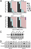
cdc14 mutants are proficient in DNA-damage checkpoints. (A and B) cdc14 mutants are properly arrested in response to DNA damage. Isogenic strains were grown to log phase at 23 °C, arrested in G1, and released at 37 °C for 2 h (A) with hydroxyurea (HU, 0.2 M) or (B) with MMS (0.01%). Cell were stained with DAPI and counted. (C) Phosphorylation of Rad53p is properly induced in cdc14 mutants in response to DNA damage. Cells were grown at 23 °C, arrested in G1 and released at 37 °C with either hydroxyurea (HU, 0.2 M) or MMS (0.01%), for 3 h. Checkpoint induction was monitored by western blotting with anti-Rad53 antibodies. (D) H2A is phosphorylated in cdc14 mutants in response to DSB. Cultures were grown in YPD to log phase at 23 °C, arrested in G1 and released in YPD at 37 °C for 3 h without any drug (37°), with hydroxyurea (HU, 0.2 M), with MMS (0.01%) or with nocodazole (Nz). The DSB presence was determined by western blotting with anti-phospho-S129 H2A antibody (64).
Fig. 4.
Fig. 4.
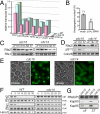
Both transcription and nuclear import defects lead to replication deficiency in cdc14. (A) Genes under control of Swi6p have partially decreased transcription in cdc14 mutants. Cells were arrested in G1 at 23 °C for 3 h, shifted at 37 °C for 30 min, and then released from G1 for 10 min. Expression levels were determined by qRT-PCR. All signals are normalized to TUB2 transcript, rDNA NTS1 - negative control. (B) Swi6p overexpression partially rescues the nucleolus segregation defect in cdc14. The cdc14 strain with Sik1p-RFP was transformed with either the pRS426gal vector (mock) or the pGAL:GST:SWI6 plasmid [derived from the library in (65)]. The cells were grown at 23 °C in plasmid-selective raffinose media, arrested with alpha factor in YPRaf for 2 h at 23 °C, galactose was added for 1 h/37 °C in the presence of alpha factor, and the cells were the released into YPRG/37 °C for 3 h. (C) RPA subunit imbalance in cdc14 mutants. Cells were arrested in G1 at 23 °C and released at 37 °C for 3 h untreated (37°), with hydroxyurea (HU, 0.2 M), with MMS (0.01%) or with nocodazole (NZ). Alpha-factor arrested cells were incubated at 37° for 1 h. Rfa2p detection: anti-GFP antibodies, Rfa1p detection: by specific antibodies. (D) Rfa2p phosphorylation is altered in cdc14 mutants even when expression is induced to wild-type level. Strains were arrested in G1 at 23 °C and then released to 37 °C or, in the presence of hydroxyurea (HU, 0.2 M), to 23 °C. Rfa2p detection as in E. The Mec1-dependent Rfa2p Ser-122 phosphorylation was detected with a specific antibody. Cdc28p levels (anti-PSTAIRE antibody): the loading control. (E) Rfa2p-GFP is largely delocalized from the nucleus in arrested cdc14 cells. Cultures were grown in YPD to log phase at 23 °C and shifted to 37 °C for 3 h. (F) Rfa1p and Rfa2p accumulate in cytoplasm in cdc14 mutants. Both cultures were arrested at 23 °C for 3 h, shifted at 37 °C for 30 min and released at 37 °C, samples were collected at indicated time points. Localization of Rfa1p and Rfa2p in nuclear (nuc) and cytoplasmic (cyt) fractions was analyzed by western blotting with specific polyclonal antibodies. Hmo1: chromatin and loading control. (G) Rfa2p does not interact with Kap60p-Kap95p importin complex in cdc14. Cells were arrested at 23 °C for 3 h, shifted at 37 °C for 30 min and split in half, with one half released from G1 at 37 °C. Rfa2p was immunoprecipitated from both G1 and anaphase cells, and analyzed with anti-Rfa2, anti-Kap60, and anti-Kap95 antibodies.

Similar articles

Cited by

References

    1. Stegmeier F, Amon A. Closing mitosis: The functions of the Cdc14 phosphatase and its regulation. Annu Rev Genet. 2004;38:203–232. - PubMed
    1. Trautmann S, et al. The S. pombe Cdc14-like phosphatase Clp1p regulates chromosome biorientation and interacts with Aurora kinase. Dev Cell. 2004;7:755–762. - PubMed
    1. Krasinska L, et al. Regulation of multiple cell cycle events by Cdc14 homologues in vertebrates. Exp Cell Res. 2007;313:1225–1239. - PubMed
    1. Bassermann F, et al. The Cdc14B-Cdh1-Plk1 axis controls the G2 DNA-damage-response checkpoint. Cell. 2008;134:256–267. - PMC - PubMed
    1. Visintin R, et al. Cfi1 prevents premature exit from mitosis by anchoring Cdc14 phosphatase in the nucleolus. Nature. 1999;398:818–823. - PubMed

Publication types

MeSH terms