Review
. 2009;85(7):217-28.
doi: 10.2183/pjab.85.217.

Hepatitis C virus utilizes lipid droplet for production of infectious virus

Affiliations
Review

Hepatitis C virus utilizes lipid droplet for production of infectious virus

Kazuya Ogawa et al. Proc Jpn Acad Ser B Phys Biol Sci. 2009.

Abstract

Hepatitis C virus (HCV) establishes a persistent infection and causes chronic hepatitis. Chronic hepatitis patients often develop hepatic cirrhosis and progress to liver cancer. The development of this pathological condition is linked to the persistent infection of the virus. In other words, viral replication/multiplication may contribute to disease pathology. Accumulating clinical studies suggest that HCV infection alters lipid metabolism, and thus causes fatty liver. It has been reported that this abnormal metabolism exacerbates hepatic diseases. Recently, we revealed that lipid droplets play a key role in HCV replication. Understanding the molecular mechanism of HCV replication will help elucidate the pathogenic mechanism and develop preventive measures that inhibit disease manifestation by blocking persistent infection. In this review, we outline recent findings on the function of lipid droplets in the HCV replication cycle and describe the relationship between the development of liver diseases and virus replication.


Figures

Fig. 1.
Fig. 1.
Structure of the HCV genome and the proteins produced by the genome. The HCV genome (upper panel) consists of a positive-sense RNA strand comprising about 10,000 nucleotides. Virus proteins are first produced as a precursor protein encoded in the largest open reading frame that comprises about 90% of the entire genome, then processed by cellular signal protease(s) followed by two virus proteases which are encoded by NS2–NS3, and NS3, respectively. 5′ one-thirds of the open reading frame encodes proteins for virus particle and the rest encodes non-structural virus proteins that function in virus-infected cells (lower panel).
Fig. 2.
Fig. 2.
HCV replication cycle. Since the detailed molecular mechanism of HCV infection/multiplication is yet to be clarified, this figure is drawn as a general view of replication of viruses in Flaviviridae, in which HCV is classified. In cells being established virus entry, most of the events for virus replication are conducted in cytoplasm.
Fig. 3.
Fig. 3.
Altered membranous structure observed in HCV genome replicating cells, in which synthesis of the positive as well as the negative HCV genome is conducted. (A) Electron micrographs of the generation of complex membranous structures (called membranous web) around the ER in HCV genome-replicating cells. The web structure is highlighted by the red circle. (The web image was from Gosert, R et al. (2003) J. Virol. 77, 5487 with permission). (B) Diagram of the distribution of NPC (non-structural proteins complex) and RC (replication complex) around the ER membrane. HCV-genome replicating cells treated with digitonin, a detergent that permeabilizes the plasma membrane but does not affect other membranes (e.g., ER-lumen and nuclear membrane)—were analyzed for virus proteins as well as HCV RNA synthesizing activity after incubation of exogenously added protease K. The majority of NS proteins were found to be sensitive to the hydrolysis; only a small portion of NS proteins were found to be resistant to the digestion because of the membrane’s protection. Further, the NS protein complex protected by membranous fraction is fully active to synthesize HCV RNA. From this result the NS protein complex exposed to outside of cytoplasm (NPC) and the NS protein complex surrounded with membrane structure (RC) are distinguished and found to have different functions (see text for details).
Fig. 4.
Fig. 4.
Characteristics and infectivity of virus particles produced into the culture supernatant. The characteristics of virus particles released from HCV(JFH1wild)-replicating cells (left) or cells replicating a mutated virus, JFH1AAA999, encoding proteins that cannot associate with lipid droplets (right) were analyzed by the sucrose density gradient centrifugation method. Dotted, red, and blue lines indicate the amount of virus particles measured by the amount of viral core protein, infectivity and viral RNA, respectively. Buoyant density of sucrose is indicated.
Fig. 5.
Fig. 5.
Membranous structures are frequently observed around the lipid droplets in JFH1-bearing cells. Electron micrographs of the lipid droplets and the surrounding area are shown. These show different images of lipid droplets in cells; a to c, lipid droplets in control HuH-7 cells, and d to f, those in JFH1 bearing HuH7 cells. Arrows indicate enriched membrane structure around the lipid droplet. Arrowhead shows rough ER membrane-like structure attached to the lipid droplet (The data was from Miyanari et al. (2007) Nat. Cell Biol. 9, 1089–1097).
Fig. 6.
Fig. 6.
Model of the association of the Core-coated lipid droplets with NPC- and RC-rich endoplasmic reticulum. Association of Core-coated lipid droplets with RC- and NPC-rich ER in HuH7 cells bearing the infectious HCV genome, JFH1 (A). Association of Core coated-lipid droplets with the lipid droplet was not observed in cells bearing the mutant JFH1, JFH1AAA999, expressing NS5A with mutations in domain-I (B), and in cells bearing the mutant JFH1, JFH1CL3B encoding NS5A with mutations in domain-III (C). Green circles represent lipid droplets. The small orange circles around the lipid droplets are the Core. The largest and the smallest circles with a mosaic represent NPC and RC, respectively. The color of NPC and RC indicates the noted mutations in NS5A in these complexes. Note that the NPC- and RC-rich ER lobes with a mutant NS5A (B and C) do not associate with the core-coated lipid droplet. However, as shown in (B) noninfectious virus particles are released into the culture medium, while NPC and RC with mutations in domain-III of NS5A do not produce virus particles, indicating the lack of virus assembly with this mutant. For NPC and RC localization, see the graphic in Fig. 3.
Fig. 7.
Fig. 7.
Structure of the HCV NS5A protein and mutant NS5A used for the analysis described in this paper., D-1- D-3 indicates domain-I - domain-III.
Fig. 8.
Fig. 8.
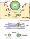
Model of the predicted role of the Core-coated lipid droplet in the production of infectious HCV. The association of the Core-coated lipid droplet with NPC-and RC-rich ER may enhance the interaction of HCV and VLDL. VLDL is generated frequently and enhanced in the micro-environment where lipid droplets associate with the ER. This increased concentration may increase the frequency of the association of HCV and VLDL. HCV/VLDL is released as an infectious particle with a low density, whereas HCV particles that are not associated with VLDL are secreted into the culture medium as noninfectious, dense particles. However, this model does not discriminate the possibility that noninfectious virus particle also associates with or is integrated with some lipoprotein like structure.
None

Similar articles

Cited by

References

    1. Lindenbach, B.D., Thiel, H.J. and Rice, C. (2007) The viruses and their replication. InField’s Virology (eds. Knipe, D.M. and Howley, P.M.). Lippincott-Raven, Philadelphia, pp. 1101–1152
    1. Tsukiyama-Kohara, K., Iizuka, N., Kohara, M. and Nomoto, A. (1992) Internal ribosome entry site within hepatitis C virus RNA. J. Virol. 66, 1476–1483 - PMC - PubMed
    1. Grakoui, A., Wychowski, C., Lin, C., Feinstone, S.M. and Rice, C.M. (1993) Expression and identification of hepatitis C virus polyprotein cleavage products. J. Virol. 67, 1385–1395 - PMC - PubMed
    1. Tanji, Y., Hijikata, M., Hirowatari, Y. and Shimotohno, K. (1994) Hepatitis C virus polyprotein processing: kinetics and mutagenic analysis of serine proteinase-dependent cleavage. J. Virol. 68, 8418–8422 - PMC - PubMed
    1. Pileri, P., Uematsu, Y., Campagnoli, S., Galli, G., Falugi, F., Petracca, R.et al. (1998) Binding of hepatitis C virus to CD81. Science 282, 938–941 - PubMed

Publication types