. 2009 Sep;37(17):5838-47.
doi: 10.1093/nar/gkp609. Epub 2009 Jul 28.

Molecular dissection of the prototype foamy virus (PFV) RNA 5'-UTR identifies essential elements of a ribosomal shunt

Affiliations

Molecular dissection of the prototype foamy virus (PFV) RNA 5'-UTR identifies essential elements of a ribosomal shunt

Mikhail Schepetilnikov et al. Nucleic Acids Res. 2009 Sep.

Abstract

The prototype foamy virus (PFV) is a nonpathogenic retrovirus that shows promise as a vector for gene transfer. The PFV (pre)genomic RNA starts with a long complex leader that can be folded into an elongated hairpin, suggesting an alternative strategy to cap-dependent linear scanning for translation initiation of the downstream GAG open reading frame (ORF). We found that the PFV leader carries several short ORFs (sORFs), with the three 5'-proximal sORFs located upstream of a structural element. Scanning-inhibitory hairpin insertion analysis suggested a ribosomal shunt mechanism, whereby ribosomes start scanning at the leader 5'-end and initiate at the downstream ORF via bypass of the central leader regions, which are inhibitory for scanning. We show that the efficiency of shunting depends strongly on the stability of the structural element located downstream of either sORFs A/A' or sORF B, and on the translation event at the corresponding 5'-proximal sORF. The PFV shunting strategy mirrors that of Cauliflower mosaic virus in plants; however, in mammals shunting can operate in the presence of a less stable structural element, although it is greatly improved by increasing the number of base pairings. At least one shunt configuration was found in primate FV (pre)genomic RNAs.


Figures
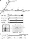
Figure 1.
Figure 1.
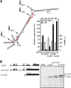
The PFV RNA leader inhibits translation of a downstream ORF. (A) Schematic representation of PFV (pre)genomic RNA. Viral ORFs are shown as shaded boxes. The secondary structure of the PFV RNA leader as predicted by the ‘M fold’ program is presented. The small open reading frames (sORFs) in the leader are named and indicated by ‘thick red lines superimposed’ on the structure. AD, sORFs; SD, splice donor; (A)n, poly(A) tail. The ‘circled in red section’ shows the 5′-proximal stem-loop (SL1). (B) Upper panel: mRNA constructs containing the chloramphenicol acetyl transferase (CAT) reporter ORF with three different leader sequences used for in vitro translation: LCAT RNA (the complete PFV leader); SL1CAT RNA; S1CAT (first 60 nt of the CaMV leader); and CAT RNA (50 vector nucleotides). Lower panels: (Left) in vitro translation of the constructs shown above in rabbit reticulocyte lysate (RRL); (right) translation efficiencies of capped (C) and uncapped (U) CAT mRNA containing either the S1 region (S1CAT RNA), or complete PFV leader (LCAT RNA) upstream of the CAT ORF. (C) 293T cells were transfected with a plasmid containing either the PFV 5′-proximal 50 nt (SL1 region; psgSL1CAT) or the PFV leader (psgLCAT) upstream of the CAT ORF. The psgGFP was used to monitor transfection efficiency. Levels of CAT expression relative to a GFP control after a 48-h incubation are indicated.
Figure 2.
Figure 2.
The PFV RNA leader re-directs expression to a downstream AUG in vivo and in vitro via ribosomal shunt. (A) Effect of insertion of a stable stem–loop structure (ΔG < –43.9 kcal/mol) at the positions indicated along the leader on in vivo expression of the CAT ORF in 293T cells. 293T cells were co-transfected with either pLCAT or the Kozak stem (ks) containing construct and psgGFP (a transfection control). Expression levels of GFP analyzed by immunoblotting using anti-GFP antibodies are shown in the panel below. mRNA levels were measured using RT-PCR and are presented with the LCAT RNA level set as 100% (white bars). The results shown represent the means of three independent experiments. (B) Representative translation in RRL of capped RNA constructs containing a short (D::CAT) or full-sized leader (LD::CAT). In LCAT RNA, ORF D overlaps CAT ORF as in the wild-type situation with GAG ORF, whereas in the capped LD::CAT and D::CAT RNAs, ORF D is in-frame with the CAT ORF.
Figure 3.
Figure 3.
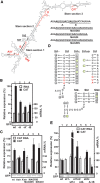
Identification of essential cis-acting shunting elements. (A) Schematic presentation of the leader indicating the main stem sections 1–3 and their mutated fragments (St1 and St2). Replacement of sORFs A and A′ by MAGDIS and variants is illustrated on the right. (B and C) Representative expression in 293T cells of DNA constructs containing alterations in the 5′-proximal sORFs: (B) [-A, -A′, -B], and (C) [wt, Astr, A′str, MAGDIS, MAGRIS and MAGDI). (D) Schematic representation of st1 and st2 of the PFV RNA leader. The sets of mutations are indicated by arrows; those introduced in st1- or in st2- are shown on the left panel in circles; the additional complementary mutations introduced in st1c or st2c to restore the formation of stem 1 are represented by squares. Mutations introduced to strengthen St1 (st1str) are shown on right panel in green. Mutations increasing base pairing in stem sections 1 and 2 are shown in green. (E) Effect of strength of base pairing in stem sections 1 and 2 on downstream CAT translation. Mutations destabilizing stem sections 1 (st1-) and 2 (st2-); restoring base pairing by compensatory mutations (st1c, st2c) or strong base pair insertion (st1str) and (st2str) were tested for their effect on CAT translation. Yields of CAT protein expressed as a percentage relative to the level of CAT detected upon transfection of LCAT mRNA are shown (black bars). mRNA levels are presented with the LCAT RNA level set as 100% (white bars). Expression levels of GFP analyzed by immunoblotting using anti-GFP antibodies are shown below (B), (C) and (E).
Figure 4.
Figure 4.
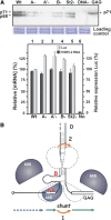
Expression of viral Gag depends on intact 5′-proximal sORFs and leader secondary structure. (A) A plasmid containing a proviral expression clone of PFV (czHSRV-2) under the control of the cytomegalovirus (CMV) immediate-early gene promoter was used to analyze the role of sORFs and secondary structure in translation initiation of the viral GAG ORF. (Left) Immunoblots of PFV GAG of lysates from 293T cells transfected with the following plasmids: either pczHSRV-2-wt or PFV leader mutants: A–, A′–, B–; st2–, or psgGAG (GAG) and non-transfected 293T cell extract (DNA-). pRL-TK containing Renilla LUC under the control of the thymidine kinase promoter was used as transfection control (LUC). Accumulation of GAG is shown after 48 h of incubation. The blots were incubated with primary antibodies against GAG and developed by staining with peroxidase-coupled goat anti-rabbit secondary antibodies. The Coomassie blue-stained gel is shown below, confirming equal loading of samples. (Right) LUC functional activity of the lysates (gray bars) described above was monitored and is shown together with mRNA levels analyzed by Q-PCR (black bars; LUC and HSRV-2 RNA values set at 100%). (B) Model showing redundancy in shunting strategy of PFV. Schematic presentation of the secondary structure stem of the PFV RNA leader with the GAG ORF. Broken outlines on the structure indicate the positions of 40S ribosomal subunits before and after shunting. Arrows show migration of scanning (dashed), translating (green) and shunting (red) ribosomes (40S and 60S subunits are shown as gray shapes, with outlines representing the subsequent path of the same 40S subunit). Scanning ribosomes enter the PFV RNA at the capped 5′-end and scan until they reach the 5′-proximal sORF start codon, where they initiate and translate this sORF. The translation event provides a specially modified shunt- and reinitiation-competent ribosome that bypasses the corresponding stem section and is ready to reinitiate close to the GAG AUG codon.
Figure 5.
Figure 5.
Comparison of primary structures of the pgRNAs of foamy viruses. The leader sequence preceding the GAG ORF is depicted by a thick line; sORFs are shown as boxes, with internal start codons indicated by vertical lines. Numbering within the leader is from the RNA 5′-end (except for SFVbab where the latter is unclear). The conserved sORFs preceding structured regions that can be used for shunting are in red. Arrows define the 5′- boundary of the elongated stem–loop structure. Leader sequences were derived from PFV proviral DNA; Simian foamy virus (SFVcpz); SFV-orangutan complete proviral genome, isolate bella (SFVora); SFV isolate OCOM1-5 long terminal repeat (SFVbab); SFV-3, (African green monkey complete genome; SFV-3agm); SFV-1 type 1 complete genome (SFV-1mac); Feline foamy virus complete genome (FeFV-1).

Similar articles

Cited by

References

    1. Kozak M. Initiation of translation in prokaryotes and eukaryotes. Gene. 1999;234:187–208. - PubMed
    1. Pestova T, Lorsch JR, Hellen CUT. The mechanism of translation initiation in eukaryotes. In: Mathews MB, Sonenberg N, Hershey JWB, editors. Translational Control in Biology and Medicine. NY: Cold Spring Harbor; 2007. pp. 87–128.
    1. Jackson R. Alternative mechanisms of initiating translation of mammalian mRNAs. Biochem. Soc. Trans. 2005;33:1231–1241. - PubMed
    1. Balvay L, Lopez LM, Sargueil B, Darlix JL, Ohlmann T. Translational control of retroviruses. Nat. Rev. Microbiol. 2007;5:128–140. - PMC - PubMed
    1. Ryabova LA, Pooggin MM, Hohn T. Translation reinitiation and leaky scanning in plant viruses. Virus Res. 2006;119:52–62. - PubMed

Publication types