. 2010 Jan;51(1):192-201.
doi: 10.1194/jlr.M900336-JLR200.

Intramembrane glycine mediates multimerization of Insig-2, a requirement for sterol regulation in Chinese hamster ovary cells

Affiliations

Intramembrane glycine mediates multimerization of Insig-2, a requirement for sterol regulation in Chinese hamster ovary cells

Peter C W Lee et al. J Lipid Res. 2010 Jan.

Abstract

Sterol-induced binding of endoplasmic reticulum (ER) membrane proteins Insig-1 and Insig-2 to SREBP cleavage-activating protein (Scap) and HMG-CoA reductase triggers regulatory events that limit cholesterol synthesis in animal cells. Binding of Insigs to Scap prevents proteolytic activation of sterol-regulatory element binding proteins (SREBPs), membrane-bound transcription factors that enhance cholesterol synthesis, by trapping Scap-SREBP complexes in the ER. Insig binding to reductase causes ubiquitination and subsequent proteasome-mediated degradation of the enzyme from ER membranes, slowing a rate-limiting step in cholesterol synthesis. Here, we report the characterization of mutant Chinese hamster ovary cells, designated SRD-20, that are resistant to 25-hydroxycholesterol, which potently inhibits SREBP activation and stimulates degradation of reductase. SRD-20 cells were produced by mutagenesis of Insig-1-deficient SRD-14 cells, followed by selection in 25-hydroxycholesterol. DNA sequencing reveals that SRD-20 cells harbor a point mutation in one Insig-2 allele that results in production of a truncated, nonfunctional protein, whereas the other allele contains a point mutation that results in substitution of glutamic acid for glycine-39. This glycine residue localizes to the first membrane-spanning segment of Insig-2 and is also present in the corresponding region of Insig-1. Mutant forms of Insig-1 and Insig-2 containing the Glu-to-Gly substitution fail to confer sterol regulation upon overexpressed Scap and reductase. These studies identify the intramembrane glycine as a key residue for normal sterol regulation in animal cells.


Figures
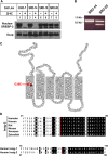
Fig. 1.
Fig. 1.
Characterization of 25-HC-resistant SRD-20 cells. A: CHO-7, SRD-14, SRD-15, and SRD-20 cells were set up on day 0 at 5 × 10⁵ cells per 100 mm dish in medium A containing 5% LPDS. On day 2, cells were switched to medium A supplemented with 5% LPDS, 10 µM sodium compactin, and 10 μM sodium mevalonate in the absence or presence of 2.5 µM 25-HC. After incubation at 37°C for 16 h, cells were harvested and subjected to cell fractionation as described in Experimental Procedures. Aliquots of the membrane (25 µg of protein/lane) and nuclear extract fractions (12 µg of protein/lane) were subjected to SDS-PAGE, the proteins were transferred to nylon membranes, and immunoblot analysis was carried out with 5 µg/ml IgG-7D4 (against SREBP-2) and 5 µg/ml IgG-9D5 (against Scap). Filters were exposed to film for 5 s to 1 min at room temperature. B: Aliquots of total RNA isolated from SRD-14 and SRD-20 cells were subjected to reverse transcription reactions; the resulting first-strand cDNA was used to amplify the Insig-2 cDNA. PCR products were subjected to electrophoresis on an agarose gel, which was subsequently stained with ethidium bromide and photographed. C: Amino acid sequence and membrane topology of human Insig-2. Glycine 39, which is found mutated to glutamic acid in SRD-20 cells, is highlighted in red. D: Comparison of amino acid residues in hamster, human, rat, mouse, xenopus, and zebrafish Insig-2. Identical residues are highlighted in black, and the conserved glycine-39 residue is highlighted in red. The lower shows the amino acid alignment between human Insig-1 and Insig-2. The residue in human Insig-1 corresponding to Gly-39 in human Insig-1 is Gly-95. All sequence alignments were performed using the CLUSTALW method (DNASTAR). GenBank accession numbers are as follows: AY112745 (human Insig-1); AF527632 (human Insig-2); AF527631 (mouse Insig-2); AF527629 (hamster Insig-2); AF527627 (zebrafish Insig-2); AY152392 (rat Insig-2); and NM_001011169 (xenopus Insig-2).
Fig. 2.
Fig. 2.
Expression of Insig-2 (G39E) and Insig-1 (G95E) in transfected CHO cells. A, B: CHO-7 cells were set up on day 0 at 5 × 10⁵ cells per 60 mm dish. On day 1, the cells were transfected with the indicated amount of wild-type or mutant Insig-1 and Insig-2 in 2 ml of medium A containing 5% LPDS. The total DNA in each dish was adjusted to 3 µg by the addition of pcDNA3 empty vector. Six hours after transfection, the cells were depleted of sterols by the direct addition of medium A containing 5% LPDS, 10 µM compactin, and 50 µM mevalonate (final concentrations). After 16 h at 37°C, cells were switched to medium A containing 5% LPDS and 10 µM compactin in the absence or presence of 2.5 µM 25-HC. Following incubation for 5 h, the cells were harvested and aliquots of the resulting membrane fractions (18 µg) were subjected to SDS-PAGE, and immunoblot analysis was carried out with 1 µg/ml monoclonal IgG-Myc (against Insigs). C: On day 0, SRD-13A cells were set up at 7 × 10⁵ cells per 60 mm dish. On day 1, the cells were transfected in 3 ml medium A containing 5% LPDS with 2 µg of pCMV-Scap and 0.1 µg of wild-type or 0.3 µg of mutant pCMV-Insig-2-Myc. After sterol depletion for 16 h at 37°C, the cells were switched to medium A containing 5% LPDS in the absence or presence of 2.5 µM 25-HC; some of the dishes also received 10 µM MG-132. Following incubation for 5 h, the cells were harvested, lysed, and subjected to SDS-PAGE. Immunoblot analysis was carried out with 1 μg/ml monoclonal IgG-9E10 and 1 μg/ml polyclonal IgG-R139 to detect Insig-2 and Scap, respectively. D, E: SRD-13A cells were set up on day 0 as in C and transfected on day 1 with 0.8 µg of pCMV-Scap and 0.05 µg of wild-type or 0.1 µg of mutant pCMV-Insig-1-Myc (E) 1.0 µg of wild-type or 3.0 µg of mutant pCMV-Insig-2-Myc (D). After incubation for 6 h 37°C, the cells were depleted of sterols. After 16 h, the cells were switched on day 2 to medium A containing 5% LPDS in the absence or presence of 1.25 µM 25-HC. After incubation for 5 h, cells were harvested, lysed, and immunoprecipitated with polyclonal anti-Myc as described in Experimental Procedures to precipitate Insig-1. Aliquots of the pellet (P; representing 0.25 dish of cells) and supernatant (S; representing 0.05 dish of cells) fractions of the immunoprecipitate were subjected to SDS-PAGE, and immunoblot analysis was carried out with 1 μg/ml polyclonal IgG-R139 (against Scap) and 1 μg/ml monoclonal IgG-9E10 (against Insig-1). All filters were exposed to film at room temperature for 3 s to 3 min.
Fig. 3.
Fig. 3.
Insig-1 (G95E) and Insig-2 (G39E) mediate sterol-regulated inhibition of SREBP-2 processing but not sterol-accelerated degradation of HMG-CoA reductase. A, B: SRD-13A cells were set up on day 0 as in Fig. 2 and transfected on day 1 with 0.5 µg of pCMV-Scap and 2 µg of pTK-HSV-SREBP-2. Cells in A were also transfected with either wild-type (0.1 µg) or mutant (0.2 µg) pCMV-Insig-1-Myc; cells in B were transfected with either wild-type (1 µg) or mutant (3 µg) pCMV-Insig-2-Myc in medium A containing 5% LPDS. The total DNA in each lane was adjusted to 5.5 µg per dish by the addition of pcDNA3 empty vector. On day 2, cells were switched to medium A containing 5% LPDS, 10 µM compactin, and 1% hydroxypropyl-β-cyclodextrin. After 1 h at 37°C, cells were washed twice with PBS and switched to medium A supplemented with 5% LPDS and 10 µM compactin in the absence or presence of 2.5 µM 25-HC. After 5 h, the cells were harvested, and membrane and nuclear extract fractions were prepared and subjected to SDS-PAGE followed by immunoblot analysis with 3 µg/ml monoclonal IgG-Myc (against Insig-1 or Insig-2) and 5 µg/ml IgG-9D5 (against Scap). The nuclear extract fractions were immunoblotted with a 1:10,000 dilution of monoclonal anti-HSV. Filters were exposed to film at room temperature for 1 to 15 s. C, D: CHO-7 cells were set up on day 0 as in Fig. 2 and transfected on day 1 with 1 µg of pCMV-HMG-Red-T7 (TM1-8) together with the indicated amount of wild-type and mutant pCMV-Insig-1-Myc (C) or pCMV-Insig-2-Myc (D). Six hours after transfection, cells were depleted of sterols for 16 h and subsequently switched to medium A containing 5% LPDS and 10 µM compactin in the absence or presence of 2.5 µM 25-HC plus 10 mM mevalonate. After 5 h, the cells were harvested; membrane fractions were isolated and subsequently immunoblotted with 1 µg/ml monoclonal IgG-T7 (against reductase) and 3 µg/ml monoclonal IgG-Myc (against Insig-1 or Insig-2). E: SRD-13A cells were set up on day 0 as in C and transfected on day 1 with 1 µg of pCMV-HMG-Red-T7 (TM1-8) and 0.01 µg of wild-type or 0.02 µg of mutant pCMV-Insig-1-Myc. After 6 h at 37°C, cells were depleted of sterols for 16 h. Following this incubation, the cells were switched to medium A containing 5% LPDS and 10 µM compactin in the absence or presence of 2.5 µM 25-HC plus 10 mM mevalonate; some of the cells also received 10 µM MG-132. After incubation for 20 min at 37°C, cells were harvested and lysed, and HMG-CoA reductase was immunoprecipitated with polyclonal anti-T7 coupled agarose beads as described in Experimental Procedures. Aliquots of the pellet (P; representing 0.25 dish of cells) and supernatant (S; representing 0.05 dish of cells) fractions of the immunoprecipitate were subjected to SDS-PAGE, and immunoblot analysis was carried out with 1 μg/ml anti-T7 (against HMG-CoA reductase) and 1 μg/ml monoclonal IgG-9E10 (against Insig-1). Filters were exposed to film at room temperature for 5 to 30 s.
Fig. 4.
Fig. 4.
Glycine-39 is required for the Insig-Insig coimmunoprecipitation. CHO-7 (left panel) or SRD-15 (right panel) cells were set up on day 0 at 5 × 10⁵ cells per 60 mm dish in medium A containing 5% LPDS. On day 1, the cells were transfected with various combinations of wild-type (0.1 µg) or mutant (0.4 µg) pCMV-Insig-1-Myc and pCMV-Insig-1-T7 in medium A supplemented with 5% LPDS. After incubation for 16 h at 37°C, the cells were harvested, lysed, and immunoprecipitated with polyclonal anti-T7 beads. The pellet (P; representing 0.25 dish of cells) and supernatant (S; representing 0.05 dish of cells) fractions of the immunoprecipitate were subjected to SDS-PAGE, and immunoblot analysis was carried out with 1 µg/ml monoclonal IgG-T7 and 3 µg/ml monoclonal IgG-Myc. Filters were exposed to film for 1 to 3 s.
Fig. 5.
Fig. 5.
Insig-1 (G95E) and Insig-2 (G39E) are defective in mediating sterol regulation of Scap and HMG-CoA reductase in SRD-15 cells. A: SRD-15 cells were set up on day 0 at 5 × 10⁵ cells per 60 mm dish in medium A supplemented with 5% LPDS. On day 1, cells were transfected with 0.5 µg of pCMV-Scap, 2 µg of pTK-HSV-SREBP-2, and either 0.1 µg wild-type pCMV-Insig-1-Myc, 0.2µg mutant pCMV-Insig-1-Myc, 1 µg wild-type pCMV-Insig-2-Myc, or 3 µg mutant pCMV-Insig-2-Myc in 5% LPDS as indicated. The total DNA in each lane was adjusted to 5.5 µg per dish by the addition of pcDNA3 empty vector. After 6 h at 37°C, cells were changed to sterol-depleting medium. On day 2, cells were switched to medium A containing 5% LPDS, 50 µM compactin, and 1% hydroxypropyl-β-cyclodextrin. After 1 h at 37°C, cells were washed twice with PBS and switched to medium A with 5% LPDS, 10 µM compactin, and the indicated concentration of 25-HC. After incubation for 5 h, cells were harvested and fractionated into membrane and nuclear extract fractions, which were subsequently subjected to SDS-PAGE. Immunoblot analysis was carried out with 3 µg/ml monoclonal IgG-Myc (against Insig-1 and Insig-2) and 5 µg/ml IgG-9D5 (against Scap). The nuclear extracts were immunoblotted with a 1:1,000 dilution of monoclonal anti-HSV IgG. Filters were exposed to film at room temperature for 3 to 15 s. B: SRD-15 cells were set up on day 0 and transfected on day 1 as described in A with 0.5 µg pCMV-Scap, 2 µg of pTK-HSV-SREBP-2, and the indicated amount of wild-type and/or mutant pCMV-Insig-1-Myc. Following sterol depletion for 16 h at 37°C, the cells were switched to medium A containing 5% LPDS, 50 µM compactin, and 1% hydroxypropyl-β-cyclodextrin. Following incubation for 1 h at 37°C, the cells were washed twice with PBS and switched to medium A with 5% LPDS and 50 µM compactin in the absence or presence of 25-HC. After 5 h, the cells were harvested; membrane and nuclear extract fraction were prepared and subsequently subjected to SDS-PAGE. Immunoblot analysis was carried out with 3 µg/ml monoclonal IgG-Myc (against Insig-1 and Insig-2) and 5 µg/ml IgG-9D5 (against Scap). The nuclear extract fractions were immunoblotted with a 1:10,000 dilution of monoclonal anti-HSV IgG. Filters were exposed to film at room temperature for 3 to 15 s.

Similar articles

Cited by

References

    1. Goldstein J. L., DeBose-Boyd R. A., Brown M. S. 2006. Protein sensors for membrane sterols. Cell. 124: 35–46. - PubMed
    1. Horton J. D., Shah N. A., Warrington J. A., Anderson N. N., Park S. W., Brown M. S., Goldstein J. L. 2003. Combined analysis of oligonucleotide microarray data from transgenic and knockout mice identifies direct SREBP target genes. Proc. Natl. Acad. Sci. USA 100: 12027–12032. - PMC - PubMed
    1. Nohturfft A., Yabe D., Goldstein J. L., Brown M. S., Espenshade P. J. 2000. Regulated step in cholesterol feedback localized to budding of SCAP from ER membranes. Cell. 102: 315–323. - PubMed
    1. DeBose-Boyd R. A., Brown M. S., Li W. P., Nohturfft A., Goldstein J. L., Espenshade P. J. 1999. Transport-dependent proteolysis of SREBP: relocation of site-1 protease from Golgi to ER obviates the need for SREBP transport to Golgi. Cell. 99: 703–712. - PubMed
    1. Goldstein J. L., Brown M. S. 1990. Regulation of the mevalonate pathway. Nature. 343: 425–430. - PubMed

Publication types

MeSH terms

Substances

LinkOut - more resources