

TCR-inducible PLZF transcription factor required for innate phenotype of a subset of gammadelta T cells with restricted TCR diversity
- PMID: 19617548
- PMCID: PMC2718370
- DOI: 10.1073/pnas.0903895106
TCR-inducible PLZF transcription factor required for innate phenotype of a subset of gammadelta T cells with restricted TCR diversity
Abstract
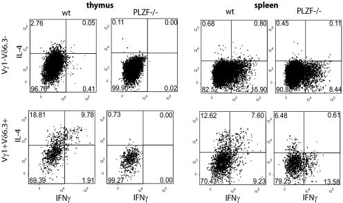
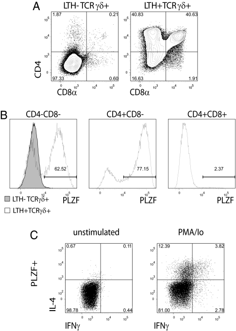
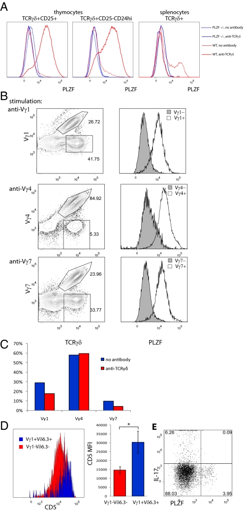
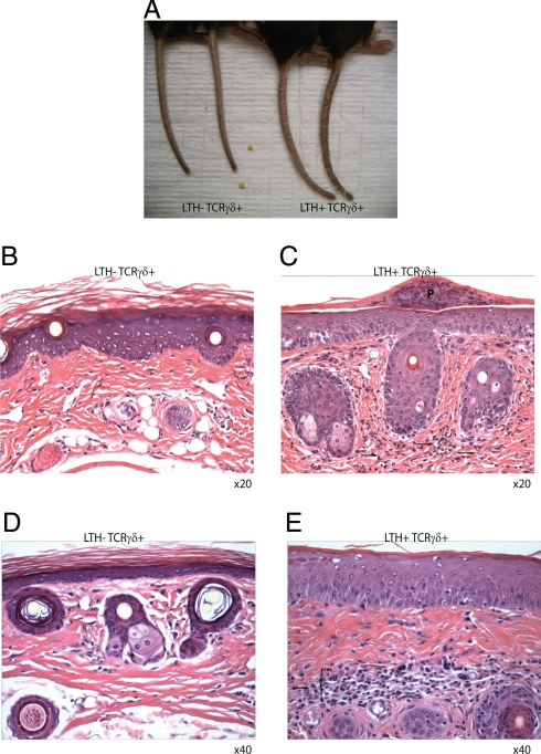
Some gammadelta and alphabeta T lymphocytes exhibit an "innate" phenotype associated with rapid cytokine responses. The PLZF transcription factor is essential for the innate phenotype of NKT cells. This report shows that PLZF is likewise responsible for the innate, NKT-like phenotype of Vgamma1+Vdelta6.3/Vdelta6.4+ cells. TCR cross-linking induced PLZF expression in all polyclonal immature gammadelta thymocytes, suggesting that agonist selection might be required for PLZF induction. Transgenic expression of Vgamma1Vdelta6.4 TCR was sufficient to support the development of large numbers of PLZF+ T cells, further supporting the importance of the TCR for PLZF induction. Interestingly, expression of this TCR transgene led to the development of spontaneous dermatitis.
Conflict of interest statement
The authors declare no conflict of interest.
Figures

Similar articles
-
Inhibitor of DNA binding 3 limits development of murine slam-associated adaptor protein-dependent "innate" gammadelta T cells.PLoS One. 2010 Feb 19;5(2):e9303. doi: 10.1371/journal.pone.0009303. PLoS One. 2010. PMID: 20174563 Free PMC article.
-
Critical role of TCR specificity in the development of Vγ1Vδ6.3+ innate NKTγδ cells.J Immunol. 2013 Aug 15;191(4):1716-23. doi: 10.4049/jimmunol.1203168. Epub 2013 Jul 12. J Immunol. 2013. PMID: 23851687
-
PLZF Controls the Development of Fetal-Derived IL-17+Vγ6+ γδ T Cells.J Immunol. 2015 Nov 1;195(9):4273-81. doi: 10.4049/jimmunol.1500939. Epub 2015 Sep 25. J Immunol. 2015. PMID: 26408661 Free PMC article.
-
When less is more: T lymphocyte populations with restricted antigen receptor diversity.J Immunol. 2014 Aug 1;193(3):975-6. doi: 10.4049/jimmunol.1401491. J Immunol. 2014. PMID: 25049427 Review. No abstract available.
-
Development of PLZF-expressing innate T cells.Curr Opin Immunol. 2011 Apr;23(2):220-7. doi: 10.1016/j.coi.2010.12.016. Epub 2011 Jan 21. Curr Opin Immunol. 2011. PMID: 21257299 Free PMC article. Review.
Cited by
-
Identification of phenotypically and functionally heterogeneous mouse mucosal-associated invariant T cells using MR1 tetramers.J Exp Med. 2015 Jun 29;212(7):1095-108. doi: 10.1084/jem.20142110. Epub 2015 Jun 22. J Exp Med. 2015. PMID: 26101265 Free PMC article.
-
Inherited human ITK deficiency impairs IFN-γ immunity and underlies tuberculosis.J Exp Med. 2023 Jan 2;220(1):e20220484. doi: 10.1084/jem.20220484. Epub 2022 Nov 3. J Exp Med. 2023. PMID: 36326697 Free PMC article.
-
Thymic signatures of tailored peripheral functions.Nat Immunol. 2012 Apr 18;13(5):431-3. doi: 10.1038/ni.2287. Nat Immunol. 2012. PMID: 22513325 No abstract available.
-
RORγt inhibition selectively targets IL-17 producing iNKT and γδ-T cells enriched in Spondyloarthritis patients.Nat Commun. 2019 Jan 2;10(1):9. doi: 10.1038/s41467-018-07911-6. Nat Commun. 2019. PMID: 30602780 Free PMC article.
-
PLZF play as an indirect facilitator of thymic retention for the innate-like T-cells to aquire innate-like functions.Cell Death Dis. 2018 Oct 11;9(10):1044. doi: 10.1038/s41419-018-1075-y. Cell Death Dis. 2018. PMID: 30310052 Free PMC article. No abstract available.
References
-
- Berg LJ. Signalling through TEC kinases regulates conventional versus innate CD8(+) T-cell development. Nat Rev Immunol. 2007;7:479–485. - PubMed
-
- Jameson J, Witherden D, Havran WL. T-cell effector mechanisms: Gammadelta and CD1d-restricted subsets. Curr Opin Immunol. 2003;15:349–353. - PubMed
-
- Hayday AC. γδ cells: A right time and a right place for a conserved third way of protection. Annu Rev Immunol. 2000;18:975–1026. - PubMed
-
- Hayday A, Tigelaar R. Immunoregulation in the tissues by γδ T cells. Nat Rev Immunol. 2003;3:233–242. - PubMed
-
- Eberl G, Littman DR. Thymic origin of intestinal αβ T cells revealed by fate mapping of RORγt+ cells. Science. 2004;305:248–251. - PubMed
Publication types
MeSH terms
Substances
Grants and funding
LinkOut - more resources
Full Text Sources
Other Literature Sources
Molecular Biology Databases
