. 2010 Jan;130(1):295-304.
doi: 10.1038/jid.2009.211.

Metalloproteinase-mediated, context-dependent function of amphiregulin and HB-EGF in human keratinocytes and skin

Affiliations

Metalloproteinase-mediated, context-dependent function of amphiregulin and HB-EGF in human keratinocytes and skin

Stefan W Stoll et al. J Invest Dermatol. 2010 Jan.

Abstract

Human keratinocytes (KCs) express multiple EGF receptor (EGFR) ligands; however, their functions in specific cellular contexts remain largely undefined. To address this issue, first we measured mRNA and protein levels for multiple EGFR ligands in KCs and skin. Amphiregulin (AREG) was by far the most abundant EGFR ligand in cultured KCs, with >19 times more mRNA and >7.5 times more shed protein than any other family member. EGFR ligand expression in normal skin was low (<8 per thousand of RPLP0/36B4); however, HB-EGF and AREG mRNAs were strongly induced in human skin organ culture. KC migration in scratch wound assays was highly metalloproteinase (MP)- and EGFR dependent, and was markedly inhibited by EGFR ligand antibodies. However, lentivirus-mediated expression of soluble HB-EGF, but not soluble AREG, strongly enhanced KC migration, even in the presence of MP inhibitors. Lysophosphatidic acid (LPA)-induced ERK phosphorylation was also strongly EGFR and MP dependent and markedly inhibited by neutralization of HB-EGF. In contrast, autocrine KC proliferation and ERK phosphorylation were selectively blocked by neutralization of AREG. These data show that distinct EGFR ligands stimulate KC behavior in different cellular contexts, and in an MP-dependent fashion.


Figures

Figure 1
Figure 1
EGFR ligand expression and shedding in human KCs and normal and organ cultured human skin. (A) Normal human KCs were plated at 5% confluence, grown as described in Material and Methods and harvested ~70–95% confluence. Relative EGFR ligand mRNA expression was analyzed by QRT-PCR. Values represent fold-change versus the control gene 36B4 times 103 (mean +/− standard error of the mean [SEM], n=8). Asterisk denotes a significant difference in AREG expression relative to other ligands with p <10−6. (B) KCs were plated in 60mm dishes and grown as described above and 24 h accumulation of shed EGFR ligands in the culture medium of near-confluent KC cultures was assayed using a Multiplex Ligand Assay as described in Material and Methods. Data are expressed as ng of shed growth factor protein per ml of KC culture medium, mean +/− SEM, n=8, *= p<0.0001 vs all other growth factors shown. Soluble HB-EGF could only be detected in 4 out of 8 samples. (C) Total RNA was isolated from full-thickness 3 mm punch biopsies of normal human skin either immediately (control) or from biopsies subjected to human skin organ culture for 4h or 24h and EGFR ligand mRNA was analyzed by QRT-PCR. Data represent fold-change versus the control gene 36B4 times 103, n= 5–8, *= p<0.05, **= p<0.007.
Figure 1
Figure 1
EGFR ligand expression and shedding in human KCs and normal and organ cultured human skin. (A) Normal human KCs were plated at 5% confluence, grown as described in Material and Methods and harvested ~70–95% confluence. Relative EGFR ligand mRNA expression was analyzed by QRT-PCR. Values represent fold-change versus the control gene 36B4 times 103 (mean +/− standard error of the mean [SEM], n=8). Asterisk denotes a significant difference in AREG expression relative to other ligands with p <10−6. (B) KCs were plated in 60mm dishes and grown as described above and 24 h accumulation of shed EGFR ligands in the culture medium of near-confluent KC cultures was assayed using a Multiplex Ligand Assay as described in Material and Methods. Data are expressed as ng of shed growth factor protein per ml of KC culture medium, mean +/− SEM, n=8, *= p<0.0001 vs all other growth factors shown. Soluble HB-EGF could only be detected in 4 out of 8 samples. (C) Total RNA was isolated from full-thickness 3 mm punch biopsies of normal human skin either immediately (control) or from biopsies subjected to human skin organ culture for 4h or 24h and EGFR ligand mRNA was analyzed by QRT-PCR. Data represent fold-change versus the control gene 36B4 times 103, n= 5–8, *= p<0.05, **= p<0.007.
Figure 2
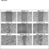
Figure 2. HB-EGF is a potent inducer of KC migration in vitro
(A) Scratch-wounded, confluent KC monolayers were incubated in basal M154 medium in the presence or absence of EGF (10 ng/ml), GM6001 (40 μM), MMPI-3 (25 μM), PD158780 (1 μM) and/or IgG225 (5 μg/ml). Scratch wounds were photographed by phase contrast microscopy after the indicated times. The results for individual panels are representative of at least two independent experiments. (B) Scratch-wounded KC monolayers were incubated in basal M154 in the presence or absence of neutralizing Abs against EGFR ligands alone or in combination (Ab cocktail) or isotype control Abs (each at 5 μg/ml) with and without EGF (10 ng/ml) for 20 h. (C) Confluent KC cultures were scratch-wounded and incubated for 18h to 24h as described above (Fig 2B). Digital images of representative areas were quantitated by measuring the scratch surface area using AxioVision-LE software (Carl Zeiss, Germany). Data are expressed as percent wound closure relative to controls at t = 0 h (% wound closure = [100 – ((scratch surface area at t = 18–24 h / surface area at t = 0 h)*100)], n=4 independent experiments, *= p ≤0.05 vs control. (D) NTERT, NTERT-TR, and NTERT or NTERT-TR stably infected with lentivirus constructs encoding transmembrane (tm) and soluble (s) AREG (NTERT-tmAREG, NTERT-sAREG) and transmembrane and soluble HB-EGF (NTERT-TR-tmHB-EGF, NTERT-TR-sHB-EGF) were grown to confluence, scratch wounded and incubated in basal KSFM medium for 36 h in the presence or absence of 40 μM GM6001 (MPI). HB-EGF expression in NTERT-TR-HB-EGF cells was induced with 1 μg/ml TET at least 3 h prior to scratch wounding. (E) Confluent cultures of NTERT and NTERT-TR with and without lentivirus-mediated expression of sAREG or sHB-EGF were scratched wounded and incubated for 18 h to 36 h in basal KSFM in the presence or absence of 40 μM GM6001 or 25 μM MMPI-3 as described above (Fig 2D). Tetracycline-induced expression of HB-EGF in NTERT-TRsHB-EGF cells is indicated by “+ TET”. Digital images of representative areas were quantitated by measuring the scratch surface area as described in Fig 2C, n=3, except N-TERT and N-TERT-TR n=2, *= p <0.05. (F) Equal amounts of RIPA cell lysates of control N-TERT or N-TERT stably infected with lentiviruses encoding AREG and HB-EGF were analyzed by ELISA for AREG or HB-EGF expression respectively, as described in Material and Methods. Data are expressed as ng of AREG or HB-EGF protein per ml of RIPA lysates, n=3 for AREG and controls and n=7–12 for HB-EGF and controls, *= p<0.001 relative to uninfected controls. (G) N-TERT-TR-sHB-EGF KC were grown and scratch wounded as described above and followed by incubation in basal KSFM medium for 24 h in the presence or absence of TET with and without 1 μg/ml mitomycin C. Results are representative of two separate experiments.
Figure 3
Figure 3
Neutralizing antibodies against AREG selectively block KC growth. KCs were incubated in basal M154 medium in the presence of blocking antibodies against AREG, BTC, EREG, HB-EGF, and TGF-α or isotype controls (each at 5 μg/ml), 40 μM GM6001 or 1 μM PD158780 with and without 1 ng/ml EGF. After an additional 7–9 days of incubation, cell growth was assessed using the 3-(4,5dimethyldiazol-2-yl)-2,5-diphenyltetrazolium (MTT) assay (Roche). KGM denotes fully supplemented KC growth medium. Each data point represents the mean OD570 reading for duplicate culture conditions. Data are expressed as percent of untreated controls, n= 3–5, with *= p<0.005 and **= p<0.0005 relative to “no treatment” controls.
Figure 4
Figure 4
Autocrine ERK phosphorylation is selectively regulated by AREG. KCs were growth factor-depleted for 48 h as described in Material and Methods and incubated for 2 h in fresh basal M154 in the presence or absence of neutralizing antibodies against EGF-like growth factors (each at 5 μg/ml) or GM6001 (40 μM), PD158780 (1 μM), or U0126 (10 μM) in the presence or absence of 100 ng/ml EGF. Equal amounts of RIPA were analyzed by western blotting with antibodies against ERK and phospho-ERK. Results are representative of three independent experiments.
Figure 5
Figure 5
HB-EGF mediates LPA-stimulated ERK phosphorylation. KCs were depleted of growth factors by incubation in basal M154 medium for 48 h followed by treatment with goat isotype control (for HB-EGF) or neutralizing Abs against EGFR ligands (each at 5 μg/ml) alone or in combination (anti-EGF-like GF), GM6001 (40 μM), PD158780 (1 μM), or U0126 (10 μM) for 1 h followed by treatment with EGF (10 ng/ml) or LPA (10 μM) for 10 or 20 min, respectively. Protein lysates were assayed for phospho-ERK and total ERK by western blotting as described in Material and Methods. The slight reduction of ERK phosphorylation in the presence of AREG Abs might be related to the component of the total signal that is due to basal ERK phosphorylation and therefore sensitive to AREG antibodies (see Fig. 4). The band underneath the phospho-ERK signal has been consistently observed with the mouse mono- but not with the rabbit polyclonal phospho-ERK Ab (Fig 4) from Cell Signaling Technologies. Its identity is unknown, however, it does not appear to be regulated in response to different treatments. The additional bands in the anti-AREG lane are due to a cross-reaction of the goat anti-mouse-HRP labeled secondary antibody with the AREG neutralizing antibody. The results shown are representative for three independent experiments.
Figure 6
Figure 6
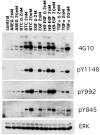
EGFR ligands differ in their ability to stimulate EGFR tyrosine phosphorylation. KCs were grown in M154 medium until ~ 40–50% confluence, growth factor depleted for 48 h, followed by growth factor treatment for 10 min at 37°C in fresh M154 as indicated. Equal amounts of RIPA lysates were analyzed by western blotting with antibodies indicated to the right of the panels. Total ERK served as a control for equal protein loading. Results are representative of three separate experiments.

Similar articles

Cited by

References

    1. Adam R, Drummond DR, Solic N, Holt SJ, Sharma RP, Chamberlin SG, et al. Modulation of the receptor binding affinity of amphiregulin by modification of its carboxyl terminal tail. Biochim Biophys Acta. 1995;1266:83–90. - PubMed
    1. Barnard JA, Graves Deal R, Pittelkow MR, DuBois R, Cook P, Ramsey GW, et al. Auto- and cross-induction within the mammalian epidermal growth factor-related peptide family. J Biol Chem. 1994;269:22817–22822. - PubMed
    1. Bhagavathula N, Nerusu KC, Fisher GJ, Liu G, Thakur AB, Gemmell L, et al. Amphiregulin and epidermal hyperplasia: amphiregulin is required to maintain the psoriatic phenotype of human skin grafts on severe combined immunodeficient mice. Am J Pathol. 2005;166:1009–1016. - PMC - PubMed
    1. Bhora FY, Dunkin BJ, Batzri S, Aly HM, Bass BL, Sidawy AN, et al. Effect of growth factors on cell proliferation and epithelialization in human skin. J Surg Res. 1995;59:236–244. - PubMed
    1. Burgess AW, Cho HS, Eigenbrot C, Ferguson KM, Garrett TP, Leahy DJ, et al. An open-and-shut case? Recent insights into the activation of EGF/ErbB receptors. Mol Cell. 2003;12:541–552. - PubMed

Publication types

MeSH terms