. 2009 Jul 10;35(1):58-69.
doi: 10.1016/j.molcel.2009.05.013.

Conformational changes associated with template commitment in ATP-dependent chromatin remodeling by ISW2

Affiliations

Conformational changes associated with template commitment in ATP-dependent chromatin remodeling by ISW2

Vamsi K Gangaraju et al. Mol Cell. .

Abstract

Distinct stages in ATP-dependent chromatin remodeling are found as ISW2, an ISWI-type complex, forms a stable and processive complex with nucleosomes upon hydrolysis of ATP. There are two conformational changes of the ISW2-nucleosome complex associated with binding and hydrolysis of ATP. The initial binding of ISW2 to extranucleosomal DNA, to the entry site, and near the dyad axis of the nucleosome is enhanced by ATP binding, whereas subsequent ATP hydrolysis is required for template commitment and causes ISW2 to expand its interactions with nucleosomal DNA to an entire gyre of the nucleosome and a short approximately 3-4 bp site on the other gyre. The histone-fold-like subunit Dpb4 associates with nucleosomal DNA approximately 15 bp from the ATPase domain as part of this change and may help to disrupt histone-DNA interactions. These additional contacts are independent of the ATPase domain tracking along nucleosomal DNA and are maintained as ISW2 moves nucleosomes on DNA.


Figures

Figure 1
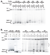
Figure 1. Template commitment of ISW2 to nucleosomes requires ATP
(A) Template commitment of ISW2 was examined with equimolar amounts of ISW2 and 70N0 nucleosomes (42 nM), ATP (800 µM), and excess competitor DNA added at the indicated times and incubated at 20°C. Samples were removed at the respective times (circled numbers) and analyzed by gel shift assay as shown on the right. In group IV ISW2 and nucleosomes were incubated together for 10 min before starting the initial start time point. Lane numbers correspond to the time points sampled and lane 1 is nucleosomes without ATP or ISW2. As nucleosomes change their translational position on DNA their electrophoretic mobility shifts from its original position (N1) to progressively more central positions (N2 and N3). (B) Template commitment assays with radioactive (r70N0) and non-radioactive (c70N0) nucleosomes were added at different reaction times to show template-committed complexes were resistant to competition by other nucleosomes. Nucleosome remodeling was initiated by incubating with 28 nM of either radiolabeled 70N0 (r70N0) or non-radiolabeled 70N0 (c70N0) and 28 nM of ISW2 at 20°C in the presence of 90 µM ATP. ISW2 at this molar concentration is completely bound with nucleosomes. Numbers above each time point indicate sample number and corresponding lane number. After each time point, the reaction was stopped and separated by 5% native gel electrophoresis.
Figure 2
Figure 2. ISW1a forms a stable, committed complex with nucleosomes upon addition of ATP
(A) Template commitment assays were done with ISW1a (42 nM) as described in Figure 1A. The location of the N1, N2, and N3 translational positions are indicated. (B) Reactions containing equimolar amounts of ISW1a and 70N0 (both 42 nM), were incubated at 20°C for the indicated times and analyzed by 5 % native gel electrophoresis the same as in Figure 1B for ISW2. Gel shift assay show the different extents of nucleosome mobilization with lane 1 being nucleosome alone and no ISW1a.
Figure 3
Figure 3. The N-terminal tail of histone H4 is required by ISW2 and ISW1a for template commitment
The experimental conditions are the same as in Figure 1A and Figure 2A, except that nucleosomes had recombinant histone H4 lacking the N-terminal tail instead of full length H4 and longer incubation times were used and reactions contained either ISW2 (A) or ISW1a (B).
Figure 4
Figure 4. Conformational changes in ISW2 while binding and hydrolyzing ATP
The interactions of ISW2 with nucleosomes (0N70) in the presence of 2 mM γ-S-ATP (A) or ADP (B) were mapped by hydroxyl radical DNA footprinting. End-positioned nucleosomes were incubated with saturating amounts of ISW2 at 30°C for 30 min and cleaved with hydroxyl radical. Overlays from the denaturing gels were plotted using Microsoft excel. The profiles for free nucleosome (black) and ISW2-nucleosome complex without (red) and with the appropriate nucleotide added (blue) are shown. Numbering of the peaks refers to the number of base pairs from the dyad axis. Shaded boxes are the sites which ISW2 protects and superhelical locations are shown on the X axis. (C) Site-directed mapping of nucleosome positions were carried out using nucleosomes that contain a unique cysteine at residue 53 of H2B. Heterogeneous nucleosomes (52 nM) were prebound with 95 nM ISW2 at 30ºC for 10 min. Remodeling was carried out at 18ºC for 5, 30 and 60 s in the presence of 90 µM of ATP and stopped with γ-S-ATP(400 µM) and SS-DNA (200 µg/µl). Mapping of the remodeled nucleosomes were carried out as described (Zofall et al., 2006). (D–F) ISW2-nucleosome complexes were DNA footprinted with ATP (90 µM), added and stopped after 5(D), 15 (E) and 90 s (F) followed by hydroxyl radical cleavage for 30 s. The profiles of ISW2 bound nucleosomes (black) are compared to those in which ATP is added (orange).
Figure 5
Figure 5. Large conformational changes in ISW2 interactions with nucleosomes occur even when translocation along DNA is blocked by gaps
DNA footprinting of ISW2-nucleosome complexes was done with either a 2 nucleotide gap in DNA (−24/−25, parts A and B) or a one nucleotide gap (−35, part C) from the dyad axis. The two nucleotide gap was designed to block DNA translocation by ISW2 and to prevent movement of the nucleosome along DNA by ISW2. The single nucleotide gap is so that nucleosomes are moved 10 bp by ISW2 and then prematurely stopped. DNA footprinting was performed and displayed as in Figure 4 with the initial regions of ISW2 bound shown as gray boxes and additional interactions that are observed in the presence of ATP as orange boxes. The extent of changes in the DNA footprint is further differentiated by light (partial) or darker (more complete) orange and gray boxes to indicate varying levels of protection. In (D) the location of the initial contacts of ISW2 (blue) and those caused by the addition of ATP (orange) with nucleosomes containing a 2 nucleotide gap 24 and 25 bp from the dyad axis are schematically shown. (E) The nucleosomal DNA sites that are strongly (red) and weakly (pink) protected by ISW2 with nucleosomes that contain a two nucleotide gap and have ATP added are highlighted in the crystal structure of the nucleosome. The histone proteins have the following color scheme: H2A (yellow), H2B (orange), H4 (green), and H3 (blue).
Figure 6
Figure 6. Dpb4 is associated with nucleosomal DNA ~15 bp in front of the ATPase domain during template commitment
(A–B) Site-specific DNA photoaffinity cross-linking was carried out under similar conditions to those in Figure 5 and Figure 6 except for photoreactive DNA. Nucleosomes were assembled with a series of photo-reactive probes spanning 17 to 67 bp from the dyad axis. DNA probes contain either a 2 (panel A) or 1 bp (panel B) gap 24/25 and 35 bp from the dyad axis, respectively, in order to determine the conformational changes of ISW2 complex during remodeling. As a control cross-linking was done at positions in the absence of gap and/or ATP for each probe position. Nucleosomes were incubated with ISW2, irradiated, and treated with DNaseI and S1 nuclease as described and the ISW2 subunits separated by 4–20% polyacrylamide SDS-PAGE. The cross-linked subunits of ISW2 complex is marked as Itc1, Isw2 and Dpb4. Bottom portion of the gel shows histone cross-linking and occasionally trace amounts of undigested probe are evident (*).
Figure 7
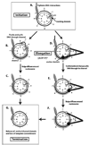
Figure 7. Model for initiation, elongation, and termination phases in ISW2 remodeling of nucleosomes
The nucleosome is represented as having its histone core in the center with only one DNA gyre (black line) evident and some extranucleosomal DNA. The initial superhelical locations on the DNA and histone core are separately labeled 1 thru 7 to track DNA movement relative to the histone octamer surface in this model. ISW2 interactions with DNA are shown in gray with the putative tracking, anchor, and channel domains labeled. The initiation (A), elongation (B–F), and termination (G) stages of ISW2 remodeling are displayed with two alternative models for elongation of the remodeling complex. In B–C the primary mode of nucleosome movement is tracking of DNA through the ATPase domain of ISW2. The other model for nucleosome movement uses large shifts in conformation represented as two black lines to help illustrate the protein structure that ties the two domains together. The distance between the domains changes as ATP is hydrolyzed due to conformational shifts.

Similar articles

Cited by

References

    1. Cairns BR. Chromatin remodeling: insights and intrigue from single-molecule studies. Nat Struct Mol Biol. 2007;14:989–996. - PMC - PubMed
    1. Corona DF, Eberharter A, Budde A, Deuring R, Ferrari S, Varga-Weisz P, Wilm M, Tamkun J, Becker PB. Two histone fold proteins, CHRAC-14 and CHRAC-16, are developmentally regulated subunits of chromatin accessibility complex (CHRAC) EMBO J. 2000;19:3049–3059. - PMC - PubMed
    1. Cramer P, Bushnell DA, Fu J, Gnatt AL, Maier-Davis B, Thompson NE, Burgess RR, Edwards AM, David PR, Kornberg RD. Architecture of RNA polymerase II and implications for the transcription mechanism. Science. 2000;288:640–649. - PubMed
    1. Cramer P, Bushnell DA, Kornberg RD. Structural basis of transcription: RNA polymerase II at 2.8 angstrom resolution. Science. 2001;292:1863–1876. - PubMed
    1. Dang W, Bartholomew B. Domain architecture of the catalytic subunit in the ISW2-nucleosome complex. Mol Cell Biol. 2007;27:8306–8317. - PMC - PubMed

Publication types

MeSH terms