. 2009 Jul;5(7):e1000506.
doi: 10.1371/journal.ppat.1000506. Epub 2009 Jul 3.

Differential gene expression patterns of EBV infected EBNA-3A positive and negative human B lymphocytes

Affiliations

Differential gene expression patterns of EBV infected EBNA-3A positive and negative human B lymphocytes

Marie L Hertle et al. PLoS Pathog. 2009 Jul.

Abstract

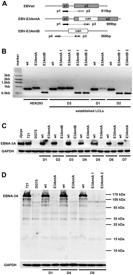
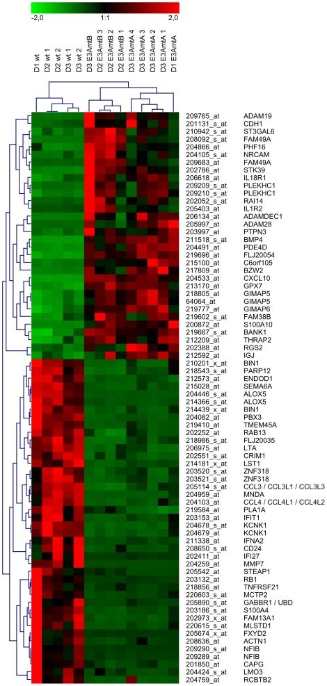
The genome of Epstein-Barr virus (EBV) encodes 86 proteins, but only a limited set is expressed in EBV-growth transformed B cells, termed lymphoblastoid cell lines (LCLs). These cells proliferate via the concerted action of EBV nuclear antigens (EBNAs) and latent membrane proteins (LMPs), some of which are rate limiting to establish a stable homeostasis of growth promoting and anti-apoptotic activities. We show here that EBV mutants, which lack the EBNA-3A gene, are impaired but can still initiate cell cycle entry and proliferation of primary human B cells in contrast to an EBNA-2 deficient mutant virus. Surprisingly, and in contrast to previous reports, these viral mutants are attenuated in growth transformation assays but give rise to permanently growing EBNA-3A negative B cell lines which exhibit reduced proliferation rates and elevated levels of apoptosis. Expression profiles of EBNA-3A deficient LCLs are characterized by 129 down-regulated and 167 up-regulated genes, which are significantly enriched for genes involved in apoptotic processes or cell cycle progression like the tumor suppressor gene p16/INK4A, or might contribute to essential steps of the viral life cycle in the infected host. In addition, EBNA-3A cellular target genes remarkably overlap with previously identified targets of EBNA-2. This study comprises the first genome wide expression profiles of EBNA-3A target genes generated within the complex network of viral proteins of the growth transformed B cell and permits a more detailed understanding of EBNA-3A's function and contribution to viral pathogenesis.


Conflict of interest statement

The authors have declared that no competing interests exist.

Figures

Figure 1
Figure 1. EBNA-3A negative viral mutants drive cell cycle entry of primary human B cells but show reduced long-term growth transformation capacity compared to EBVwt.
(A) Cell cycle entry of primary human B cells after infection with EBVwt and EBV-E3AmtA was analyzed by thymidine incorporation assays. 2×10⁵ B cells were plated on lethally irradiated MRC5 feeder layer and infected with 3000 GRUs of EBVwt, EBV-E3AmtA or EBVΔE2 or left uninfected. At day 0, 2, 4, 6, 8 and 14 p.i. cells were pulsed with [3H]-thymidine and analyzed for thymidine incorporation. Cultures with feeder only were set up in parallel and show the background levels of [3H]-thymidine measurements. Results are given as means from 6 single values and represent one of three independent experiments. (B) 2×10⁵ B cells derived from the same donor as analyzed in (A) were plated without feeder cells and infected with 3000 GRUs of EBVwt, EBV-E3AmtA or EBVΔE2 or left uninfected. At day 0, 2, 4, 8 and 14 p.i. cells were pulsed with [3H]-thymidine and analyzed for thymidine incorporation. Results are given as means from 6 single values. (C) The growth transformation efficiency of EBV-E3AmtA is weakly impaired but not abolished. To assess the growth transformation capacity of EBV-E3AmtA, the number of GRUs required to sustain B cell proliferation in a single well of a 96-well cluster plate was determined for EBVwt and EBV-E3AmtA in limiting dilution assays. Briefly, primary B cells derived from 4 individual donors were infected with serial dilutions of normalized viral supernatants and plated on lethally irradiated MRC5 feeder layer in groups of 48 cultures per degree of dilution. The percentage of wells per group with proliferating cells was determined 5 weeks p.i. with EBVwt and EBV-E3AmtA. The results are given as the mean percentage of proliferating cultures per group and the standard deviations are shown as error bars. The horizontal line at 63% (30 out of 48 wells plated) indicates the zero term of the Poisson equitation and identifies the average number of GRUs necessary to establish one proliferating B cell culture. Control infections with EBVΔE2 were set up in parallel for each donor but never gave rise to proliferating cultures.
Figure 2
Figure 2. EBNA-3A negative viral mutants give rise to permanently growing lymphoblastoid B cell lines.
(A) Diagram of the EBNA-3A gene locus in the EBVwt genome and the two distinct recombinant viral mutants. In order to generate EBV-E3AmtA a canamycin resistance gene (can) cassette was inserted into the second exon of EBNA-3A, while the entire coding sequence of EBNA-3A was replaced by a canamycin cassette in order to generate EBV-E3AmtB. The position of primers (p) used for analysis of the EBNA-3A locus in wt or modified EBV genomes and the expected product sizes are depicted. (B) LCLs established from 3 individual donors (D1–D3) by infection of B cells with EBVwt, EBV-E3AmtA or EBV-E3AmtB virus stocks and the corresponding HEK293 virus producing helper cell lines were tested for the correct state of the modified EBNA-3A gene locus by PCR. (C) EBNA-3A negative LCLs are not co-infected with EBV type II. Wt and EBNA-3A negative LCLs established from 7 individual donors (D1–D7) were analyzed for EBNA-3A expression by immunoblotting using a polyclonal α-EBNA-3A antibody detecting both, EBV type I and II encoded EBNA-3A. The EBV type II infected cell line Jijoye, the EBV type I infected cell line 721, and the EBV-negative cell line DG75 were included as controls. (D) The disruption of the EBNA-3A ORF by the canamycin cassette does not lead to expression of a truncated EBNA-3A protein. LCLs established by infection with EBV-E3AmtA were inspected for expression of a truncated EBNA-3A protein by immunoblotting using a monoclonal α-EBNA-3A antibody detecting an epitope within the first 50 amino acids of EBNA-3A. The respective wt LCLs, the EBV-positive cell line 721 and the EBV-negative cell line DG75 were included as controls. GAPDH immunodetection was used to control for equal loading of the lanes.
Figure 3
Figure 3. EBNA-3A negative LCLs proliferate at reduced rates and exhibit higher levels of apoptotic cells.
(A) EBNA-3A negative LCLs proliferate at reduced rates. Three independent wt and EBNA-3A negative LCLs derived from two individual donors were seeded at an initial density of 2×10⁵ cells per ml and viable cell counts were determined over a period of three weeks. Results are given as total numbers of viable cells corrected for the expansion of the cultures over time. The data are shown as mean values of triplicates. (B) EBNA-3A negative LCLs show reduced S-phase entry compared to wt LCLs. The cell cycle status of wt and EBNA-3A negative LCLs derived from two different donors was determined with the thymidine analogue BrdU, which was added to the respective cultures for 2 hrs prior to FACS analysis. The incorporated BrdU was stained with an APC-coupled anti-BrdU antibody and total DNA was counterstained with 7-AAD. Cells were inspected for G0/G1, S and G2/M phases of the cell cycle and for sub G1 DNA content by FACS analysis. (C) EBNA-3A negative LCLs exhibit higher levels of apoptotic cells compared to wt LCLs. The fraction of apoptotic cells in cultures of wt and EBNA-3A negative LCLs was determined by FACS analysis after staining of cells with Cy5-coupled Annexin V and 7-AAD. Cells that stain positive for Annexin V-Cy5 but negative for 7-AAD are in early apoptosis, while cells that stain positive for both are either in the end stage of apoptosis or dead.
Figure 4
Figure 4. EBNA-3A negative LCLs exhibit protein expression levels of EBV nuclear antigens similar to wt LCLs.
Wt and EBNA-3A negative LCLs derived from 5 individual donors were analyzed for expression levels of EBNA-3A, -3C, -2 and -1 by immunoblotting. The EBV-positive cell line 721 and the EBV-negative cell line DG75 were used as positive and negative control, respectively. GAPDH immunodetection was used to control for equal loading of the lanes.
Figure 5
Figure 5. EBV infected B cells show no EBNA-3A dependent changes in C-promoter usage and expression patterns of latent membrane proteins.
LCLs established from 7 different donors by infection of B cells with EBVwt or either EBV-E3AmtB (D2, D3) or EBV-E3AmtA (D1, D4–D7) were analyzed for (A) C-promoter usage and transcript levels of (B) LMP1, (C) LMP2A and (D) LMP2B by real-time RT-PCR. Mean values of triplicates are expressed as relative units after internal normalization for 18S rRNA levels.
Figure 6
Figure 6. Expression profiles of wt and EBNA-3A negative LCLs.
Shown are 74 probe sets displaying at least 4-fold changes in expression levels with significance p≤0.01 in EBNA-3A negative LCLs compared to wt LCLs. Vertical columns represent data obtained for each individual cell line by hybridization to a single microarray, while horizontal rows represent data obtained for a particular probe set across all cell lines. After normalization of expression values on a scale ranging from −2.0 to 2.0 for each probe set, an unsupervised hierarchical clustering analysis was performed, using Pearson correlation as a measure for similarity between genes and complete linkage as a clustering allocation algorithm. High expression values are represented by red, low expression values by green and medium values by black.
Figure 7
Figure 7. EBNA-3A negative LCLs exhibit higher expression levels of p16.
Total cellular protein extracts of EBNA-3A positive and negative LCLs derived from 7 donors were analyzed for p14 and p16 protein expression by western blot analysis. HeLa cell extracts served as positive control for both proteins. GAPDH immunodetection was used to control for equal loading of the lanes.

Similar articles

Cited by

References

    1. Tomkinson B, Robertson E, Kieff E. Epstein-Barr virus nuclear proteins EBNA-3A and EBNA-3C are essential for B-lymphocyte growth transformation. J Virol. 1993;67:2014–2025. - PMC - PubMed
    1. Maruo S, Johannsen E, Illanes D, Cooper A, Kieff E. Epstein-Barr Virus nuclear protein EBNA3A is critical for maintaining lymphoblastoid cell line growth. J Virol. 2003;77:10437–10447. - PMC - PubMed
    1. Lee W, Hwang YH, Lee SK, Subramanian C, Robertson ES. An Epstein-Barr virus isolated from a lymphoblastoid cell line has a 16-kilobase-pair deletion which includes gp350 and the Epstein-Barr virus nuclear antigen 3A. J Virol. 2001;75:8556–8568. - PMC - PubMed
    1. Kempkes B, Pich D, Zeidler R, Sugden B, Hammerschmidt W. Immortalization of human B lymphocytes by a plasmid containing 71 kilobase pairs of Epstein-Barr virus DNA. J Virol. 1995;69:231–238. - PMC - PubMed
    1. Bain M, Watson RJ, Farrell PJ, Allday MJ. Epstein-Barr virus nuclear antigen 3C is a powerful repressor of transcription when tethered to DNA. J Virol. 1996;70:2481–2489. - PMC - PubMed

Publication types

MeSH terms

Substances