. 2009 Sep;83(18):9258-72.
doi: 10.1128/JVI.00355-09. Epub 2009 Jul 1.

Protective and pathologic roles of the immune response to mouse hepatitis virus type 1: implications for severe acute respiratory syndrome

Affiliations

Protective and pathologic roles of the immune response to mouse hepatitis virus type 1: implications for severe acute respiratory syndrome

Aaruni Khanolkar et al. J Virol. 2009 Sep.

Abstract

Intranasal mouse hepatitis virus type 1 (MHV-1) infection of mice induces lung pathology similar to that observed in severe acute respiratory syndrome (SARS) patients. However, the severity of MHV-1-induced pulmonary disease varies among mouse strains, and it has been suggested that differences in the host immune response might account for this variation. It has also been suggested that immunopathology may represent an important clinical feature of SARS. Little is known about the host immune response to MHV-1 and how it might contribute to some of the pathological changes detected in infected mice. In this study we show that an intact type I interferon system and the adaptive immune responses are required for controlling MHV-1 replication and preventing morbidity and mortality in resistant C57BL/6J mice after infection. The NK cell response also helps minimize the severity of illness following MHV-1 infection of C57BL/6J mice. In A/J and C3H/HeJ mice, which are highly susceptible to MHV-1-induced disease, we demonstrate that both CD4 and CD8 T cells contribute to morbidity during primary infection, and memory responses can enhance morbidity and mortality during subsequent reexposure to MHV-1. However, morbidity in A/J and C3H/HeJ mice can be minimized by treating them with immune serum prior to MHV-1 infection. Overall, our findings highlight the role of the host immune response in contributing to the pathogenesis of coronavirus-induced respiratory disease.


Figures

FIG. 1.
FIG. 1.
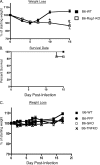
Protective role of type I IFN during intranasal MHV-1 infection of B6 mice. B6 wild-type (WT) mice were treated with either 200 μg of purified mouse IgG2a or anti-mouse NK1.1 (clone PK136) monoclonal antibody at 48 and 24 h prior to intranasal MHV-1 infection (105 PFU/mouse). Untreated B6-IFN-αβR-ΚΟ mice were also included in this analysis. A total of 5 to 10 mice were evaluated in each group for weight loss and survival for 10 days following infection. (A) Changes in body weight after infection were measured at the indicated time points. Statistically significant differences between the mean weights of B6 mice treated with mouse IgG2a and the other two groups of mice at each time point were determined using an unpaired Student's t test and are indicated by an asterisk. (B) Kaplan-Meier survival curves depict the percentage of surviving mice in each group at the indicated time points following infection.
FIG. 2.
FIG. 2.
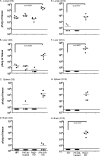
MHV-1 replication is controlled by both innate and adaptive immune responses. Virus titers were measured by plaque assays in the lungs, livers, spleens, and brains of wild-type (WT) B6 mice, anti-NK1.1-treated B6 mice, B6-Rag1-KO mice, and B6-IFN-αβR-KO mice 3 days (D3) and 10 days (D10) after intranasal infection with MHV-1 (105 PFU/mouse). All B6-IFN-αβR-KO mice succumbed to infection prior to day 10. Five mice per group were analyzed at each time point, and data are represented as the number of PFU/g of tissue from individual mice. Statistically significant differences in titers between wild-type B6 mice and the rest of the experimental groups were determined using an unpaired Student's t test and are indicated by an asterisk. The limit of detection is indicated in each graph by the horizontal line parallel to the x axis; mouse groups are indicated on the x axes of panels D and H, and values in graphs above are aligned similarly.
FIG. 3.
FIG. 3.
Susceptibility to MHV-1 infection is enhanced in Rag1-KO mice. B6 wild-type (WT), B6-PFP, B6-GKO, B6-TNFKO, and B6-Rag1-KO mice were intranasally infected with 105 PFU/mouse of MHV-1. A total of 5 to 10 mice were evaluated in each group, and mice were followed for weight loss (A and C) and survival (B) for 15 days following infection. Statistically significant differences in the mean weights of mice between the two groups were determined using an unpaired Student's t test and are indicated by an asterisk. Survival data are represented by Kaplan-Meier curves indicating the percentage of mice that survived the infection.
FIG. 4.
FIG. 4.
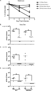
Passive immunization can control infection and morbidity following intranasal MHV-1 infection in susceptible strains of mice. A/J and C3H/HeJ mice (5 mice/group) received 300 μl of serum pooled from either naïve syngeneic donors or from syngeneic donor mice that had been infected intranasally 3 to 6 months previously with MHV-1. One day later all mice were intranasally infected with 1.5 × 10⁴ PFU/mouse of MHV-1. (A) Weight loss in recipient mice was measured at the time points indicated. Statistically significant differences between the weights of mice treated with naïve serum and those treated with immune serum were determined using an unpaired Student's t test and are indicated by an asterisk. (B to D) Mice were euthanized on day 4 (D4) after infection, and virus titers were determined by plaque assays in lungs, livers, and spleens and are represented as the number of PFU/g. Statistically significant differences between virus titers in mice treated with naïve serum (NS) and those treated with immune serum (IS) were determined using an unpaired Student's t test and are indicated by an asterisk. The limit of detection is indicated in each graph by the horizontal line parallel to the x axis. The limit of detection in the spleens of C3H/HeJ mice is lower than that in the spleens A/J mice and is indicated by the dashed line.
FIG. 5.
FIG. 5.
Reduced morbidity of MHV-1-infected A/J and C3H/HeJ mice following depletion of CD4 and CD8 T cells. A/J or C3H/HeJ mice (5 mice/group) were treated with 400 μg of either purified rat IgG or rat anti-mouse CD4 (clone GK1.5) or rat anti-mouse CD8 (clone 2.43) or both rat anti-mouse CD4 and CD8 1 day prior to, on the day of, and 7 days after intranasal infection with 5 × 10³ PFU/mouse of MHV-1. (A and C) Changes in body weight were monitored at the indicated time points. Statistically significant differences in the mean weights of mice treated with the control antibody and mice treated with both anti-CD4 and anti-CD8 antibodies were determined using an unpaired Student's t test and are indicated by an asterisk (*). Statistically significant differences between the mean weights of mice treated with the control antibody and the group of mice treated with the anti-CD4 antibody are indicated by a plus (+) symbol. No statistically significant differences were observed between the mean weights of mice treated with the control antibody and the groups of mice treated with the anti-CD8 antibody. These weight loss data are representative of two independent experiments. (B and D) Penh was determined using a whole-body plethysmograph at the indicated time points. Data are shown as means ± SEM, and statistically significant differences between the Penh values of control mice and mice depleted of both CD4 CD8 T cells were determined using an unpaired Student's t test and are indicated by an asterisk (*). Statistically significant differences between the Penh values of mice treated with the control antibody and the groups of mice treated with the anti-CD4 antibody are indicated by a plus (+) symbol. No statistically significant differences were observed between the Penh values of mice treated with the control antibody and the groups of mice treated with the anti-CD8 antibody. α, anti.
FIG. 6.
FIG. 6.
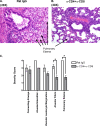
T-cell-dependent development of pulmonary pathology in MHV-1-infected mice. Whole lungs were harvested at day 10 postinfection with MHV-1 from C3H/HeJ mice treated with either rat IgG or anti-CD4 plus anti-CD8 monoclonal antibody as described in Materials and Methods. Hematoxylin and eosin sections were prepared as described in Materials and Methods (A and B). Slides were evaluated for fibrin deposition in alveoli and the development of pulmonary edema according to the severity grading scale described in the Materials and Methods (C). Data are shown as means ± SEM, and statistically significant differences between control mice and mice treated with both anti-CD4 and anti-CD8 monoclonal antibodies were determined using an unpaired Student's t test and are indicated by an asterisk. α, anti.
FIG. 7.
FIG. 7.
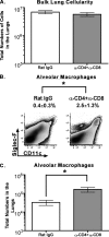
Increased numbers of alveolar macrophages in the lungs of MHV-1-susceptible mice treated with T-cell-depleting antibodies. Lungs were harvested following perfusion from MHV-1-infected C3H/HeJ mice that were treated with either rat IgG or both anti-CD4 and anti-CD8 monoclonal antibodies as described in Materials and Methods. Single-cell suspensions were prepared, and bulk lung cells were counted using trypan blue exclusion (A). The single-cell suspensions were subsequently stained with a panel of monoclonal antibodies as described in Materials and Methods, examined by flow cytometry for the presence of alveolar macrophages (CD11c+ Siglec-F+), and enumerated by multiplying the percentage of CD11c+ Siglec-F+ cells by the numbers of cells measured in the bulk lung cell population (B and C). Results are representative of two experiments (n = 3 to 5 per group). Data are shown as means ± SEM, and statistically significant differences between control mice and mice treated with both anti-CD4 and anti-CD8 monoclonal antibodies were determined using an unpaired Student's t test and are indicated by an asterisk. α, anti.
FIG. 8.
FIG. 8.
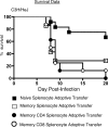
Memory splenocytes induce enhanced morbidity and mortality following intranasal MHV-1 infection. Naïve C3H/HeJ hosts received 2 × 10⁷ splenocytes/mouse by intravenous injection from either naïve C3H/HeJ donors (filled squares; n = 12) or C3H/HeJ mice that had been immunized with MHV-1 50 to 224 days previously (open squares; n = 8). The data shown for bulk naïve and memory splenocyte adoptive transfers are representative of three independent experiments. Additionally, separate groups of naïve recipients also received physiologically relevant input numbers of either purified bulk CD4(4.3 × 10⁶ cells/mouse; filled circles; n = 8) or bulk CD8 (1.8 × 10⁶ cells/mouse; open circles; n = 8) T cells obtained from donor mice that had been immunized with MHV-1 >200 days previously. The following day the recipient mice were intranasally challenged with 5 × 10³ PFU/mouse of MHV-1. Survival data for the four groups of mice are represented by Kaplan-Meier curves indicating the percentages of mice that survived the challenge.

Similar articles

Cited by

References

    1. Badovinac, V. P., S. E. Hamilton, and J. T. Harty. 2003. Viral infection results in massive CD8+ T cell expansion and mortality in vaccinated perforin-deficient mice. Immunity 18:463-474. - PubMed
    1. Badovinac, V. P., and J. T. Harty. 2006. Programming, demarcating, and manipulating CD8+ T-cell memory. Immunol. Rev. 211:67-80. - PubMed
    1. Bergmann, C. C., B. Parra, D. R. Hinton, C. Ramakrishna, K. C. Dowdell, and S. A. Stohlman. 2004. Perforin and gamma interferon-mediated control of coronavirus central nervous system infection by CD8 T cells in the absence of CD4 T cells. J. Virol. 78:1739-1750. - PMC - PubMed
    1. Bisht, H., A. Roberts, L. Vogel, A. Bukreyev, P. L. Collins, B. R. Murphy, K. Subbarao, and B. Moss. 2004. Severe acute respiratory syndrome coronavirus spike protein expressed by attenuated vaccinia virus protectively immunizes mice. Proc. Natl. Acad. Sci. USA 101:6641-6646. - PMC - PubMed
    1. Buchholz, U. J., A. Bukreyev, L. Yang, E. W. Lamirande, B. R. Murphy, K. Subbarao, and P. L. Collins. 2004. Contributions of the structural proteins of severe acute respiratory syndrome coronavirus to protective immunity. Proc. Natl. Acad. Sci. USA 101:9804-9809. - PMC - PubMed

Publication types

MeSH terms

Substances