

Involvement of vps33a in the fusion of uroplakin-degrading multivesicular bodies with lysosomes
- PMID: 19566896
- PMCID: PMC4494113
- DOI: 10.1111/j.1600-0854.2009.00950.x
Involvement of vps33a in the fusion of uroplakin-degrading multivesicular bodies with lysosomes
Abstract
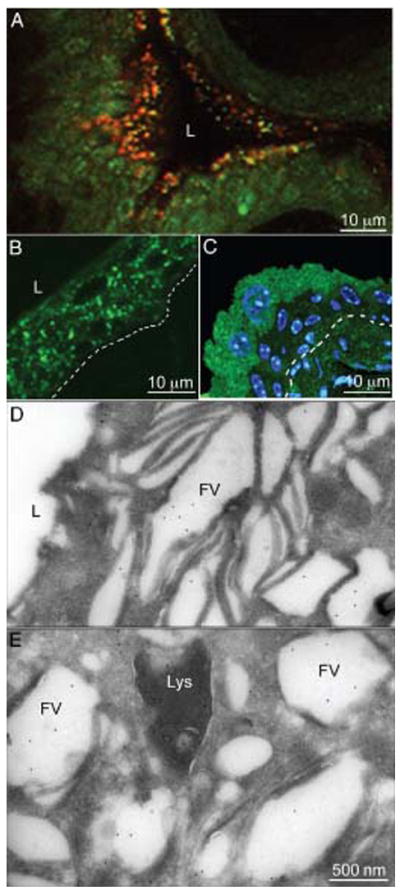
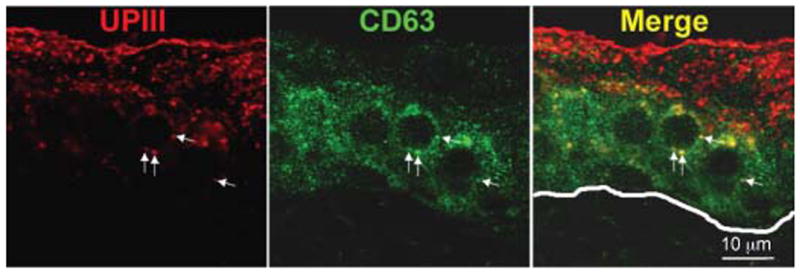
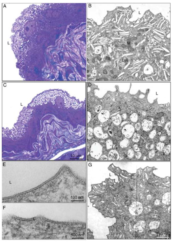
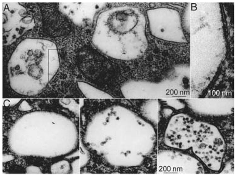
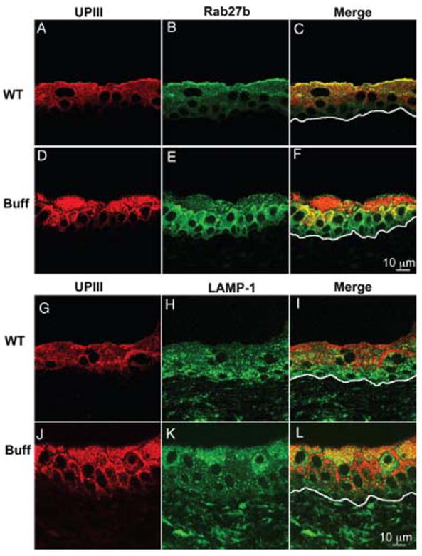
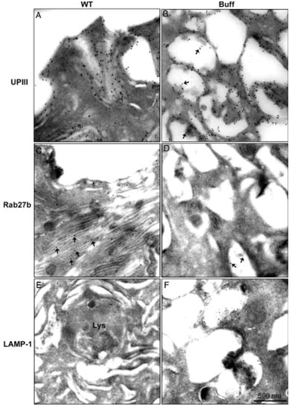
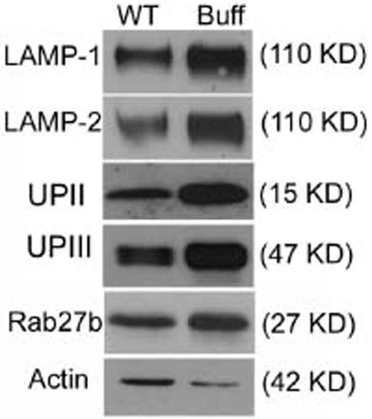
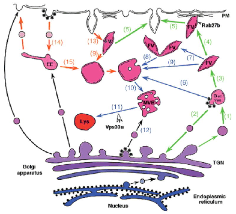
The apical surface of the terminally differentiated mouse bladder urothelium is largely covered by urothelial plaques, consisting of hexagonally packed 16-nm uroplakin particles. These plaques are delivered to the cell surface by fusiform vesicles (FVs) that are the most abundant cytoplasmic organelles. We have analyzed the functional involvement of several proteins in the apical delivery and endocytic degradation of uroplakin proteins. Although FVs have an acidified lumen and Rab27b, which localizes to these organelles, is known to be involved in the targeting of lysosome-related organelles (LROs), FVs are CD63 negative and are therefore not typical LROs. Vps33a is a Sec1-related protein that plays a role in vesicular transport to the lysosomal compartment. A point mutation in mouse Vps33a (Buff mouse) causes albinism and bleeding (Hermansky-Pudlak syndrome) because of abnormalities in the trafficking of melanosomes and platelets. These Buff mice showed a novel phenotype observed in urothelial umbrella cells, where the uroplakin-delivering FVs were almost completely replaced by Rab27b-negative multivesicular bodies (MVBs) involved in uroplakin degradation. MVB accumulation leads to an increase in the amounts of uroplakins, Lysosomal-associated membrane protein (LAMP)-1/2, and the activities of beta-hexosaminidase and beta-glucocerebrosidase. These results suggest that FVs can be regarded as specialized secretory granules that deliver crystalline arrays of uroplakins to the cell surface, and that the Vps33a mutation interferes with the fusion of MVBs with mature lysosomes thus blocking uroplakin degradation.
Figures
Similar articles
-
SNX31: a novel sorting nexin associated with the uroplakin-degrading multivesicular bodies in terminally differentiated urothelial cells.PLoS One. 2014 Jun 10;9(6):e99644. doi: 10.1371/journal.pone.0099644. eCollection 2014. PLoS One. 2014. PMID: 24914955 Free PMC article.
-
Mitochondrial lipid droplet formation as a detoxification mechanism to sequester and degrade excessive urothelial membranes.Mol Biol Cell. 2019 Nov 15;30(24):2969-2984. doi: 10.1091/mbc.E19-05-0284. Epub 2019 Oct 2. Mol Biol Cell. 2019. PMID: 31577526 Free PMC article.
-
MAL facilitates the incorporation of exocytic uroplakin-delivering vesicles into the apical membrane of urothelial umbrella cells.Mol Biol Cell. 2012 Apr;23(7):1354-66. doi: 10.1091/mbc.E11-09-0823. Epub 2012 Feb 9. Mol Biol Cell. 2012. PMID: 22323295 Free PMC article.
-
Formation of asymmetric unit membrane during urothelial differentiation.Mol Biol Rep. 1996;23(1):3-11. doi: 10.1007/BF00357068. Mol Biol Rep. 1996. PMID: 8983014 Review.
-
Uroplakins as markers of urothelial differentiation.Adv Exp Med Biol. 1999;462:7-18; discussion 103-14. doi: 10.1007/978-1-4615-4737-2_1. Adv Exp Med Biol. 1999. PMID: 10599409 Review. No abstract available.
Cited by
-
Urothelial endocytic vesicle recycling and lysosomal degradative pathway regulated by lipid membrane composition.Histochem Cell Biol. 2013 Feb;139(2):249-65. doi: 10.1007/s00418-012-1034-0. Epub 2012 Oct 12. Histochem Cell Biol. 2013. PMID: 23064746
-
Temporally and spatially controllable gene expression and knockout in mouse urothelium.Am J Physiol Renal Physiol. 2010 Aug;299(2):F387-95. doi: 10.1152/ajprenal.00185.2010. Epub 2010 Apr 28. Am J Physiol Renal Physiol. 2010. PMID: 20427471 Free PMC article.
-
Loss of the Sec1/Munc18-family proteins VPS-33.2 and VPS-33.1 bypasses a block in endosome maturation in Caenorhabditis elegans.Mol Biol Cell. 2014 Dec 1;25(24):3909-25. doi: 10.1091/mbc.E13-12-0710. Epub 2014 Oct 1. Mol Biol Cell. 2014. PMID: 25273556 Free PMC article.
-
Cell biology and physiology of the uroepithelium.Am J Physiol Renal Physiol. 2009 Dec;297(6):F1477-501. doi: 10.1152/ajprenal.00327.2009. Epub 2009 Jul 8. Am J Physiol Renal Physiol. 2009. PMID: 19587142 Free PMC article. Review.
-
SNX31: a novel sorting nexin associated with the uroplakin-degrading multivesicular bodies in terminally differentiated urothelial cells.PLoS One. 2014 Jun 10;9(6):e99644. doi: 10.1371/journal.pone.0099644. eCollection 2014. PLoS One. 2014. PMID: 24914955 Free PMC article.
References
Publication types
MeSH terms
Substances
Grants and funding
- P30 CA016056/CA/NCI NIH HHS/United States
- R01 EY012104-08/EY/NEI NIH HHS/United States
- CA-16056/CA/NCI NIH HHS/United States
- GM-43583/GM/NIGMS NIH HHS/United States
- R01 DK039753/DK/NIDDK NIH HHS/United States
- 52206/PHS HHS/United States
- R01 DK039753-19/DK/NIDDK NIH HHS/United States
- R01 EY012104/EY/NEI NIH HHS/United States
- R01 HL051480-10/HL/NHLBI NIH HHS/United States
- R01 GM043583/GM/NIGMS NIH HHS/United States
- P01 DK052206/DK/NIDDK NIH HHS/United States
- HL-51480/HL/NHLBI NIH HHS/United States
- DK-52206/DK/NIDDK NIH HHS/United States
- DK-39753/DK/NIDDK NIH HHS/United States
- R01 HL031698/HL/NHLBI NIH HHS/United States
- P01 DK052206-12/DK/NIDDK NIH HHS/United States
- HL-31698/HL/NHLBI NIH HHS/United States
- EY-12104/EY/NEI NIH HHS/United States
- R01 HL051480/HL/NHLBI NIH HHS/United States
LinkOut - more resources
Full Text Sources
Miscellaneous
