. 2009 Jun 30;7(6):e1000138.
doi: 10.1371/journal.pbio.1000138. Epub 2009 Jun 30.

Eps8 regulates axonal filopodia in hippocampal neurons in response to brain-derived neurotrophic factor (BDNF)

Affiliations

Eps8 regulates axonal filopodia in hippocampal neurons in response to brain-derived neurotrophic factor (BDNF)

Elisabetta Menna et al. PLoS Biol. .

Erratum in

Abstract

The regulation of filopodia plays a crucial role during neuronal development and synaptogenesis. Axonal filopodia, which are known to originate presynaptic specializations, are regulated in response to neurotrophic factors. The structural components of filopodia are actin filaments, whose dynamics and organization are controlled by ensembles of actin-binding proteins. How neurotrophic factors regulate these latter proteins remains, however, poorly defined. Here, using a combination of mouse genetic, biochemical, and cell biological assays, we show that genetic removal of Eps8, an actin-binding and regulatory protein enriched in the growth cones and developing processes of neurons, significantly augments the number and density of vasodilator-stimulated phosphoprotein (VASP)-dependent axonal filopodia. The reintroduction of Eps8 wild type (WT), but not an Eps8 capping-defective mutant, into primary hippocampal neurons restored axonal filopodia to WT levels. We further show that the actin barbed-end capping activity of Eps8 is inhibited by brain-derived neurotrophic factor (BDNF) treatment through MAPK-dependent phosphorylation of Eps8 residues S624 and T628. Additionally, an Eps8 mutant, impaired in the MAPK target sites (S624A/T628A), displays increased association to actin-rich structures, is resistant to BDNF-mediated release from microfilaments, and inhibits BDNF-induced filopodia. The opposite is observed for a phosphomimetic Eps8 (S624E/T628E) mutant. Thus, collectively, our data identify Eps8 as a critical capping protein in the regulation of axonal filopodia and delineate a molecular pathway by which BDNF, through MAPK-dependent phosphorylation of Eps8, stimulates axonal filopodia formation, a process with crucial impacts on neuronal development and synapse formation.


Conflict of interest statement

The authors have declared that no competing interests exist.

Figures

Figure 1
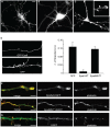
Figure 1. Eps8 is localized to sites of active actin polymerization in hippocampal neurons, and its removal increases neurite number.
(A–D) Eps8 localization in hippocampal neurons. Three days in vitro (3DIV) cultured hippocampal neurons were stained with anti-Eps8 antibody alone (A and A′) or in combination with either FITC-conjugated phalloidin to label F-actin (B), or anti-fascin (C) as indicated. The arrow in (A) indicates the axonal growth cone. In the inset (A′), the arrowheads indicate axonal protrusions (arrowheads). The arrowheads indicate Eps8- and actin- (B), or Eps8- and fascin-rich (C) filopodia protruding from the axon. (D) Actin rich axonal protrusions are also immunopositive for VASP. (E) Eps8, its binding partner Abi1, and the growth cone markers GAP43 and actin, are enriched in preparations of growth cone particles (GCPs) relative to low-speed supernatant brain homogenates (LSS). GCP preparations, described in Materials and Methods, were immunoblotted with the indicated antibodies. The immunoblot is a representative example of five independent experiments. (F) Lysates from wild-type (WT) and eps8 / (KO) hippocampi were immunoblotted with anti-Eps8 antibody. Molecular weight markers are indicated. (G and H) Cultured hippocampal neurons (1DIV) prepared from WT (G) or eps8 null (H) mice were fixed and stained with anti–beta-tubulin antibody. (I) Quantification of the number of neurites/neuron. Hippocampal neurons were prepared as described in (G and H). The difference between the numbers of neurites of eps8 / WT neurons is statistically significant (Mann-Whitney rank sum test, p≤0.001). Data are expressed as mean±standard deviation (SD); n = 897 examined WT neurons, and 760 examined eps8 KO neurons. Bars indicate 5 µm for (A, B, C, and D); 3 µm for a′; and 10 µm for (G and H). Double asterisks (**) in (F and G) indicate p<0.001.
Figure 2
Figure 2. Genetic removal of eps8 causes increased number of axonal filopodia in hippocampal neurons.
(A and B) Cultured hippocampal neurons (3DIV) from WT and eps8 / mice were fixed and stained with synaptobrevin/VAMP2 antibody. The selective sorting of synaptobrevin/VAMP2 immunoreactivity to a single process reveals that only one neurite is the putative axon. Bar indicates 10 µm. (A′ and B′) Still images of phase-contrast videos of the axonal shaft of cultured hippocampal neurons (3DIV) from WT and eps8 / mice (see Video S1). Arrowheads highlight axonal filopodia. (C) Representative examples of axonal shafts of WT and eps8 / neurons, labeled with an antibody against synaptobrevin/VAMP2. Bar indicates 10 µm. (D and E) Filopodia protruding from axons were costained with anti-synaptobrevin/VAMP2 antibody and phalloidin (D) to detect synaptic vesicle and filamentous actin, respectively, or with anti-fascin and phalloidin (E), as indicated. Bar indicates 7 µm. (F and G) Neurons from eps8 / mice display a significantly higher density of axonal filopodia. (F) Quantification of the percentage of neurons with filopodia in WT and eps8 / neurons. Neurons with more than 0.04 filopodia/µm were considered as filopodia-bearing neurons (Student t-test, p≤0.001). (G) Quantification of the density of axonal filopodia in WT and eps8 null hippocampal neurons (Mann-Whitney rank sum test, p≤0.001). n = 424 examined WT neurons and 621 examined eps8 KO neurons. Data are expressed as the mean±SD.
Figure 3
Figure 3. The regulation of filopodia density depends on Eps8 actin barbed-end capping activity.
(A–D) Primary eps8 / hippocampal neurons (3DIV) were infected with either WT Eps8-EGFP (A and C) or control EGFP (B) lentiviral vectors, then fixed and processed for epifluorescence. Neurons expressing Eps8 ([D] left top panel), but not those expressing EGFP ([D] left bottom panel), show a reduction in the density of axonal filopodia ([D] right panel; number of examined neurons: 120 Eps8wt-EGFP, 55 EGFP; Kruskal-Wallis one-way analysis of variance, p<0.001) Data are expressed as the mean±SEM. Of note, occasionally high levels of ectopically expressed Eps8 induced the presence of flattened structures, sometimes with club-like endings, ([C] and inset in [E]). (E–G) Primary eps8 / hippocampal neurons (3DIV) infected with either Eps8wt-EGFP (E) or Eps8MUT1-EGFP (number of examined neurons: 31 [F]), an Eps8 mutant devoid of actin-capping activity (Figure S3), or control EGFP (G) lentiviral vectors were fixed and processed for epifluorescence or stained with Texas Red phalloidin, as indicated. Merged images are also shown. Eps8MUT1 neurons do not display flattened membrane protrusions and are morphologically undistinguishable from EGFP-infected neurons (see also quantitation [D], right panel). Bar indicates 10 µm for (A, B, and C); 3 µm for the inset in (C), 5 µm for (D), and 8 µm for (E and G).
Figure 4
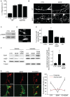
Figure 4. BDNF induces filopodia and regulates Eps8 intracellular localization through MAPK activation.
(A and B) BDNF fails to increase the number of filopodia in eps8 / neurons. Wild-type (WT) and eps8 / (KO) hippocampal neurons (3DIV) treated with BDNF (100 ng/ml for 20 min) or mock-treated (CTR), were fixed and stained to detect F-actin (B). Arrowheads highlight axonal filopodia. The density of filopodia was calculated as in Figure 2F. Data are expressed as the mean±SD. Kruskal-Wallis one-way ANOVA on ranks, Dunn's methods for comparison among groups: there is a significant difference between “WT Ctr” and “WT BDNF” (p<0.05), whereas there is no difference among “WT BDNF”, “eps8 KO Ctr,” and “eps8 KO BDNF” (p>0.05). (B) Example of axonal filopodia induced by BDNF in WT and eps8 / hippocampal neurons fixed and stained as described above. Bar indicates 5 µm. (C) Lysates of GCPs treated with BDNF (100 ng/ml for 20 min) were immunoblotted with the indicated antibodies to detect phosphorylated MAPK and NR1 subunit of NMDA receptor, used as loading control. (D) Hippocampal neurons treated with BDNF (100 ng/ml for 20 min) (BDNF) or mock-treated (CTR) were stained with anti-Synapsin I antibody (Syn). Bar indicates 2 µm. (E) Hippocampal neurons were incubated with PD98059 or vehicle as control, and treated with BDNF (100 ng/ml for 20 min) (BDNF) or mock-treated (CTR). Cells were fixed and processed as above. Neurons with axonal filopodia (neurons with more than 0.04 filopodia/µm were considered as positive) were quantified. BDNF treatment almost doubles the percentage of neurons with filopodia, whereas pretreatment with PD98059 prevents this effect (one-way ANOVA, post hoc Tukey test, double asterisks [**] indicate p≤0.001). Data are expressed as the mean±SD. (F) GCPs treated (BDNF) or not (CTR) with BDNF, prior or not PD98059 exposure, were fractionated using 1% Triton X100. Triton X100–soluble (sol) and -insoluble (ins) fractions were immunoblotted with the indicated antibodies. Synaptotagmin I (Stg) and tubulin were used as controls. Right graph: quantification of the Eps8 distribution to Triton X100–soluble fraction was performed by densitometry and normalized using synaptotagmin I (one-way ANOVA, post hoc Tukey test, p≤0.001). (G) Left: vehicle or PD98059-incubated hippocampal neurons (3DIV) were mock-treated or treated for 20 min with 100 ng/ml BDNF, as indicated. Cells were fixed and stained with anti-Eps8 antibody (red) and FITC-phalloidin to detect F-actin (green). Merged images are shown. Note that Eps8 immunoreactivity is virtually lost from filopodia upon BDNF treatment and that PD98059 prevents this relocalization. Right graph: quantitation of Eps8 fluorescence intensity measured in filopodia and axonal shafts under the different experimental conditions. Error bars represent SEM. Quantitation has been carried out by ImageJ software on 30 images from three independent experiments, filopodia are identified by labeling with phalloidin (one-way ANOVA, p<0.001 in filopodia and p>0.05 in axonal shaft). Bar indicates 5 µm. Note that Eps8 immunoreactivity is significantly reduced from filopodia upon BDNF treatment, and restored by PD98059.
Figure 5
Figure 5. Eps8 is phosphorylated by MAPK.
(A) 2-D immunoblot analysis of Eps8 from synaptosomes mock-treated (CTR) or treated with BDNF (BDNF) or pretreated with PD98059 before BDNF stimulation (PD+BDNF), or treated with BDNF followed by incubation with alkaline phosphatase (BDNF+AP). Note the appearance, upon BDNF application, of additional and more intense Eps8 immunopositive spots with more acidic isoelectric points (spots: 6–9). These spots disappear by preincubating synaptosomes with alkaline phosphatase. Insets show the analysis of synaptosomes treated as above, resolved by monodimensional SDS-PAGE and immunoblotted with anti–phospho-MAPK (upper band) or anti-ribophorin (lower band), the latter used as loading control. (B) 2-D immunoblot analysis of Eps8 in GCPs in the absence (CTR) or the presence of BDNF. (C) Recombinant purified His-Eps8 (1–821) or recombinant GST-fused Eps8 fragments (5 µg) were incubated with 10 µCi [γ-32P]-ATP in the presence (+) or absence (−) of purified MAPK (20 ng) in kinase buffer (see Materials and Methods), resolved by SDS-PAGE, stained with Coomassie Blue (lower panel), and subjected to autoradiography (upper panel). The red arrow indicates the shift of Eps8 due to phosphorylation. (D) Recombinant purified GST-Eps8 535–821 wild-type (WT) or S624A, T628A (SATA) mutant (5 µg) were incubated with 10 µCi [γ-32P]-ATP in the presence (+) or absence (−) of purified MAPK (20 ng) in kinase buffer (MB), resolved by SDS-PAGE, stained with Coomassie Blue (lower panel) and subjected to autoradiography (upper panel). In (C and D), MBP (myelin basic protein) was used as a control substrate.
Figure 6
Figure 6. The phosphomimetic Eps8 (Eps8-SETE) mutant has a 10-fold reduced barbed-end capping activity with respect to Eps8 WT or phosphoimpaired Eps8 (Eps8-SATA) mutant when in complex with Abi1.
(A) The indicated, increasing concentrations of equimolar amounts of purified His–Eps8 WT, SATA, and SETE (Eps8, SATA, and SETE) and GST-Abi1 were incubated with 2.5 µM G-actin (10% pyrenyl-labeled) in polymerization buffer. Actin polymerization was initiated by spectrin–actin seeds as described in Materials and Methods. GST was used as a control. (B) The relative inhibition of barbed-end polymerization rate at the indicated concentrations of the Eps8-WT:Abi1, Eps8-SATA:Abi1, and Eps8-SETE:Abi1 complexes is shown. Under suboptimal, nonsaturating conditions (100 nM concentrations), the Eps8-SETE:Abi1 complex is less effective in inhibiting barbed-end growth than Eps8 WT or Eps8-SATA:Abi1 complexes. Three independent experiments were performed. Error bars indicate SEM. (C) The relative inhibition of barbed-end polymerization at the various, indicated concentrations of the WT Eps8:Abi1 or Eps8-SETE:Abi1 complexes. The concentrations of Eps8-WT:Abi1 and Eps8-SETE:Abi1 at which half-maximal inhibition (IC50) was observed were 60 and 200 nM, respectively, which correspond to a concentration of the respective complexes of: Eps8(WT):Abi1 = 0.15 nM; Eps8-SETE:Abi1 = 1.5 nM (based on calculated thermodynamic constant of association between Eps8 and Abi1 (K d = 35 µM [72]). The data shown are representative of four independent experiments with similar results. (D) Eps8 phosphomutants bind Abi1 similar to Eps8 WT. Lysates of cells expressing GFP-Eps8 WT or SATA or SETE (indicated at the top) were incubated with immobilized GST, or GST-PIN1, used as negative control, or GST-Abi1 (asterisk [*] in the figure). The lower bands on the GST-Abi1 lanes are likely premature termination or degradative products of the Abi1 moiety of the fusion protein. Lysates and bound proteins were immunoblotted with the indicated antibodies (on the right). IVB, in vitro bound.
Figure 7
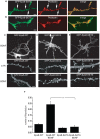
Figure 7. Eps8-SATA is preferentially localized along actin-rich structures and inhibits BDNF-induced filopodia.
(A and B) Hippocampal neurons (3DIV) were transfected by calcium phosphate with GFP-Eps8-SATA (A), GFP-Eps8-SETE (B) and examined 20 h after transfection. Cells were fixed and stained with TRITC phalloidin (red) and processed for epifluorescence to detect F-actin and Eps8 (green), respectively. Merged images are shown. Bar indicates 5 µm. (C–E) BDNF induces dispersion of WT Eps8, but not of Eps8-SATA or Eps8-SETE. Hippocampal neurons (3DIV) transfected with GFP-Eps8-WT (C), GFP-Eps8-SATA (D), or GFP-Eps8-SETE (E) were examined 20 h after transfection, upon stimulation with 100 ng/ml BDNF (BDNF) or mock-treatment (CTR). Cells were fixed and processed for epifluorescence as described above. Bar indicates 10 µm in the upper panels and 5 µm in the magnified axons shown in the lower panels. (F) Quantification of the density of filopodia in cultured hippocampal neurons (3DIV) transfected with GFP-Eps8-WT (n = 26) or GFP-Eps8-SATA (n = 34) and analyzed 20 h after transfection, either untreated or upon 20-min exposure to BDNF (100 ng/ml). Data are expressed as the mean±SD (Student t-test, double asterisks [**] indicate p≤0.001).

Comment in

  • The finer points of filopodia.
    Lundquist EA. Lundquist EA. PLoS Biol. 2009 Jun 30;7(6):e1000142. doi: 10.1371/journal.pbio.1000142. Epub 2009 Jun 30. PLoS Biol. 2009. PMID: 19564901 Free PMC article.

Similar articles

Cited by

References

    1. Cingolani LA, Goda Y. Actin in action: the interplay between the actin cytoskeleton and synaptic efficacy. Nat Rev Neurosci. 2008;9:344–356. - PubMed
    1. Jontes JD, Buchanan J, Smith SJ. Growth cone and dendrite dynamics in zebrafish embryos: early events in synaptogenesis imaged in vivo. Nat Neurosci. 2000;3:231–237. - PubMed
    1. Toni N, Buchs PA, Nikonenko I, Bron CR, Muller D. LTP promotes formation of multiple spine synapses between a single axon terminal and a dendrite. Nature. 1999;402:421–425. - PubMed
    1. Engert F, Bonhoeffer T. Dendritic spine changes associated with hippocampal long-term synaptic plasticity. Nature. 1999;399:66–70. - PubMed
    1. Koleske AJ. Do filopodia enable the growth cone to find its way? Sci STKE. 2003;2003:pe20. - PubMed

Publication types

MeSH terms