. 2009 Aug 7;284(32):21209-17.
doi: 10.1074/jbc.M109.018374. Epub 2009 Jun 15.

X-linked inhibitor of apoptosis protein and its E3 ligase activity promote transforming growth factor-{beta}-mediated nuclear factor-{kappa}B activation during breast cancer progression

Affiliations

X-linked inhibitor of apoptosis protein and its E3 ligase activity promote transforming growth factor-{beta}-mediated nuclear factor-{kappa}B activation during breast cancer progression

Jason R Neil et al. J Biol Chem. .

Abstract

The precise sequence of events that enable mammary tumorigenesis to convert transforming growth factor-beta (TGF-beta) from a tumor suppressor to a tumor promoter remains incompletely understood. We show here that X-linked inhibitor of apoptosis protein (xIAP) is essential for the ability of TGF-beta to stimulate nuclear factor-kappaB (NF-kappaB) in metastatic 4T1 breast cancer cells. Indeed whereas TGF-beta suppressed NF-kappaB activity in normal mammary epithelial cells, those engineered to overexpress xIAP demonstrated activation of NF-kappaB when stimulated with TGF-beta. Additionally up-regulated xIAP expression also potentiated the basal and TGF-beta-stimulated transcriptional activities of Smad2/3 and NF-kappaB. Mechanistically xIAP (i) interacted physically with the TGF-beta type I receptor, (ii) mediated the ubiquitination of TGF-beta-activated kinase 1 (TAK1), and (iii) facilitated the formation of complexes between TAK1-binding protein 1 (TAB1) and IkappaB kinase beta that enabled TGF-beta to activate p65/RelA and to induce the expression of prometastatic (i.e. cyclooxygenase-2 and plasminogen activator inhibitor-1) and prosurvival (i.e. survivin) genes. We further observed that inhibiting the E3 ubiquitin ligase function of xIAP or expressing a mutant ubiquitin protein (i.e. K63R-ubiquitin) was capable of blocking xIAP- and TGF-beta-mediated activation of NF-kappaB. Functionally xIAP deficiency dramatically reduced the coupling of TGF-beta to Smad2/3 in NMuMG cells as well as inhibited their expression of mesenchymal markers in response to TGF-beta. More importantly, xIAP deficiency also abrogated the formation of TAB1.IkappaB kinase beta complexes in 4T1 breast cancer cells, thereby diminishing their activation of NF-kappaB, their expression of prosurvival/metastatic genes, their invasion through synthetic basement membranes, and their growth in soft agar. Collectively our findings have defined a novel role for xIAP in mediating oncogenic signaling by TGF-beta in breast cancer cells.


Figures

FIGURE 1.
FIGURE 1.
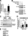
Increased xIAP expression alters MEC response to TGF-β. A, cell detachment activated apoptotic signaling and reduced xIAP expression in NMuMG cells. In contrast, this same cellular condition suppressed apoptotic stimuli and elevated xIAP expression in metastatic 4T cells. Data are from a representative experiment that was performed three times with similar results. B, stable xIAP expression in NMuMG cells significantly potentiated basal and TGF-β1 (5 ng/ml)-stimulated pSBE-luciferase activity. Data are the mean (n = 3) luciferase activities relative to untreated NMuMG-YFP cells (*, p < 0.05; Student's t test). C, stable xIAP expression in NMuMG cells significantly enhanced basal NF-κB-driven luciferase activity. In contrast to YFP-expressing cells, TGF-β1 (5 ng/ml) treatment of xIAP-expressing cells increased their expression of luciferase driven by NF-κB. Data are the mean (n = 3) luciferase activities relative to untreated NMuMG-YFP cells (*, p < 0.05; Student's t test). D, detergent-solubilized whole-cell extracts prepared from NMuMG-YFP and NMuMG-xIAP cells were immunoprecipitated (IP) with anti-TAB1 antibodies and immunoblotted for IKKβ as shown, and whole-cell extracts were immunoblotted with antibodies against TAK1, TAB1, xIAP, IKKβ, and β-actin as indicated (left panels). Additionally whole-cell extracts from YFP- or xIAP-expressing cells were incubated with biotinylated NF-κB oligonucleotides to isolate active p65 (p65 oligo; right panels). Images are from a representative experiment that was performed three times with identical results. PARP, poly(ADP-ribose) polymerase. The error bars indicate ± S.E.
FIGURE 2.
FIGURE 2.
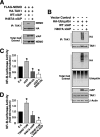
xIAP interacts with TβR-I and enables autocrine TGF-β signaling to NF-κB in malignant MECs. A, transient expression of T204D-TβR-I in 4T1 cells stimulated significant activation of NF-κB-driven luciferase activity. Data are the mean (n = 3) luciferase activities relative to corresponding untransfected cells (*, p < 0.05; Student's t test). B, human 293T cells were transiently transfected either with xIAP or with xIAP together with T204D-TβR-I as indicated. Afterward detergent-solubilized whole-cell extracts were prepared and immunoprecipitated (IP) with anti-HA antibodies followed by immunoblotting with antibodies against xIAP or TβR-I as shown. Differences in protein loading were monitored by reprobing stripped membranes with β-actin antibodies. Images are from a representative experiment that was performed three times with identical results. C, control (i.e. non-silencing (Non-Sil)) or xIAP-deficient (i.e. xIAP shRNA) 4T1 cells were stimulated with TGF-β1 (5 ng/ml) for 0–15 min as indicated and subsequently immunoprecipitated with anti-xIAP antibodies followed by immunoblotting for TβR-I as shown. Additionally whole-cell extracts also were immunoblotted for xIAP, TβR-I, and β-actin as indicated. Data are from a representative experiment that was performed at least three times with similar results. D, NMuMG and 4T1 cells were transiently transfected overnight with pNF-κB-luciferase and pCMV-β-gal prior to administration of a TβR-I antagonist (100 ng/ml) for 24 h as indicated. Data are the mean (n = 3) luciferase activities relative to untreated NMuMG cells (*, p < 0.05; Student's t test). E, administration of a TβR-I antagonist (100 ng/ml for 24 h) to 4T1 cells decreased the phosphorylation and activation of TAK1 as determined by immunoblotting for phospho-TAK1 (pTAK1). Differences in protein loading were monitored by reprobing stripped membranes with TAK1 and β-actin antibodies as shown. Data are from a representative experiment that was performed three times with similar results. Inh, inhibitor. The error bars indicate ± S.E.
FIGURE 3.
FIGURE 3.
xIAP-deficient MEFs exhibit reduced TGF-β signaling. A, WT and xIAP-deficient (xIAP−/−) MEFs were transiently transfected with pSBE-luciferase and β-galactosidase followed by overnight stimulation with TGF-β1 (5 ng/ml) prior to measuring luciferase and β-galactosidase activities. Data are the mean (n = 3) luciferase activities relative to untreated WT MEFs (*, p < 0.05; Student's t test). B, quiescent WT and xIAP-deficient (xIAP−/−) MEFs were incubated in the absence or presence of TGF-β1 (5 ng/ml) as indicated and subsequently immunoblotted with anti-phospho-Smad3 (p-Smad3) antibodies as shown. Differences in protein loading were monitored by reprobing the stripped membranes with antibodies against Smad3 and β-actin as indicated. Data are from a representative experiment that was performed at least three times with similar results. C, WT and xIAP-deficient (xIAP−/−) MEFs were transiently transfected with pNF-κB-luciferase and β-galactosidase and subsequently processed for determination of luciferase and β-galactosidase activities as above. Data are the mean (n = 3) luciferase activities relative to untreated WT MEFs (*, p < 0.05; Student's t test). D, quiescent WT and xIAP-deficient (xIAP−/−) MEFs were stimulated with TGF-β1 (5 ng/ml) as indicated and subsequently immunoprecipitated (IP) with anti-TAK1 antibodies followed by immunoblotting for phospho-TAK1 (p-TAK1) as shown. Additionally whole-cell extracts also were immunoblotted for TAK1 and β-actin as indicated. Data are from a representative experiment that was performed at least three times with similar results. The error bars indicate ± S.E.
FIGURE 4.
FIGURE 4.
xIAP ubiquitinates TAK1 and facilitates its interaction with IKKγ/NEMO and activation of NF-κB. A, human 293T cells were transiently transfected with IKKγ/NEMO (FLAG-tagged), TAK1 (HA-tagged), and WT or mutant (H467A) xIAP as indicated. Afterward detergent-solubilized whole-cell extracts were prepared and immunoprecipitated (IP) with TAK1 antibodies. The resulting immunocomplexes were probed with antibodies against IKKγ/NEMO or HA as shown. Direct immunoblot analysis of an aliquot of the total cell extract was performed to monitor the levels of xIAP and IKKγ/NEMO expression and differences in protein loading. Images are from a representative experiment that was performed three times with identical results. B, human 293T cells were transiently transfected with ubiquitin (HA-tagged) together with either WT or mutant (H467A) xIAP as indicated. Afterward TAK1 immunocomplexes were isolated and immunoblotted with antibodies against HA or TAK1 as shown. Direct immunoblot analysis of an aliquot of the total cell extract was performed to monitor the levels of HA, ubiquitin, xIAP, TAK1, and β-actin expression and differences in protein loading. Images are from a representative experiment that was performed three times with identical results. C, human 293T cells were transiently transfected with pNF-κB-luciferase and β-galactosidase together with either empty vector (E.V.), xIAP, or K63R-ubiquitin (Ub) as indicated. Afterward luciferase and β-galactosidase activities present in whole-cell extracts were measured. Data are the mean ± S.E. (n = 3) luciferase activities relative to 293T cells transfected with empty vector (*, p < 0.05; Student's t test). D, 4T1 cells were transiently transfected with pNF-κB-luciferase and β-galactosidase together with either empty vector (E.V.), T204D-TβR-I, and H467A-xIAP as indicated. Afterward luciferase and β-galactosidase activities present in whole-cell extracts were measured. Data are the mean (n = 3) luciferase activities relative to 4T1 cells transfected with empty vector (*, p < 0.05; Student's t test). The error bars indicate ± S.E.
FIGURE 5.
FIGURE 5.
xIAP deficiency alters TGF-β signaling and activation of mesenchymal gene expression in NMuMG cells. A and B, control (i.e. non-silencing (Non-Sil)) and xIAP-deficient (i.e. xIAP shRNA) NMuMG cells were transiently transfected with β-galactosidase and either pSBE-luciferase (A) or NF-κB-luciferase (B) followed by overnight stimulation with TGF-β1 (5 ng/ml) prior to measuring luciferase and β-galactosidase activities. Data are the mean (n = 3) luciferase activities relative to untreated control cells (*, p < 0.05; Student's t test). C, parental (i.e. empty vector (E.V.)) and xIAP-expressing NMuMG cells were stimulated with TGF-β1 (5 ng/ml) for 24 h at which point detergent-solubilized whole-cell extracts were prepared and immunoblotted with antibodies against E-cadherin, Cox-2, or xIAP as indicated. Differences in protein loading were monitored by reprobing stripped membranes with β-actin antibodies. Data are from a representative experiment that was performed three times with similar results. D, bright field images of control (i.e. non-silencing (Non-Sil)) and xIAP-deficient (i.e. xIAP shRNA) NMuMG cells before and after their stimulation with TGF-β1 (5 ng/ml for 24 h) were captured from a representative experiment that was performed three times with identical results. E, control (i.e. non-silencing (Non-Sil)) and xIAP-deficient (i.e. xIAP shRNA) NMuMG cells were stimulated with TGF-β1 (5 ng/ml) for 24 h at which point detergent-solubilized whole-cell extracts were prepared and immunoblotted with antibodies against Cox-2, N- and E-cadherins, or xIAP as indicated. Differences in protein loading were monitored by reprobing stripped membranes with β-actin antibodies. Data are from a representative experiment that was performed three times with similar results. The error bars indicate ± S.E.
FIGURE 6.
FIGURE 6.
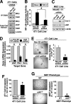
xIAP deficiency impairs the activation of NF-κB and tumorigenic behavior of malignant MECs. A, detergent-solubilized whole-cell extracts prepared from control (i.e. non-silencing (Non-Sil)) or xIAP-deficient (i.e. xIAP shRNA) 4T1 cells were immunoprecipitated (IP) with anti-TAB1 antibodies followed by immunoblotting for IKKβ or TAB1 as shown. Additionally whole-cell extracts also were immunoblotted for IKKβ, xIAP, TAB1, and β-actin as indicated. Data are from a representative experiment that was performed three times with identical results. B, control (i.e. non-silencing (Non-Sil)) or xIAP-deficient (i.e. xIAP shRNA) 4T1 cells were transiently transfected with pNF-κB-luciferase and β-galactosidase and subsequently stimulated overnight with TGF-β1 (5 ng/ml) prior to measuring luciferase and β-galactosidase activities. Data are the mean (n = 2) luciferase activities relative to unstimulated control NMuMG cells (*, p < 0.05; Student's t test). C, detergent-solubilized whole-cell extracts prepared from control (i.e. non-silencing (Non-Sil)) or xIAP-deficient (i.e. xIAP shRNA) NMuMG and 4T1 cells were immunoblotted with antibodies against Cox-2, xIAP, and β-actin as indicated. Images are from a representative experiment that was performed three times with identical results. D, total RNA was isolated from control (i.e. non-silencing (Non-Sil)) and xIAP-deficient (i.e. xIAP shRNA) 4T1 cells and subjected to semiquantitative real time PCR to monitor expression of xIAP, plasminogen activator inhibitor-1 (PAI-1), and survivin (Surv) as indicated. Data are the mean (n = 3) -fold changes in gene expression relative to control cells. E, the growth of control (i.e. non-silencing (Non-Sil)) and xIAP-deficient (i.e. xIAP shRNA) 4T1 cells in soft agar was quantified after 14 days in culture. Data and images are from a single experiment that was performed three times with identical results (*, p < 0.05; Student's t test). F, control (i.e. non-silencing (Non-Sil)) and xIAP-deficient (i.e. xIAP shRNA) 4T1 cells were induced by 2% serum to invade through synthetic basement membranes. Data are the mean (n = 3) invasion relative to that of control 4T1 cells (*, p < 0.05; Student's t test). G, WT and xIAP-deficient (xIAP−/−) MEFs were induced by 2% serum to invade through synthetic basement membranes. Data are the mean (n = 3) invasion relative to that of WT MEFs (*, p < 0.05; Student's t test). The error bars indicate ± S.E.

Similar articles

Cited by

References

    1. Galliher A. J., Neil J. R., Schiemann W. P. (2006) Future Oncol. 2, 743–763 - PubMed
    1. Gordon K. J., Blobe G. C. (2008) Biochim. Biophys. Acta 1782, 197–228 - PubMed
    1. Lin W. W., Karin M. (2007) J. Clin. Investig. 117, 1175–1183 - PMC - PubMed
    1. Nagarajan R. P., Chen F., Li W., Vig E., Harrington M. A., Nakshatri H., Chen Y. (2000) Biochem. J. 348, 591–596 - PMC - PubMed
    1. López-Rovira T., Chalaux E., Rosa J. L., Bartrons R., Ventura F. (2000) J. Biol. Chem. 275, 28937–28946 - PubMed

Publication types