. 2009 Jun 1;185(5):827-39.
doi: 10.1083/jcb.200812121.

GNL3L stabilizes the TRF1 complex and promotes mitotic transition

Affiliations

GNL3L stabilizes the TRF1 complex and promotes mitotic transition

Qubo Zhu et al. J Cell Biol. .

Abstract

Telomeric repeat binding factor 1 (TRF1) is a component of the multiprotein complex "shelterin," which organizes the telomere into a high-order structure. TRF1 knockout embryos suffer from severe growth defects without apparent telomere dysfunction, suggesting an obligatory role for TRF1 in cell cycle control. To date, the mechanism regulating the mitotic increase in TRF1 protein expression and its function in mitosis remains unclear. Here, we identify guanine nucleotide-binding protein-like 3 (GNL3L), a GTP-binding protein most similar to nucleostemin, as a novel TRF1-interacting protein in vivo. GNL3L binds TRF1 in the nucleoplasm and is capable of promoting the homodimerization and telomeric association of TRF1, preventing promyelocytic leukemia body recruitment of telomere-bound TRF1, and stabilizing TRF1 protein by inhibiting its ubiquitylation and binding to FBX4, an E3 ubiquitin ligase for TRF1. Most importantly, the TRF1 protein-stabilizing activity of GNL3L mediates the mitotic increase of TRF1 protein and promotes the metaphase-to-anaphase transition. This work reveals novel aspects of TRF1 modulation by GNL3L.


Figures

Figure 1.
Figure 1.
Interaction between TRF1 and GNL3L is mediated by the homodimerization domain of TRF1 and the GTP-binding domain of GNL3L. (A) Cell lysates were extracted from HEK293 cells cotransfected with Myc-tagged TRF1 and HA-tagged nucleostemin (NS), GNL3L (G3L), or Ngp-1. Protein complexes were immunoprecipitated (IP) by anti-tag antibodies and immunoblotted (IB) by the indicated antibodies. TRF1 interacts with NS and GNL3L, but not with Ngp-1. Arrows mark the precipitated TRF1. (B) Endogenous coIP confirmed that TRF1 and GNL3L co-reside in the same protein complex immunoprecipitated by anti-GNL3L antibody (α-G3L) from HeLa cell lysates. Pre-im: preimmune serum. (C) None of the NS family proteins interacts with TIN2. (D1, E1) Diagrams of TRF1 and GNL3L deletion mutants. Gray lines mark the deleted regions; numbers indicate amino acid positions. Domain abbreviations: A, acidic; HBD, homodimerization; UD, undefined; B, basic; C1 and C2, coiled-coil-1 and -2; G, GTP-binding; I, intermediate. GST pull-down assays demonstrated that the HBD domain of TRF1 (D2) and the G domain of GNL3L (E2) are required for the interaction of these two proteins. Agarose-retained (Ret) and supernatant fractions (Sup) are indicated.
Figure 2.
Figure 2.
GNL3L promotes TRF1 homodimerization, and its binding to TRF1 is competed by TIN2. (A) Agarose-bound GST-TRF1 was incubated with cell lysates containing a fixed amount of Myc-tagged TRF1, mixed with increasing amounts of wild-type GNL3L (A1, G3L), the G3dG (A2), G3dBC (A3), or N166I mutant (A4). Wild-type GNL3L enhances the pull-down efficiency of Myc-tagged TRF1 by GST-TRF1 in a dose-dependent manner. Deleting the TRF1-interacting G domain (G3dG) completely abolishes this activity. (B) The coIP efficiency between FLAG- and Myc-tagged TRF1 was examined in control knockdown (shScr) and GNL3L knockdown (shG3-a) HEK293 cells by immunoprecipitation with anti-Myc antibody and immunoblotting with anti-FLAG antibody. The results confirmed that endogenous GNL3L promotes TRF1 homodimerization. (C) A small effect of GNL3L was observed on promoting the binding between Myc-tagged TIN2 and GST-TRF1. (D) This finding was supported by coIP of FLAG-tagged TRF1 and Myc-tagged TIN2 in the control (shScr) and GNL3L (shG3-a) knockdown HEK293 cells. (E) TIN2 binding significantly reduced the pull-down of GNL3L by GST-TRF1. (F) Protein complexes were immunoprecipitated from HEK293 cells cotransfected with GNL3L (HA), TIN2 (Myc), and TRF1 (FLAG) by anti-HA (F1) or anti-Myc antibody (F2). GNL3L, TIN2, and TRF1 proteins were detected by anti-tag antibodies in the IP or input fraction. The results showed that TRF1 does not bind GNL3L and TIN2 simultaneously. (G) Double and triple coIP of TRF1 (FLAG), TERT (Myc), and GNL3L (HA) showed that the coIP efficiency between TERT and GNL3L or between TRF1 and GNL3L is not affected by the coexpression of TRF1 or TERT1, respectively. (H) The ability of GNL3L to promote TRF1 homodimerization, measured by the GST-TRF1 pull-down of Myc-tagged TRF1 and compared with that in Fig. 2 A1, is not changed by TERT overexpression.
Figure 3.
Figure 3.
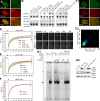
GNL3L increases telomeric retention of TRF1 in living cells. (A) Lack of nucleolar or telomeric colocalization of GFP-fused TRF1 (green) and HA-tagged GNL3L (red) in HeLa cells was shown by confocal analyses. The rectangular area is enlarged and shown on the right. Bars: 10 µm (left) and 2.5 µm (right). (B; left) ChIP assays showed that immunoprecipitation of HA-tagged GNL3L (G3L-HA/α-HA) does not copurify telomeric DNAs (Tel-ChIP) more than the vector-transfected, α-HA–precipitated sample (Ctrl/α-HA), the GNL3L-transfected, IgG-precipitated sample (G3L-HA/IgG), or the copurified Alu sequence (Alu-ChIP). (Middle) Coexpression of GNL3L increases the association between TRF1 and telomeric DNAs (TRF1-Myc+G3L/α-Myc) compared with the TRF1 alone or TRF1+G3dG transfections. (Right) GNL3L does not affect TRF2 binding to telomeric DNAs. All experiments were repeated three times. (C1) The C-terminally GFP-fused TRF1 (TRF1-gfp) colocalizes with the endogenous TRF2 in HeLa cells. High magnification of the indicated area (rectangle) is shown on the right. Bars: 10 µm (left) and 2 µm (right). (C2) Quantification of TRF1-gfp and TRF2 colocalization. All pixels are plotted based on their red (X-axis) and green (Y-axis) fluorescence intensities, and pseudocolored based on the event frequency, with red representing the highest and blue representing the lowest frequency. (D; left) FRAP analyses showed that GNL3L knockdown in HeLa cells increases the recovery rate and plateau level of TRF1-gfp at the telomere (red; P < 0.0001). Such phenotype can be rescued by the shG3-a–resistant full-length GNL3L (orange) but not by the shG3-a–resistant G3dG (green). RFI, relative fluorescence index. Error bars represent SEM shown on one side indicated by arrows. Top arrows mark the bleaching event. (Right) Time-sequenced images are shown with labels indicating the bleached telomere (yellow circles and arrows) and intervals between image acquisition and the bleaching pulse (in seconds). Bar: 2 µm. (E) The telomeric retention time of TRF1 was increased by overexpression of GNL3L (G3L) or G3dBC (P < 0.0001) but not by overexpression of G3dG. (F) GNL3L knockdown has no effect on the telomeric FRAP of TRF1-gfp in U2OS cells. (G) EMSA was performed using a (TTAGGG)6 probe and nuclear extracts from HEK293 cells expressing the indicated recombinant proteins (G2). A specific TRF1-probe complex was identified in lane 3 (black arrow), which could be competed by excess nonlabeled probes (lane 4) and supershifted by anti-Myc antibody (lane 5, gray arrow). Coexpression of GNL3L had no effect on the TRF1-probe complex (lane 9 and 10).
Figure 4.
Figure 4.
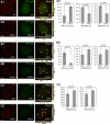
GNL3L negatively regulates APB formation in U2OS cells. (A) APB formation in U2OS cells was scored by colocalization of TRF1-gfp and PML bodies in the control (A1) or GNL3L-overexpression U2OS cells (A2). Serial confocal images of 1-µm thickness were collected at 0.5-µm intervals and reconstructed along the X-Y, X-Z, and Y-Z planes for individual cell. Arrows indicate overlapped spots. (A3) Coexpression of GNL3L increases the percentage of APB-minus cells, and decreases the ratios of APB/PML bodies and APB/TRF1+ foci. (B) Colocalization of TRF1+ foci and PML bodies was measured in control (shScr, B1) or GNL3L knockdown (shG3-a, B2) U2OS cells. (B3) GNL3L knockdown increases colocalization of TRF1 foci and PML bodies. (C) GNL3L knockdown by siG3-2 promotes APB formation in U2OS cells, as scored by colocalization of TRF2+ foci and PML bodies.
Figure 5.
Figure 5.
GNL3L stabilizes TRF1 protein by preventing its ubiquitylation and binding to FBX4. (A) Knocking down the endogenous GNL3L expression by siG3-1 or siG3-2 in HeLa cells reduces the endogenous TRF1 protein level, which can be rescued by an siG3-2–resistant GNL3L (G3L-siR). (B) The same result was confirmed in H1299 cells by using the Doxycycline (Dox)-inducible shRNAmir knockdown approach. Western analyses show a time-dependent reduction of GNL3L protein in the Dox-treated GNL3L knockdown (shG3) cells, but not in the Dox-treated shScr cells. Specifically, GNL3L depletion decreases the protein level of TRF1 but not that of TRF2. (C) Overexpression of GNL3L (HA) increases the protein level of TRF1 (Myc) in a dose-dependent manner after normalization by the protein level of GFP cotransfected in the same sample. (D) The GNL3L’s ability to increase TRF1 protein is abolished by deleting its TRF1-interactive G domain and by the N166I mutation, but not by the BC domain deletion. (E) TRF1 protein stability was measured by a protein degradation assay in control (Ctrl) and GNL3L-overexpressing (G3L, HA-tagged) HEK293 cells. The TRF1 protein amounts were measured from three experiments, adjusted based on their α-tubulin (Tub) amounts, and expressed as percentages of the TRF1 protein amount at the 0-h time point. (F) GNL3L depletion by shG3-a increases the protein degradation of TRF1 (top two panels), which can be rescued by coexpression of an shG3-a–resistant full-length GNL3L but not an shG3-a–resistant G3dG mutant (bottom panel). (G) Overexpression of the wild-type (lane 4) or G3dBC mutant (lane 5) of GNL3L decreases TRF1 ubiquitylation compared with the control sample (lane 3), whereas the G3dG (lane 6) and N166I mutants (lane 7) have no such effect. (H) GNL3L depletion by shG3-a (lane 4) increases the ubiquitylation of TRF1, which can be reversed by overexpression of an shG3-a–resistant GNL3L (G3L-shR, lane 6). (I) CoIP experiments showed that GNL3L (HA) binding to TRF1 (FLAG) decreases the coIP efficiency between TRF1 and FBX4 (Myc).
Figure 6.
Figure 6.
GNL3L stabilizes TRF1 protein during mitosis and promotes the metaphase-to-anaphase transition. (A) The protein amounts of endogenous TRF1 (left) and exogenously expressed TRF1 (Myc-tagged, right) were measured in the nontreated (Ctrl), S phase-synchronized (S), and M phase-synchronized HEK293 cells. (B) GNL3L depletion by siG3-1 and siG3-2 abolishes the mitotic increase of TRF1 protein. This effect of siG3-2 can be reversed by coexpression of a siG3-2–resistant GNL3L (G3L-siR). (C) In addition to the increase in protein level, the binding efficiency between GNL3L and TRF1 is also increased during mitosis. To control for the mitotic increase of TRF1 and GNL3L proteins, their input amounts in each sample were adjusted to the same before coIP. (D) PI-labeled cell cycle analyses showed that GNL3L knockdown in HeLa (D1) or HEK293 cells (D2) increases the percentage of G2/M phase cells. A mild decrease in the G1 cell percentage and an increase in the sub-G1 cell percentage were also noticed. (E) The percentages of cells at different mitotic stages were scored by anti–phospho-Histone H3 (pH3) staining in control and GNL3L knockdown HeLa (E1) and HEK293 cells (E2). Over 1.6 × 10⁴ cells from eight independent experiments were collected for each sample. The results showed that GNL3L depletion triggers cell cycle arrest at the metaphase-to-anaphase transition, and this effect can be rescued by coexpression of the siG3-2–resistant GNL3L (G3L-siR) or TRF1. Cells cotransfected with siG3-2 and G3L-siR (in both HeLa and HEK293 cells) or with siScr and TRF1 (in HeLa cells only) showed decreased prophase cell percentages. *, P < 0.01; **, P < 0.001; ***, P < 0.0001. (F) Silencing GNL3L expression increases the amount of phospho-cdc2 (Tyr15), supporting a role of GNL3L in the prophase entry of dividing cells as well.
Figure 7.
Figure 7.
Knockdown of GNL3L or TRF1 increases the number of cells with multipolar spindle. (A) Distribution of endogenous TRF1 protein at different stages of mitosis is shown by high-resolution confocal analyses of anti-TRF1 (TRF-78) staining in H2B-GFP HeLa cells. Bar: 5 µm. (B1 and B2) GNL3L or TRF1 knockdown significantly increases the number of cells with multipolar spindle, shown by α-tubulin immunostaining (Tub). Y-axis shows the percentage of metaphase cells with multipolar spindle. *, P < 0.01; **, P < 0.001. The centromeres in these cells, labeled by anti–CENP-A antibody, are aligned along the multipolar metaphase plate. TRF1 knockdown efficiency was shown in B3. (C) Models of GNL3L-mediated regulation of TRF1 protein modification and function in mitotic transition (C1) and telomere maintenance (C2).

Similar articles

Cited by

References

    1. Bilaud T., Brun C., Ancelin K., Koering C.E., Laroche T., Gilson E. 1997. Telomeric localization of TRF2, a novel human telobox protein.Nat. Genet. 17:236–239 - PubMed
    1. Broccoli D., Smogorzewska A., Chong L., de Lange T. 1997. Human telomeres contain two distinct Myb-related proteins, TRF1 and TRF2.Nat. Genet. 17:231–235 - PubMed
    1. Bryan T.M., Englezou A., Gupta J., Bacchetti S., Reddel R.R. 1995. Telomere elongation in immortal human cells without detectable telomerase activity.EMBO J. 14:4240–4248 - PMC - PubMed
    1. Canudas S., Houghtaling B.R., Kim J.Y., Dynek J.N., Chang W.G., Smith S. 2007. Protein requirements for sister telomere association in human cells.EMBO J. 26:4867–4878 - PMC - PubMed
    1. Chang W., Dynek J.N., Smith S. 2003. TRF1 is degraded by ubiquitin-mediated proteolysis after release from telomeres.Genes Dev. 17:1328–1333 - PMC - PubMed

Publication types

MeSH terms