. 2009 Aug 15;18(16):3075-89.
doi: 10.1093/hmg/ddp249. Epub 2009 May 28.

In vivo and in vitro effects of two novel gamma-actin (ACTG1) mutations that cause DFNA20/26 hearing impairment

Affiliations

In vivo and in vitro effects of two novel gamma-actin (ACTG1) mutations that cause DFNA20/26 hearing impairment

Matías Morín et al. Hum Mol Genet. .

Abstract

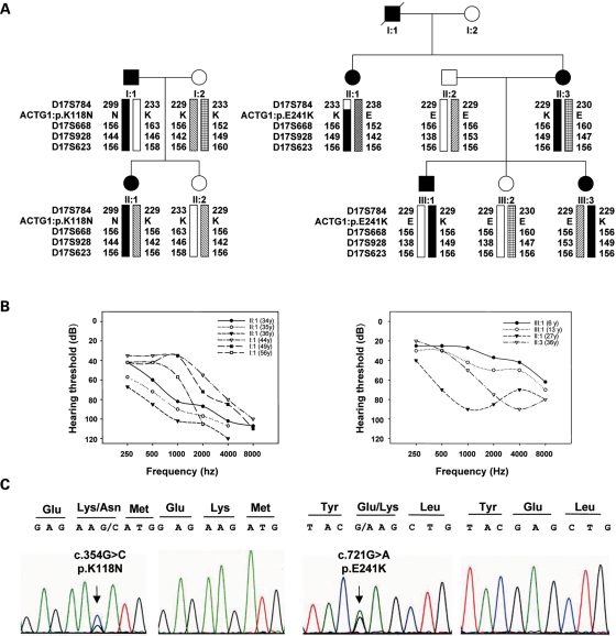
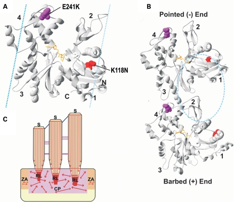
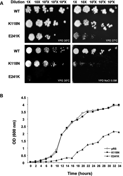
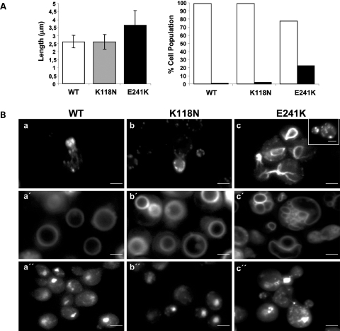
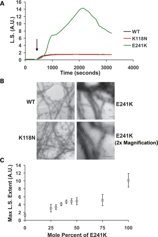
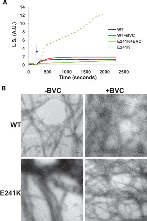
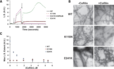
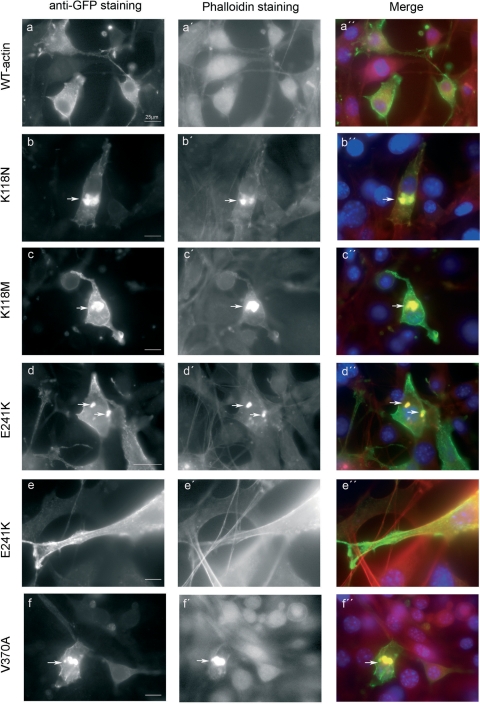
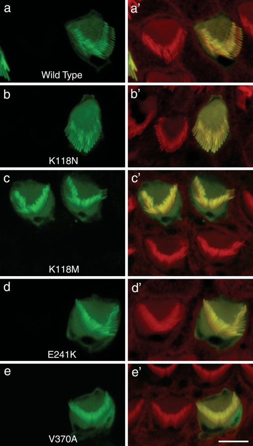
Here we report the functional assessment of two novel deafness-associated gamma-actin mutants, K118N and E241K, in a spectrum of different situations with increasing biological complexity by combining biochemical and cell biological analysis in yeast and mammalian cells. Our in vivo experiments showed that while the K118N had a very mild effect on yeast behaviour, the phenotype caused by the E241K mutation was very severe and characterized by a highly compromised ability to grow on glycerol as a carbon source, an aberrant multi-vacuolar pattern and the deposition of thick F-actin bundles randomly in the cell. The latter feature is consistent with the highly unusual spontaneous tendency of the E241K mutant to form bundles in vitro, although this propensity to bundle was neutralized by tropomyosin and the E241K filament bundles were hypersensitive to severing in the presence of cofilin. In transiently transfected NIH3T3 cells both mutant actins were normally incorporated into cytoskeleton structures, although cytoplasmic aggregates were also observed indicating an element of abnormality caused by the mutations in vivo. Interestingly, gene-gun mediated expression of these mutants in cochlear hair cells results in no gross alteration in cytoskeletal structures or the morphology of stereocilia. Our results provide a more complete picture of the biological consequences of deafness-associated gamma-actin mutants and support the hypothesis that the post-lingual and progressive nature of the DFNA20/26 hearing loss is the result of a progressive deterioration of the hair cell cytoskeleton over time.


Figures

Figure 1.
Figure 1.
(A) Pedigree of the Spanish families S840 (left) and S582 (right). Affected members are shown in black. Haplotypes are represented by bars, with the haplotype associated with hearing loss in black. The relative order of ACTG1 gene and the microsatellite markers within the DFNA20/A26 genetic interval is indicated. The numbers beside the bars are the allelic sizes for each microsatellite marker and the mutations identified in the families are notated as ACTG1: p.K118N and p.E241K, respectively. (B) Audiograms showing the air conduction values obtained from affected patients of S840 (I:1 and II:1) and S582 (II:1, II:3 and III:1) families. The age of the patients from which each audiometric record was obtained is indicated. Each graph point represents the average hearing loss for the right and left ears. (C) Electropherograms of the mutations c.354G>C and c.721G>A identified in exons 3 and 4 of ACTG1 in S840 and S582 families, respectively. The arrow points to the precise nucleotide that is mutated. An affected subject and a control are depicted.
Figure 2.
Figure 2.
Locations of the K118N and E241K γ-actin mutations in yeast actin. (A) Front view of the crystal structure of the yeast actin monomer (43), modified from Protein Data Bank code 1YAG using Swiss-PdbViewer, version 4.01. The positions of the mutations are modelled in space-fill and colour-highlighted as follows: K118N, red and E241K, purple. ATP is modelled in ball-stick and coloured orange. Numbers denote the actin subdomains. G-actin monomers are characterized by a cleft that divides it into two similar domains (4,7), the small outer domain (subdomains 1 and 2) and the larger inner domain (subdomains 3 and 4). The names relate to the position of each domain in the F-actin helix where the inner domain is closer to the helix axis. N and C mark the respective termini. Cyan dashed lines denote the general vicinity on the actin surface to which tropomyosin can occupy (29,30). (B) Model illustrating the longitudinal contacts between two neighbouring monomers within the same filament strand based on the Holmes filament model (7). The pointed (−) and barbed (+) ends are indicated. Each monomer is labelled as in (A) with regard to the mutations, ADP and subdomains. Cyan dashed circle denote the general vicinity on the actin surface to which cofilin binds (22). (C) Molecular architecture of the hair cell's (HC) actin cytoskeleton. Actin filaments are denoted by red arrows. The barbed (subdomains 1 and 3) and pointed (subdomains 2 and 4) ends of the filaments are represented by circles and arrowheads, respectively. CP, cuticular plate; S, stereocilia; ZA, zonula adherens; RL, rootlets.
Figure 3.
Figure 3.
(A) Comparison of the growth of WT and mutant yeast strains on YPD medium at 30°C (a), YPD at 37°C (b), YPG medium with glycerol as a sole carbon source (c) and on YPD + NaCl 0.5 m at 30°C (d). The experiments were repeated three times with similar results. (B) Growth curves of cells expressing either Wt or mutant yeast actins. Cells were grown in YNB medium at 30°C on a shaking platform.
Figure 4.
Figure 4.
(A) Measure of the length of cells expressing Wt and mutants yeast actins (left). Difference was statistically significant between Wt and E241K mutant based upon Student t-test analysis with a P-value lower than 0.001. Error bars represent SD. Assessment of vacuole phenotype (right). White bars and black bars represent the percentage of cells with 1–4 vacuoles and with more than 4 vacuoles, respectively. Results based on the measurement of a population of at least 200 cells. (B) Fluorescence microscopy of cells expressing WT, K118N and E241K yeast actins. The cytoskeleton was visualized after staining fixed cells (at a stage of the cell cycle in which the bud represents roughly one-third of the mother cell) with rhodamine–phalloidin (a–c). At this stage, two abnormal cytoskeleton patterns were observed in the majority of E241K cells. They consisted of, in addition to patches, abnormally distributed thick-actin cables (c) or in randomly distributed actin patches that are not confined to the bud as expected at this stage of the cell cycle (inset). Vacuoles were observed following exposure the cells to dye FM4-64 (a′–c′). Nuclear and mitochondrial DNAs were visualized following staining of the cells with Hoechst (a″–c″). The largest bright spot in each cell is the nucleus and the cytoplasmic spots correspond to mtDNA. Scale bars = 2 µm.
Figure 5.
Figure 5.
(A) Comparison of the polymerization kinetics of WT and mutant actins. Polymerization of 4.8 µm actin was initiated by the addition of magnesium and potassium chloride (denote by arrow) as described in Materials and Methods, and the increase in light scattering (LS) was monitored as a function of time at 25°C. Shown are representative plots from experiments performed at least three times with three independent actin preparations. AU stands for arbitrary units. (B) Effects of mutations on filament morphology. Samples of the actin were prepared as described in Materials and Methods. Shown are electron micrographs of WT, K118N and E241K filaments. Note the high degree of filament bundling of the E241K filaments. Scale bar = 100 nm. (C) Effect of increasing the mole percent of E241K on light scattering caused by actin polymerization. Shown is the relationship between the various percentages of E241K mutant actin in the samples and the maximum extent in light scattering signal value attained. The open circles represent the average light scattering value and the error bars are the standard deviation in the measurements from five independent trials using three different actin preparations.
Figure 6.
Figure 6.
Effects of BVC on E241K mutant filament bundling. (A) Effects of the absence (dashed lines) or presence (solid lines) of bovine cardiac tropomyosin has on the polymerization kinetics and extents as measured by the light scattering of samples containing either WT and E241K actin when it is added prior to inducing polymerization at 25°C. Arrow denotes induction of polymerization by the addition of salts (Fig. 5). (B) Electron micrographs of WT and E241K filaments either in the absence (left) or presence of BVC (right). Scale bar = 100 nm.
Figure 7.
Figure 7.
Effects of mutations on filament sensitivity to cofilin severing. (A) The change in light scattering as a function of time was monitored for samples containing 4.8 µm of either WT, K118N or E241K actin. Polymerization was induced by the addition of salts (denoted by first arrow) and once the samples reached steady state a stoichiometric amount of yeast cofilin or an equivalent amount of buffer was added to the samples (denoted by the second arrow). Dashed green line represents an E241K control sample to which buffer was added instead of cofilin. (B) Electron micrograph which shows the effect the post-polymerization addition of a stoichiometric amount of cofilin has on E241K filaments. Scale bar = 100 nm. (C) Each point represents the average maximum light scattering value obtained for samples containing either 4.8 µm WT, K118N or E241K actin and the following concentrations of yeast cofilin: 0, 0.48, 0.96, 1.92, 3.2, 4.8 and 5.4 µm, respectively. Error bars denote range determined from two independent trials with two different preparations of actin.
Figure 8.
Figure 8.
Expression of wild-type and γ-actin mutants in NIH3T3 fibroblasts. Wild-type actin (A) was used as a control and was incorporated into stress fibres, ruffles, lamellipodia and into the cortical network beneath the plasma membrane and generally co-localized with the endogenous filamentous actin, as visualized with phalloidin (A′, A″). The mutant K118N localized less to stress fibres and lamellipodia (B′, B″). The mutant protein accumulated in a big cytoplasmic clump (arrow) in the cytoplasm that stained with phalloidin in 50% of cells, (B). The incorporation of the E241K into the stress fibres, ruffles and lamellipodia appears normal (D and E), but in 20% of cells, a big or several small aggregates were visualized that co-localized with phalloidin (D′, D″). Both the K118M and the V370A were incorporated into stress fibres, ruffles and lamellipodia (C and F). However, in 30% of cells expressing K118M and in 20% expressing V370A, the mutant proteins accumulated in a big cytoplasmic clump that co-localized with phalloidin (C′, C″ and F′, F″). Scale bar = 25 µm.
Figure 9.
Figure 9.
Gene-gun mediated expression of γ-actin mutants into mouse cochlear hair cells. Cochlear explants of mice sacrificed at post-natal day 2–3 (P2–P3) were transfected with the same constructs as in Figure 8, maintained in medium and fixed 24 h after transfection. Cells expressing any of the four γ-actin mutants assayed, K118N, K118M, E241K and V370A, displayed a normal wild-type appearance, with no appreciable alteration in the incorporation of actin or the morphology of the stereocilia. Confocal microscopy revealed that at 24 h after transfection, the wild-type γ-actin-GFP (green) and mutant actins were incorporated along the entire length of the stereocilia bundle and into the randomly organized filaments of the cuticular plate. In all cases, co-localization with rhodamine–phalloidin staining (red) was observed. The stereocilia of the actin-GFP transfected cells maintained their lengths as judged by comparing with the stereocilia of neighbouring non-transfected cells (red) in each panel. Scale bars = 2.5 µm.

Similar articles

Cited by

References

    1. Hudspeth A.J. How the ear's works work. Nature. 1989;341:397–404. - PubMed
    1. Tilney L.G., Egelman E.H., DeRosier D.J., Saunder J.C. Actin filaments, stereocilia, and hair cells of the bird cochlea. II. Packing of actin filaments in the stereocilia and in the cuticular plate and what happens to the organization when the stereocilia are bent. J. Cell Biol. 1983;96:822–834. - PMC - PubMed
    1. Hirokawa N., Tilney L.G. Interactions between actin filaments and between actin filaments and membranes in quick-frozen and deeply etched hair cells of the chick ear. J. Cell Biol. 1982;95:249–261. - PMC - PubMed
    1. Kabsch W., Mannherz H.G., Suck D., Pai E.F., Holmes K.C. Atomic structure of the actin: DNase I complex. Nature. 1990;347:37–44. - PubMed
    1. De La Cruz E.M. Cofilin binding to muscle and non-muscle actin filaments: isoform-dependent cooperative interactions. J. Mol. Biol. 2005;346:557–564. - PubMed

Publication types