. 2009 Jul 10;284(28):18707-14.
doi: 10.1074/jbc.M109.017483. Epub 2009 May 21.

CAPS activity in priming vesicle exocytosis requires CK2 phosphorylation

Affiliations

CAPS activity in priming vesicle exocytosis requires CK2 phosphorylation

Mari Nojiri et al. J Biol Chem. .

Abstract

CAPS (Ca(2+)-dependent activator protein for secretion) functions in priming Ca(2+)-dependent vesicle exocytosis, but the regulation of CAPS activity has not been characterized. Here we show that phosphorylation by protein kinase CK2 is required for CAPS activity. Dephosphorylation eliminated CAPS activity in reconstituting Ca(2+)-dependent vesicle exocytosis in permeable and intact PC12 cells. Ser-5, -6, and -7 and Ser-1281 were identified by mass spectrometry as the major phosphorylation sites in the 1289 residue protein. Ser-5, -6, and -7 but not Ser-1281 to Ala substitutions abolished CAPS activity. Protein kinase CK2 phosphorylated CAPS in vitro at these sites and restored the activity of dephosphorylated CAPS. CK2 is the likely in vivo CAPS protein kinase based on inhibition of phosphorylation by tetrabromo-2-benzotriazole in PC12 cells and by the identity of in vivo and in vitro phosphorylation sites. CAPS phosphorylation by CK2 was constitutive, but the elevation of Ca(2+) in synaptosomes increased CAPS Ser-5 and -6 dephosphorylation, which terminates CAPS activity. These results identify a functionally important N-terminal phosphorylation site that regulates CAPS activity in priming vesicle exocytosis.


Figures

FIGURE 1.
FIGURE 1.
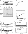
Phosphorylation is essential for CAPS activity. A, CAPS was immunoprecipitated from extracts of 32P-labeled PC12 cells and analyzed on 7.5% SDS gels for Western blotting with CAPS antibody (Ab, left lane) or by autoradiography for 32P (right lane). B, recombinant CAPS purified from Sf9 cells (right lane) was incubated with λ-protein phosphatase (λPP, left lane) and analyzed by electrophoresis on native 6% polyacrylamide gels and Coomassie staining. Bands from the bottom to top correspond to native phosphorylated dimer (phospho), dephosphorylated dimer (dephospho), and higher molecular weight tetramer. C, native phosphorylated (○) but not dephosphorylated CAPS (●) restores Ca2+-triggered release of [3H]norepinephrine from permeable PC12 cells. Permeable cells were prepared as described under “Experimental Procedures” and incubated for 3 min at 30 °C with 10 μm Ca2+ plus the indicated concentrations of CAPS. Mean values of duplicate determinations with the indicated ranges correspond to the CAPS-dependent component of norepinephrine release with maximal value set at 100% for wild type CAPS.
FIGURE 2.
FIGURE 2.
CAPS is phosphorylated at N- and C-terminal Ser residues. A, MALDI-TOF spectra of CAPS phosphopeptides. Phosphopeptides enriched from CAPS tryptic peptides by Ga(III)- immobilized metal ion affinity chromatography were incubated without (upper panel) or with (lower panel) alkaline phosphatase before MALDI-TOF to detect mass shifts of −80 Da (HPO3) or multiples of −80 Da. B, MALDI-QTOF MS/MS spectrum of triply phosphorylated CAPS residues 1–21 (2622.9 atomic mass units (amu)) with a summary of Ser phosphorylations detected by neutral loss of H3PO4 by β-elimination (β-elim). C, schematic figure of CAPS indicating phosphorylation sites relative to other functional domains. Functional domains correspond to C2 (protein kinase C C2-like), PH, and MH (munc 13 homology).
FIGURE 3.
FIGURE 3.
Phosphorylation of Ser-5, -6, and -7 is essential for CAPS activity. A, reconstituting activity of S5A, S6A, S7A, S5A/S6A/S7A, and S1281A CAPS mutants. Each mutant was expressed in COS-1 cells as a C-terminal Myc-His6-tagged protein and purified. [3H]Norepinephrine release from permeable PC12 cells was measured as in Fig. 1C. Maximal release promoted by wild-type CAPS was set as 100%, and release with Ca2+ alone was set as 0%. Values are the means of duplicate determinations with indicated ranges. WT, wild type. B, reconstituting activity of S5E/S6E CAPS was compared with that of the S5A/S6A mutant. Dephosphorylation of S5E/S6E (S5-6E*) reduced activity. C, dominant negative activity of S5A/S6A/S7A mutant CAPS. The effect of adding the mutant CAPS proteins to wild-type CAPS proteins in 1:1 or 5:1 ratio on norepinephrine release from permeable PC12 cells was measured by rotating disk electrode assay. D, detection of phosphorylated CAPS with phosphospecific antibody (Ab). Phosphospecific antibody generated against CAPS 1–19 with phospho-Ser-5 and -6 (upper panel) and a general CAPS antibody (lower panel) were used in Western blotting with PC12 cell lysate (lane 1), recombinant phosphorylated CAPS (lane 2), and dephosphorylated CAPS (lane 3). E, comparison of CAPS N-terminal amino acid sequences indicates conservation of Ser-5, -6, and -7.
FIGURE 4.
FIGURE 4.
Phosphoacceptor Ser-5, -6, and -7 residues are essential for in vivo CAPS activity in evoked vesicle exocytosis. A, CAPS was down-regulated in PC12 cells by transfection of an short hairpin RNA plasmid (lower panel) resulting in a 5-fold reduction in the number of exocytic events evoked by 56 mm K+ depolarization (upper panel). GAPDH, glyceraldehyde-3-phosphate dehydrogenase; WT, wild type. B, transfection with a pcDNA3.1-CAPS-tag red fluorescent protein harboring silent mutations enabled re-expression of wild-type or S5A/S6A/S7A CAPS in down-regulated PC12 cells (lower panel). Wild-type CAPS expression restored evoked exocytosis, whereas S5A/S6A/S7A CAPS expression failed to do so (upper panel). Error bars indicate S.E. for n = 13–17 cells. AFM, arbitrary fluorescence units.
FIGURE 5.
FIGURE 5.
CK2 phosphorylates CAPS in vitro and restores activity. A, in vitro phosphorylation of CAPS by Ca2+/calmodulin-dependent protein kinase II, CK1, CK2, glycogen synthase kinase 3, protein kinase A (PKA), and protein kinase C (PKC). Dephosphorylated CAPS was incubated with the indicated enzymes in the presence of [32P]ATP. Proteins separated by 7.5% SDS-PAGE were analyzed by phosphorimaging (upper panel) and Coomassie G250 staining (lower panel). B, the approximate stoichiometry of phosphorylation of 20 μg of dephospho-CAPS by 50 units of CK2 was determined at the indicated incubation times at 30 °C. C, CAPS proteins were analyzed by 6% acrylamide native gel electrophoresis and staining with Coomassie G250. Phosphorylated dimer, dephosphorylated dimer, and tetramer are indicated by arrowheads from bottom to top. D, activity of CAPS phosphorylated by CK1 or CK2. Dephosphorylated CAPS (●) incubated with either CK1 (□) or CK2 (■) plus MgATP were re-purified and tested for activity in Ca2+-dependent [3H]norepinephrine release from permeable PC12 cells. Maximal release promoted by native phosphorylated CAPS (○) was set as 100%, and release with Ca2+ alone was set as 0%. Values are the mean of duplicate determination with indicated ranges. E and F, native phosphorylated CAPS (upper panels) and dephosphorylated CAPS incubated with CK2 and MgATP (lower panels) were compared by MALDI-TOF after trypsin digestion. MALDI-TOF spectra corresponding to CAPS 1–21 (E) and CAPS 1280–1309 (F) are shown.
FIGURE 6.
FIGURE 6.
CK2 phosphorylates CAPS in vivo. A, CAPS phosphorylation was inhibited in PC12 cells treated with a specific CK2 inhibitor. PC12 cells were labeled with [32P]orthophosphate for 1 h in the absence or presence of 60 μm tetrabromo-2-benzotriazole (TBB). CAPS immunoprecipitates were analyzed by 7.5% SDS-PAGE and autoradiography or Western blotting with a general CAPS antibody (Ab). Densitometric scanning of autoradiogram and Western blot was used to assess relative phosphorylation. Values are the mean of triplicate determinations with the indicated S.D. Tetrabromo-2-benzotriazole inhibited CAPS phosphorylation by 70%. B, CAPS dephosphorylation was promoted by Ca2+ influx in synaptosomes. Mouse brain synaptosomes were incubated under control conditions (♦) or stimulated with high K+ (▲) for the indicated times. Synaptosomes were hypotonically lysed, and supernatant fractions were analyzed by Western blotting with Ser(P)-5 and -6 phosphospecific antibody or general CAPS antibody. For the graphic representation the signal at the zero time point is set as 100%, and values are the mean of duplicate determination with indicated ranges.

Similar articles

Cited by

References

    1. Malsam J., Kreye S., Söllner T. H. (2008) Cell. Mol. Life Sci. 65, 2814–2832 - PMC - PubMed
    1. Walent J. H., Porter B. W., Martin T. F. (1992) Cell 70, 765–775 - PubMed
    1. Ann K., Kowalchyk J. A., Loyet K. M., Martin T. F. (1997) J. Biol. Chem. 272, 19637–19640 - PubMed
    1. Grishanin R. N., Kowalchyk J. A., Klenchin V. A., Ann K., Earles C. A., Chapman E. R., Gerona R. R., Martin T. F. (2004) Neuron 43, 551–562 - PubMed
    1. Speese S., Petrie M., Schuske K., Ailion M., Ann K., Iwasaki K., Jorgensen E. M., Martin T. F. (2007) J. Neurosci. 27, 6150–6162 - PMC - PubMed

Publication types