Comparative Study
. 2009 Jun 30;48(25):5984-93.
doi: 10.1021/bi900270w.

Redox-dependent dynamics of a dual thioredoxin fold protein: evolution of specialized folds

Affiliations
Comparative Study

Redox-dependent dynamics of a dual thioredoxin fold protein: evolution of specialized folds

Andrea Hall et al. Biochemistry. .

Abstract

An enzyme system protecting bacteria from oxidative stress includes the flavoprotein AhpF and the peroxiredoxin AhpC. The N-terminal domain of AhpF (NTD), with two fused thioredoxin (Trx) folds, belongs to the hyperthermophilic protein disulfide oxidoreductase family. The NTD is distinct in that it contains a redox active a fold with a CxxC sequence and a redox inactive b fold that has lost the CxxC motif. Here we characterize the stability, the (15)N backbone relaxation, and the hydrogen-deuterium exchange properties of reduced [NTD-(SH)(2)] and oxidized (NTD-S(2)) NTD from Salmonella typhimurium. While both NTD-(SH)(2) and NTD-S(2) exhibit similar equilibrium unfolding transitions and order parameters, R(ex) relaxation terms are quite distinct with considerably more intermediate time scale motions in NTD-S(2). Hydrogen exchange protection factors show that the slowly exchanging core corresponds to residues in the b fold in both NTD-(SH)(2) and NTD-S(2). Interestingly, folded-state dynamic fluctuations in the catalytic a fold are significantly increased for residues in NTD-S(2) compared to NTD-(SH)(2). Taken together, these data demonstrate that oxidation of the active site disulfide does not significantly increase stability but results in a dramatic increase in conformational heterogeneity in residues primarily in the redox active a fold. Differences in dynamics between the two folds of the NTD suggest that each evolved a specialized function which, in the a fold, couples redox state to internal motions which may enhance catalysis and specificity and, in the b fold, provides a redox insensitive stable core.


Figures
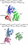
Figure 1
Figure 1
Two pathways for the reduction of peroxides by flavoproteins. A) The bacterial AhpF/AhpC system (also known as the alkyl hydroperoxide reductase system) uses the two enzymes AhpF and AhpC to reduce hydrogen peroxide and organic hydroperoxides using NADH. A ribbon diagram of AhpF (pdb code 1HYU), depicting only one subunit of the dimer, is shown. B) The TrxR/Trx/Prx system is analogous to the AhpF/AhpC system. Ribbon diagrams of TrxR (pdb code 1TDE) and Trx (pdb code 2TRX) are shown. A comparison of the proteins in the two systems reveals that AhpF consists of a fused TrxR-like domain and a Trx-like domain. To highlight this homology, the same coloring scheme is used in both A) and B) with red and blue shades for the FAD binding (FAD) and the NADH binding redox-active disulfide-containing (NADH/SS) domains of the TrxR(-like) protein, and green shades for the Trx(-like) protein/domain. The N-terminal domain in AhpF (NTD) is made up of two interacting Trx folds and acts like an appended substrate for the TrxR-like domain of AhpF (linker region is colored cyan). The bound FAD molecule and redox active disulfides in both A) and B) are shown as ball and stick with disulfide sulfur atoms colored yellow.
Figure 2
Figure 2
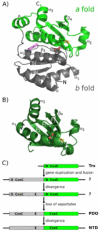
Comparison of the NTD and EcTrx. A) Ribbon diagram of the NTD (pdb code 1ZYN). The two Trx folds are colored in grey (b fold) and green (a fold). The redox active cysteines (Cys129 and Cys132 with sulfur atoms colored yellow), conserved glutamic acid (Glu86 with oxygen atoms colored red) and fluorescence reporter (Trp96 with side chain colored purple) are shown as ball and stick. Secondary structure elements are labeled; conserved secondary structure elements of the Trx fold are labeled with subscripts. Helix α2 of the Trx fold is not present in the b fold of the NTD. The only secondary structure element in the NTD that is not in the conserved Trx fold is an α-helix, labeled α, that is present at the beginning of both the a and b folds. The letters, N and C mark the N and C-termini of the NTD. B) Ribbon diagram of EcTrx (pdb code 2TRX) in the same orientation as the a fold of the NTD in A). The redox active cysteines and conserved aspartic acid are shown as in A) and only the conserved structural elements of the Trx fold are labeled. A structure-based sequence alignment of EcTrx on the a and b folds of the NTD gives a 1.3 Å (for 73 Cα atoms) and 1.5 Å (for 51 Cα atoms) RMSD with 15% and 16 % sequence identity, respectively, while alignment of the two NTD folds gives an RMSD of 1.3 Å (for 46 Cα atoms) with 14% sequence identity. C) The proposed evolution of the NTD begins with an ancestral Trx-like protein that has an active site aspartic acid and redox active disulfide (D and CxxC in top green bar). Gene duplication and fusion (53) creates a hypothetical ancestral protein that has two folds and two active sites (each containing a buried aspartic acid). Sequence divergence introduces a buried glutamic acid in the b fold (shown as an E). With the addition of this charged group to the active site, it has been speculated that the oxidoreductase activity of the protein could be rescued after the loss of the buried aspartate in the a fold (5). The active site CxxC motif in the b fold seen in the evolutionary intermediate, protein disulfide oxidoreductase (PDO), is lost with time. Fold coloring is as in A).
Figure 3
Figure 3
Global, cooperative denaturation of the NTD by urea. The reversible unfolding of NTD-S2 (triangles) and NTD-(SH)2 (circles) in urea was followed by fluorescence (grey shapes) and CD (black shapes). Solid curves are global fits of the unfolding data to a two-state unfolding model.
Figure 4
Figure 4
Select data from 15N backbone relaxation experiments and model free analyses. Shown are R2, S2 and Rex data for NTD-S2 (left panels) and NTD-(SH)2 (right panels) plotted per residue. Secondary structure elements with labels shown above the plots are colored by fold as in Figure 2A. Additional relaxation data are shown as Supplemental Information.
Figure 5
Figure 5
H/D exchange protection factors for the NTD-S2 are lower than for NTD-(SH)2. Overlay of protection factors for NTD-S2 (black bars) and NTD-(SH)2 (grey bars). Residues for which there is no bar or only one bar (all grey or all black) are those amides that were not measured due to overlap or unassigned peaks. Blue bars are shown for the estimated protection factors of the slowest exchanging residues (21-26, 35, 37, 41, 53, 65-68, 92, 93, 95, and 96). For residues where a protection factor was measured in both redox states, a grey and black bar are overlaid and the color on top indicates which form is more protected (more slowly exchanging). Residues 77-80, 83, 90, 121-125, 136, 137, 141-143, 148, 152-156, 174-176, 179-181 and 191 are 5 to 100 fold more protected in NTD-(SH)2 while only residues 42, 44, 110 and 190 are more protected in NTD-S2. The residues that are significantly faster exchanging in NTD-S2 (shorter bars) cluster in α1, β1, β2, β3 and β4 of the a fold and in β4 and near Glu86 of the b fold. Secondary structure elements are shown with labels above the plot and are colored by fold as in Figure 2A. The positions of active site Cys129 and Cys132 are indicated. Protection factors are computed for exchange rate constants determined at pH 6.2 and 25 °C.
Figure 6
Figure 6
Different intermediate exchange motions for NTD-S2 and NTD-(SH)2. Model free analysis results for intermediate timescale motions (Rex, Figure 4 bottom graphs) are plotted on the crystal structure. Comparison of A) NTD-S2 and B) NTD-(SH)2 reveals an increase in Rex motions across the entire protein for NTD-S2 (green, yellow and red colors). The same coloring scale is used for both panels and the redox active disulfide is shown as ball and stick in both structures. Residues with Rex less than 2 s-1 or with no measurements are colored grey.
Figure 7
Figure 7
The two Trx folds of the NTD display different protection from exchange. A) Data shown in Figure 5 are mapped on the crystal structure. The slowest exchanging residues (blue) are located in the b fold while residues with significant differences in protection factor with oxidation (colored yellow to red by magnitude of difference) are located around the active site and found primarily in the a fold. The redox active disulfide is shown as ball and stick and residues with no measurements or with a difference less than 0.5 in protection factor are colored grey. B) The slow exchanging residues (blue) of EcTrx are located in the core of the Trx fold (data from Jeng and Dyson, 1995). The redox active disulfide is shown as ball and stick with sulfur atoms colored yellow. EcTrx is aligned with the b fold of the NTD.
Figure 8
Figure 8
Inferred energy landscapes for the NTD and EcTrx. Schematic energy wells illustrate changes in energy landscapes that accompany oxidation: the width of the wells represents qualitative conformational heterogeneity as inferred from H/D exchange behavior and the roughness at the bottom of the wells represents qualitative intermediate timescale motions as inferred from Rex (see text). The reduced states of the NTD and EcTrx (grey wells) are shown with the same energy landscape that has the bottom of the well arbitrarily set to zero. The oxidized states (black wells) are drawn to represent the inferred quantitative changes in thermodynamics relative to the reduced state. The dotted line marks the expected 1.2 kcal/mol stability increase for the oxidized form due to the formation of the disulfide bond (43). Oxidation of the NTD slightly increases stability and causes increased internal fluctuations (wider well) of rapidly interconverting states on an intermediate timescale (rougher well). Oxidation of EcTrx increases stability (49) and decreases the conformational heterogeneity (51, 52) (narrower well) without large changes in intermediate timescale motions (50) (similar roughness).
Figure 9
Figure 9
The a and b folds show similar mobility in crystal structures. The B-factors along the chain from NTD-S2 (pdb code 1ZYN) for the a fold (green) and b fold (grey) are mapped onto the b fold by a structure-based sequence alignment. Secondary structure elements are indicated - helix α2 is present only in the a fold and is colored green. Other NTD crystal forms (pdb codes 1ZYP and 1HYU) behave similarly suggesting that crystal packing in not a major influence. Minor local differences in B-factor between the two folds occur in regions where structural elements have different solvent accessibilities due to the fold-fold packing.

Similar articles

Cited by

References

    1. Jacobson FS, Morgan RW, Christman MF, Ames BN. An alkyl hydroperoxide reductase from Salmonella typhimurium involved in the defense of DNA against oxidative damage. Purification and properties. J Biol Chem. 1989;264:1488–1496. - PubMed
    1. Niimura Y, Poole LB, Massey V. Amphibacillus xylanus NADH oxidase and Salmonella typhimurium alkyl-hydroperoxide reductase flavoprotein components show extremely high scavenging activity for both alkyl hydroperoxide and hydrogen peroxide in the presence of S. typhimurium alkyl-hydroperoxide reductase 22-kDa protein component. J Biol Chem. 1995;270:25645–25650. - PubMed
    1. Poole LB, Ellis HR. Flavin-dependent alkyl hydroperoxide reductase from Salmonella typhimurium. 1. Purification and enzymatic activities of overexpressed AhpF and AhpC proteins. Biochemistry. 1996;35:56–64. - PubMed
    1. Poole LB. Bacterial defenses against oxidants: mechanistic features of cysteine-based peroxidases and their flavoprotein reductases. Arch Biochem Biophys. 2005;433:240–254. - PubMed
    1. Wood ZA, Poole LB, Karplus PA. Structure of intact AhpF reveals a mirrored thioredoxin-like active site and implies large domain rotations during catalysis. Biochemistry. 2001;40:3900–3911. - PubMed

Publication types