. 2009 Jul;20(14):3422-35.
doi: 10.1091/mbc.e09-02-0175. Epub 2009 May 20.

Focal adhesion kinase signaling regulates the expression of caveolin 3 and beta1 integrin, genes essential for normal myoblast fusion

Affiliations

Focal adhesion kinase signaling regulates the expression of caveolin 3 and beta1 integrin, genes essential for normal myoblast fusion

Navaline L Quach et al. Mol Biol Cell. 2009 Jul.

Abstract

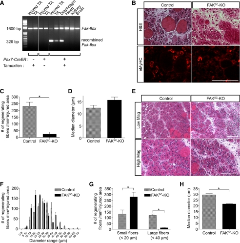
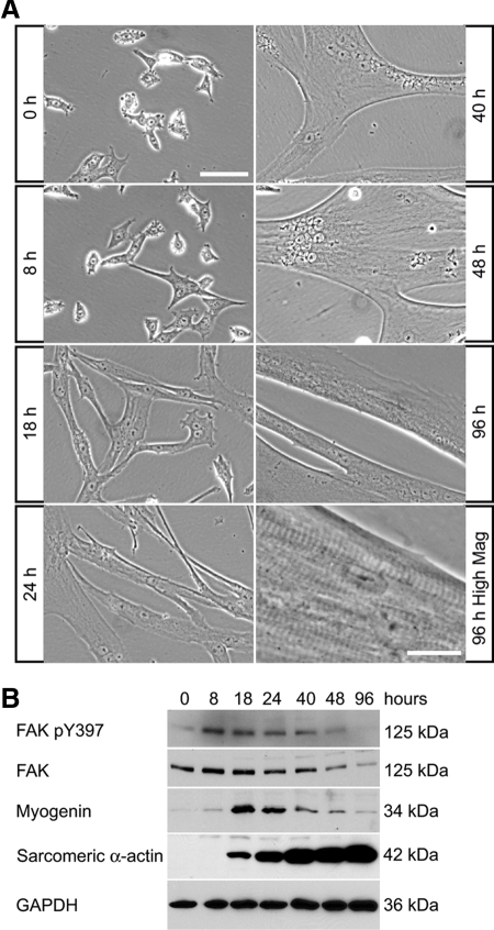
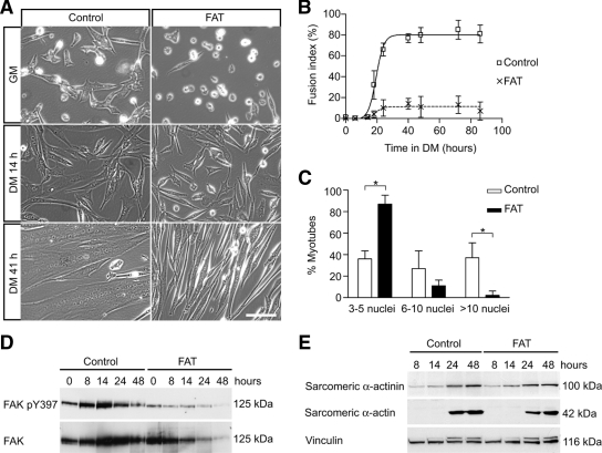
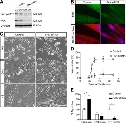
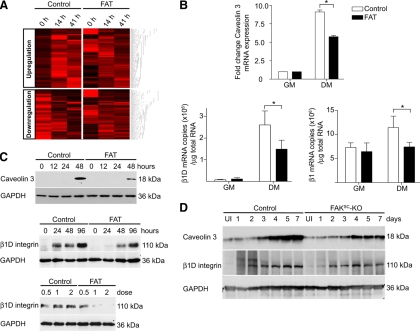
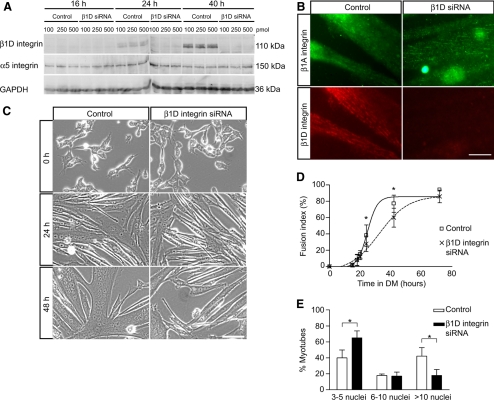
An essential phase of skeletal myogenesis is the fusion of mononucleated myoblasts to form multinucleated myotubes. Many cell adhesion proteins, including integrins, have been shown to be important for myoblast fusion in vertebrates, but the mechanisms by which these proteins regulate cell fusion remain mostly unknown. Here, we focused on the role of focal adhesion kinase (FAK), an important nonreceptor protein tyrosine kinase involved in integrin signaling, as a potential mediator by which integrins may regulate myoblast fusion. To test this hypothesis in vivo, we generated mice in which the Fak gene was disrupted specifically in muscle stem cells ("satellite cells") and we found that this resulted in impaired myotube formation during muscle regeneration after injury. To examine the role of FAK in the fusion of myogenic cells, we examined the expression of FAK and the effects of FAK deletion on the differentiation of myoblasts in vitro. Differentiation of mouse primary myoblasts was accompanied by a rapid and transient increase of phosphorylated FAK. To investigate the requirement of FAK in myoblast fusion, we used two loss-of-function approaches (a dominant-negative inhibitor of FAK and FAK small interfering RNA [siRNA]). Inhibition of FAK resulted in markedly impaired fusion but did not inhibit other biochemical measures of myogenic differentiation, suggesting a specific role of FAK in the morphological changes of cell fusion as part of the differentiation program. To examine the mechanisms by which FAK may be regulating fusion, we used microarray analysis to identify the genes that failed to be normally regulated in cells that were fusion defective due to FAK inhibition. Several genes that have been implicated in myoblast fusion were aberrantly regulated during differentiation when FAK was inhibited. Intriguingly, the normal increases in the transcript of caveolin 3 as well as an integrin subunit, the beta1D isoform, were suppressed by FAK inhibition. We confirmed this also at the protein level and show that direct inhibition of beta1D subunit expression by siRNA inhibited myotube formation with a prominent effect on secondary fusion. These data suggest that FAK regulation of profusion genes, including caveolin 3 and the beta1D integrin subunit, is essential for morphological muscle differentiation.


Figures

Figure 1.
Figure 1.
Targeted disruption of Fak in satellite cells impairs skeletal muscle regeneration. (A) Tissue genotyping. In 2-mo-old Pax7-CreER,Fakflox/flox mice, recombination at the Fak locus was detected in skeletal muscles only after tamoxifen treatment. (B) Control (Fakflox/flox) and FAKSC-KO (Pax7-CreER,Fakflox/flox) mice treated with tamoxifen were injured by BaCl2 injection in tibialis anterior muscles. Serial sections of 3-d regenerating muscles were stained with H&E (top) and immunostained with an antibody to eMyHC. Bar, 100 μm. (C) The numbers of regenerating myofibers expressing eMyHC were measured in control and FAKSC-KO muscles 3 d after injury (n = 3; *p < 0.05). (D) The diameters of regenerating myofibers expressing eMyHC were measured in control and FAKSC-KO muscles 3 d after injury (n = 3). (E) Muscle regeneration was analyzed 5 d after injury by H&E staining in control and FAKSC-KO muscles. Bar, 100 μm. (F) The numbers of regenerating myofibers (containing centrally located nuclei) were measured in control and FAKSC-KO muscles 5 d after injury and expressed as a histogram plot (n = 3). (G) The average numbers of either small (<20 μm) or large (>40 μm) regenerating fibers in control and FAKSC-KO muscles were determined 5 d after injury (n = 3; *p < 0.05). (H) The median diameters of regenerating myofibers were measured in control and FAKSC-KO muscles 5 d after injury (n = 3; *p < 0.05).
Figure 2.
Figure 2.
Expression of FAK during myogenic differentiation in primary myoblast cultures. (A) Mouse primary myoblasts were cultured in DM and observed by phase contrast microscopy. Multinucleated myotubes formed within 48 h of initiation of differentiation. Myotube maturation is associated with the appearance of aligned sarcomeric striations and myotube contractions (seen at high magnification at 96 h). Bar, 100 μm (except for 96 h, high-magnification panel, 20 μm). (B) Primary myoblasts were cultured in DM then analyzed by Western blots. The levels of FAK and phosphorylated FAK were high during early differentiation then decreased as myotubes mature. Myogenin was transiently up-regulated during early differentiation, whereas sarcomeric α-actin increased dramatically with myogenic differentiation.
Figure 3.
Figure 3.
Inhibition of FAK by its dominant-negative FAT inhibits myoblast fusion without blocking the expression of muscle terminal differentiation genes. (A) Myoblasts infected with adenoviruses expressing GFP (“control”) or FAT-GFP (“FAT”) were induced to differentiate to determine the effect of FAK inhibition on myogenesis. The morphology of the cells was analyzed by phase contrast microscopy. Myoblast fusion was impaired in FAT-expressing cultures that were composed mostly of mono- or binucleated cells, even after 3 d in DM. Bar, 100 μm. (B) Quantitative analysis of morphological differentiation by determination of the fusion index. The fusion index in FAT-expressing cultures was dramatically decreased compared with control cultures. (C) Quantitative analysis of myonuclear content after 48 h of differentiation. This analysis demonstrates a decreased number of nuclei in FAT-expressing myotubes, with the proportion of myotubes with three to five nuclei being increased, and the proportion of myotubes with >10 nuclei being decreased compared with control cultures (*p < 0.05). (D) Myoblasts expressing GFP (“control”) and FAT-GFP (“FAT”) were cultured in DM and analyzed by Western blots. A transient up-regulation of phosphorylated FAK and FAK protein was detected during early differentiation in controls but not in FAT-expressing cultures. (E) Expression of myogenic markers. Sarcomeric α-actinin and α-actin were induced normally in FAT-expressing cells, suggesting that the expression of muscle terminal differentiation proteins was not blocked when FAK was inhibited (representative blot of at least 3 independent experiments). Unlike FAK, the expression of focal adhesion protein vinculin did not seem to change appreciably during myogenic differentiation. Note that the muscle-specific isoform, metavinculin, which is of slightly greater molecular weight and thus runs just above vinculin and is recognized by the same antibody, increased during muscle differentiation and is similarly increased in FAT-expressing and control cells.
Figure 4.
Figure 4.
Down-regulation of FAK protein expression by siRNA inhibits myoblast fusion. (A) Myoblasts were transfected with control siRNA or FAK siRNA, cultured in DM for 48 h and then analyzed by Western blots to determine the efficacy of FAK knockdown by siRNA. The levels of phosphorylated FAK and FAK protein were dramatically down-regulated in FAK siRNA-treated cells. GAPDH was used as a loading control. (B) Immunofluorescence of siRNA-treated myotubes with an anti-FAK antibody revealed the presence of FAK at focal adhesions in control cells but not in FAK siRNA-treated cells. Coimmunostaining for sarcomeric α-actinin showed expression of this marker of muscle terminal differentiation in FAK-inhibited cells. Nuclei are labeled with DAPI. Bar, 20 μm. (C) Myoblasts transfected with control or FAK siRNA were cultured in DM to determine the effect of FAK down-regulation on myoblast fusion. Myoblast fusion was impaired in FAK siRNA-treated cultures in which most cells were elongated but remained mono- or binucleated. Bar, 100 μm. (D) Determination of the fusion index in control and FAK siRNA-treated cultures confirmed the defect of myoblast fusion when FAK was inhibited. (E) Determination of myonuclear content in control and FAK siRNA-treated cultures. Cells transfected with siRNA oligonucleotides were cultured in DM for 48 h. The proportion of myotubes containing few nuclei was increased whereas the proportion of myotubes containing a large number of nuclei was decreased in FAK siRNA-treated cultures (*p < 0.05).
Figure 5.
Figure 5.
Identification of genes that failed to be normally regulated in FAT-expressing cells by microarray analysis and confirmation for two candidate genes. (A) Hierarchical clustering of the 1919 genes that failed to be up-regulated in cells with inhibited FAK signaling, and 1614 genes that failed to be down-regulated. (B) Quantitative real-time RT-PCR confirmed the inhibition of caveolin 3, β1D integrin subunit, and total β1 integrin transcript up-regulation in FAT-expressing cultures within 48 h after the switch to DM (*p < 0.05). (C) FAT-expressing myoblasts were cultured in DM then analyzed by Western blots to determine the effect of FAK inhibition on caveolin 3 and β1D integrin subunit at the protein level. The induction of caveolin 3 and β1D subunit upon differentiation was delayed in FAT-expressing cultures compared with control cultures. Myoblasts infected with different doses of Ad-GFP (control) or Ad-FAT-GFP (FAT) also were analyzed 48 h after differentiation. Inhibition of expression of the β1D subunit by FAT expression was dose dependent. (D) Protein expression of caveolin 3 and β1D integrins was analyzed in uninjured (UI) and regenerating muscles of control and FAKSC-KO mice. Caveolin 3 and β1D integrin up-regulation failed to occur normally in regenerating FAKSC-KO muscles.
Figure 6.
Figure 6.
Down-regulation of β1D subunit by siRNA inhibits myoblast fusion. (A) siRNA was used to down-regulate the level of β1D integrins expression during myogenic differentiation to study the effect on cell fusion. Myoblasts were transfected with different amounts of siRNA oligonucleotides (100, 250, or 500 pmol/well) and then cultured in DM. Analysis by Western blots demonstrates an effective inhibition of β1D integrin up-regulation upon differentiation at all doses tested. The α5 integrin subunit, used here as a control, remained unchanged. (B) Immunofluorescence of β1A and β1D integrin subunits in cells treated with siRNA and cultured for 48 h in DM. In control myotubes, both isoforms were localized at focal adhesions. In cultures treated with β1D subunit siRNA, only the β1A isoform was detected. Bar, 20 μm. (C) Myoblasts treated by siRNA were induced to differentiate to determine the effect of β1D subunit inhibition on morphological differentiation. Cells treated with β1D subunit siRNA formed myotubes of a smaller size. Bar, 100 μm. (D) Determination of fusion index in siRNA-treated cultures. Fusion was delayed in β1D siRNA-treated cultures but ultimately reached a similar plateau to that seen in control cultures, suggesting that primary fusion was relatively normal in the absence of the β1D isoform (*p < 0.05). (E) Determination of myonuclear content in siRNA-treated cultures. Cells transfected with siRNA were cultured in DM for 72 h. The proportion of myotubes containing a large number of nuclei was decreased in β1D siRNA-treated cultures suggesting a selective inhibition of secondary fusion (*p < 0.05).

Similar articles

Cited by

References

    1. Abmayr S. M., Balagopalan L., Galletta B. J., Hong S. J. Cell and molecular biology of myoblast fusion. Int. Rev. Cytol. 2003;225:33–89. - PubMed
    1. Abmayr S. M., Zhuang S., Geisbrecht E. R. Myoblast fusion in Drosophila. Methods Mol. Biol. 2008;475:75–97. - PubMed
    1. Andres V., Walsh K. Myogenin expression, cell cycle withdrawal, and phenotypic differentiation are temporally separable events that precede cell fusion upon myogenesis. J. Cell Biol. 1996;132:657–666. - PMC - PubMed
    1. Baudoin C., Goumans M. J., Mummery C., Sonnenberg A. Knockout and knockin of the beta1 exon D define distinct roles for integrin splice variants in heart function and embryonic development. Genes Dev. 1998;12:1202–1216. - PMC - PubMed
    1. Beggs H. E., Schahin-Reed D., Zang K., Goebbels S., Nave K. A., Gorski J., Jones K. R., Sretavan D., Reichardt L. F. FAK deficiency in cells contributing to the basal lamina results in cortical abnormalities resembling congenital muscular dystrophies. Neuron. 2003;40:501–514. - PMC - PubMed

Publication types

MeSH terms

LinkOut - more resources