

Listeria monocytogenes delivery of HPV-16 major capsid protein L1 induces systemic and mucosal cell-mediated CD4+ and CD8+ T-cell responses after oral immunization
- PMID: 19435416
- PMCID: PMC2763599
- DOI: 10.1089/vim.2008.0071
Listeria monocytogenes delivery of HPV-16 major capsid protein L1 induces systemic and mucosal cell-mediated CD4+ and CD8+ T-cell responses after oral immunization
Abstract
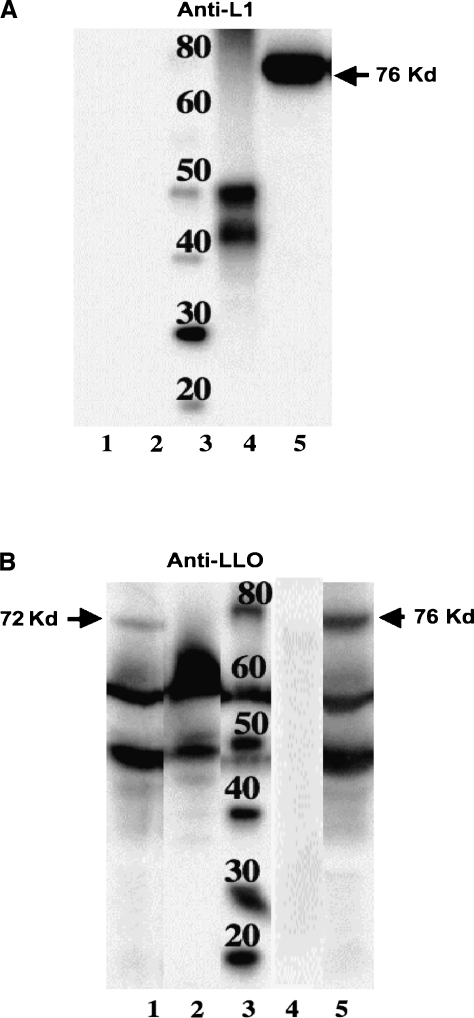
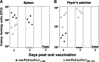
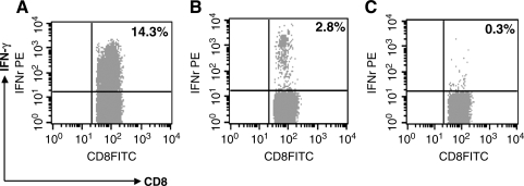
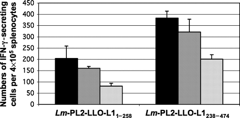
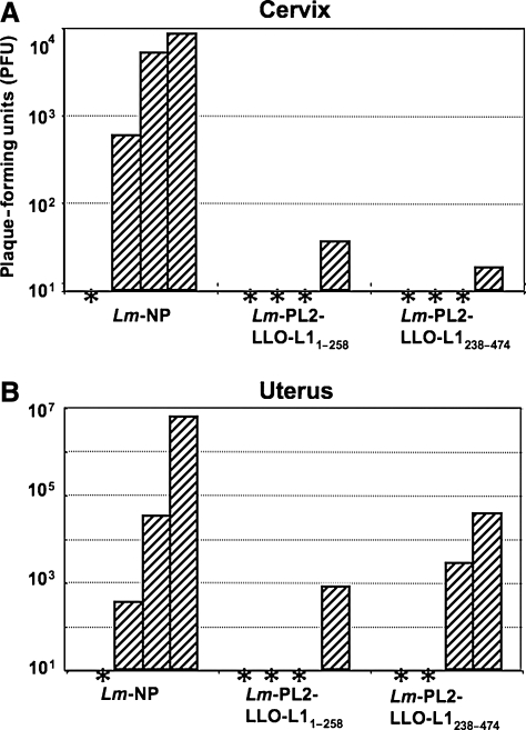
Neutralizing antibodies are thought to be required at mucosal surfaces to prevent human papillomavirus (HPV) transmission. However, the potential for cell-mediated immunity in mediating protection against HPV infection has not been well explored. We generated recombinant Listeria monocytogenes (Lm) constructs that secrete listeriolysin O (LLO) fused with overlapping N-terminal (LLO-L1(1-258)) or C-terminal (LLO-L1(238-474)) fragments of HPV type 16 major capsid protein L1 (HPV-16-L1). Oral immunization of mice with either construct induced IFN-gamma-producing CD8+ and CD4+ T cells in the spleen and in the Peyer's patches with the C-terminal construct. Oral immunization with both constructs resulted in diminished viral titers in the cervix and uterus of mice after intravaginal challenge with vaccinia virus expressing HPV-16-L1.
Figures
Similar articles
-
Two Listeria monocytogenes vaccine vectors that express different molecular forms of human papilloma virus-16 (HPV-16) E7 induce qualitatively different T cell immunity that correlates with their ability to induce regression of established tumors immortalized by HPV-16.J Immunol. 2001 Dec 1;167(11):6471-9. doi: 10.4049/jimmunol.167.11.6471. J Immunol. 2001. PMID: 11714814
-
Recombinant Listeria vaccines containing PEST sequences are potent immune adjuvants for the tumor-associated antigen human papillomavirus-16 E7.Cancer Res. 2004 Dec 15;64(24):8821-5. doi: 10.1158/0008-5472.CAN-04-1958. Cancer Res. 2004. PMID: 15604239
-
Regression of established human papillomavirus type 16 (HPV-16) immortalized tumors in vivo by vaccinia viruses expressing different forms of HPV-16 E7 correlates with enhanced CD8(+) T-cell responses that home to the tumor site.J Virol. 2001 Oct;75(20):9654-64. doi: 10.1128/JVI.75.20.9654-9664.2001. J Virol. 2001. PMID: 11559797 Free PMC article.
-
Developments in L2-based human papillomavirus (HPV) vaccines.Virus Res. 2017 Mar 2;231:166-175. doi: 10.1016/j.virusres.2016.11.020. Epub 2016 Nov 23. Virus Res. 2017. PMID: 27889616 Free PMC article. Review.
-
Therapeutic options for treatment of human papillomavirus-associated cancers - novel immunologic vaccines: ADXS11-001.Gynecol Oncol Res Pract. 2017 Jul 14;4:10. doi: 10.1186/s40661-017-0047-8. eCollection 2017. Gynecol Oncol Res Pract. 2017. PMID: 28725449 Free PMC article. Review.
Cited by
-
HPV16L1-attenuated Shigella recombinant vaccine induced strong vaginal and systemic immune responses in guinea pig model.Hum Vaccin Immunother. 2014;10(12):3491-8. doi: 10.4161/hv.36084. Hum Vaccin Immunother. 2014. PMID: 25483698 Free PMC article.
-
Co-administration with DNA encoding papillomavirus capsid proteins enhances the antitumor effects generated by therapeutic HPV DNA vaccination.Cell Biosci. 2015 Jun 25;5:35. doi: 10.1186/s13578-015-0025-y. eCollection 2015. Cell Biosci. 2015. PMID: 26113972 Free PMC article.
-
Dendritic cell vaccine therapy for colorectal cancer.Pharmacol Res. 2021 Feb;164:105374. doi: 10.1016/j.phrs.2020.105374. Epub 2020 Dec 28. Pharmacol Res. 2021. PMID: 33348026 Free PMC article. Review.
-
Trial Watch: DNA vaccines for cancer therapy.Oncoimmunology. 2014 Jan 1;3(1):e28185. doi: 10.4161/onci.28185. Epub 2014 Apr 1. Oncoimmunology. 2014. PMID: 24800178 Free PMC article. Review.
-
New approaches to prophylactic human papillomavirus vaccines for cervical cancer prevention.Antivir Ther. 2012;17(3):425-34. doi: 10.3851/IMP1941. Epub 2011 Nov 7. Antivir Ther. 2012. PMID: 22293302 Free PMC article. Review.
References
-
- Ho GY. Burk RD. Klein S, et al. Persistent genital human papillomavirus infection as a risk factor for persistent cervical dysplasia. J Natl Cancer Inst. 1995;87:1365–1371. - PubMed
-
- Nobbenhuis MA. Walboomers JM. Helmerhorst TJ, et al. Relation of human papillomavirus status to cervical lesions and consequences for cervical-cancer screening: a prospective study. Lancet. 1999;354:20–25. - PubMed
-
- Wallin KL. Wiklund F. Angstrom T, et al. Type-specific persistence of human papillomavirus DNA before the development of invasive cervical cancer. N Engl J Med. 1999;341:1633–1638. - PubMed
-
- Zhou J. Sun XY. Davies H. Crawford LV. Park D. Frazer IH. Definition of linear antigenic regions of the HPV16 L1 capsid protein using synthetic virion-like particles. Virology. 1992;189:592–529. - PubMed
Publication types
MeSH terms
Substances
Grants and funding
LinkOut - more resources
Full Text Sources
Other Literature Sources
Research Materials
Miscellaneous
