. 2009 May 14;27(22):2994-3006.
doi: 10.1016/j.vaccine.2009.02.103. Epub 2009 Mar 10.

Role of cell signaling in poxvirus-mediated foreign gene expression in mammalian cells

Affiliations

Role of cell signaling in poxvirus-mediated foreign gene expression in mammalian cells

Ningjie Hu et al. Vaccine. .

Abstract

Poxviruses have been extensively used as a promising vehicle to efficiently deliver a variety of antigens in mammalian hosts to induce immune responses against infectious diseases and cancer. Using recombinant vaccinia virus (VV) and canarypox virus (ALVAC) expressing enhanced green fluorescent protein (EGFP) or multiple HIV-1 gene products, we studied the role of four cellular signaling pathways, the phosphoinositide-3-OH kinase (PI3K), extracellular signal-regulated kinase (ERK), p38 mitogen-activated protein kinase (p38 MAPK), and c-Jun N-terminal kinase (JNK), in poxvirus-mediated foreign gene expression in mammalian cells. In nonpermissive infection (human monocytes), activation of PI3K, ERK, p38 MAPK, and JNK was observed in both VV and ALVAC and blocking PI3K, p38 MAKP, and JNK pathways with their specific inhibitors significantly reduced viral and vaccine antigen gene expression. Whereas, blocking the ERK pathway had no significant effect. Among these cellular signaling pathways studied, PI3K was the most critical pathway involved in gene expression by VV- or ALVAC-infected monocytes. The important role of PI3K in poxvirus-mediated gene expression was further confirmed in mouse epidermal cells stably transfected with dominant-negative PI3K mutant, as poxvirus-mediated targeted gene expression was significantly decreased in these cells when compared with their parental cells. Signaling pathway activation influenced gene expression at the mRNA level rather than virus binding. In permissive mammalian cells, however, VV DNA copies were also significantly decreased in the absence of normal function of the PI3K pathway. Poxvirus-triggered activation of PI3K pathway could be completely abolished by atazanavir, a new generation of antiretroviral protease inhibitors (PIs). As a consequence, ALVAC-mediated EGFP or HIV-1 gag gene expression in infected primary human monocytes was significantly reduced in the presence of atazanavir. These findings implicate that antiretroviral therapy (ART), also known as highly active antiretroviral therapy (HAART), may negatively impact the efficacy of live poxvirus vector-based vaccines and should be carefully considered when administering such live vaccines to individuals on ART.


Figures
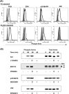
Figure 1
Figure 1
Poxvirus exposure induces phosphorylation of PI3K, ERK, p38 MAPK, and JNK. Isolated primary human monocytes from PBMCs were exposed to VV or ALVAC at 10 MOI for up to 1 h at 37°C in a 5% CO2 incubator. Cells were then subjected to intracellular staining and flow cytometric analysis (Fig. 1A) or Western blot analysis (Fig. 1B) with specific Abs for the phospho-PI3K, phospho-ERK, phospho-p38 MAPK, and phospho-JNK. In some cases, the monocytes were preincubated with signal transduction pathway inhibitors for 15 min before exposure to VV or ALVAC. (A) Gray histogram represents background without staining. Histogram in a thick line represents positive staining with specific Abs for the phospho-PI3K, phospho-ERK, phospho-p38 MAPK, or phospho-JNK, whereas histogram in a thin line represents negative background staining with isotype-matched control antibodies. (B) Western blot results of signaling pathway activation induced by ALVAC exposure: left side shows the detection of phosphorylated forms of Akt, ERK, p38 MAPK, and JNK, and right side shows the same plots probed for total Akt, ERK, p38 MAPK, and JNK proteins, respectively, to demonstrate equal loading of samples. Monocytes that had been incubated with DMSO alone were used as controls for the inhibitors, as signal transduction pathway inhibitors were all dissolved in DMSO. Results are representative of three independent experiments with different donors and similar Western blot results were also obtained from VV-exposed monocytes (data not shown). D, DMSO control.
Figure 2
Figure 2
Cellular signaling pathways are involved in poxvirus-mediated protein expression. Primary human peripheral monocytes (up to 1 × 10⁶ cells) were pretreated with specific inhibitors of LY294002, PD98059, SB203580, or SP600125 at 10 μM for 15 min at 37°C for blocking PI3K, ERK, p38 MAPK, or JNK, respectively. DMSO treatment was used as a negative control in every experiment. Cells were then infected with EGFP-VV, EGFP-ALVAC (vCP1540), or vCP1452 at an MOI of 10 for 8 h at 37°C in a 5% CO2 incubator. After infection, cells infected with EGFP-VV or vCP1540 were harvested for surface staining with anti-human CD14APC Ab, and then subjected to flow cytometric analysis to determine virus-mediated EGFP expression. Cells infected with vCP1452 were lysed for measuring HIV Gag p24 expression. (A) Cells were infected with EGFP-VV or vCP1540 and the infection was determined by analysis of EGFP-positive cells. The number in each dot plot shows the percentage of EGFP-positive monocytes in a representative experiment from five independent experiments with different donors. (B) Pooled data are mean ± SD of percentage of EGFP-positive monocytes, presenting five independent experiments with different donors. (C) HIV-1 p24 ELISA results from cells infected with vCP1452. Pooled data are mean ± SD of HIV-1 p24 levels, presenting five independent experiments with different donors. LY, LY294002; PD, PD98059; SB, SB203580; SP, SP600215.
Figure 2
Figure 2
Cellular signaling pathways are involved in poxvirus-mediated protein expression. Primary human peripheral monocytes (up to 1 × 10⁶ cells) were pretreated with specific inhibitors of LY294002, PD98059, SB203580, or SP600125 at 10 μM for 15 min at 37°C for blocking PI3K, ERK, p38 MAPK, or JNK, respectively. DMSO treatment was used as a negative control in every experiment. Cells were then infected with EGFP-VV, EGFP-ALVAC (vCP1540), or vCP1452 at an MOI of 10 for 8 h at 37°C in a 5% CO2 incubator. After infection, cells infected with EGFP-VV or vCP1540 were harvested for surface staining with anti-human CD14APC Ab, and then subjected to flow cytometric analysis to determine virus-mediated EGFP expression. Cells infected with vCP1452 were lysed for measuring HIV Gag p24 expression. (A) Cells were infected with EGFP-VV or vCP1540 and the infection was determined by analysis of EGFP-positive cells. The number in each dot plot shows the percentage of EGFP-positive monocytes in a representative experiment from five independent experiments with different donors. (B) Pooled data are mean ± SD of percentage of EGFP-positive monocytes, presenting five independent experiments with different donors. (C) HIV-1 p24 ELISA results from cells infected with vCP1452. Pooled data are mean ± SD of HIV-1 p24 levels, presenting five independent experiments with different donors. LY, LY294002; PD, PD98059; SB, SB203580; SP, SP600215.
Figure 3
Figure 3
Cellular signaling pathways are involved in viral mRNA expression, not in poxvirus binding to monocytes. Primary human peripheral monocytes were pretreated with specific inhibitors of LY294002, PD98059, SB203580, or SP600125 at 10 μM for 15 min at 37°C for blocking PI3K, ERK, p38 MAPK, or JNK, respectively. DMSO treatment was used as a negative control in every experiment. Cells were then exposed to parental VV or parental ALVAC at an MOI of 10 for 1 h at 37°C in a 5% CO2 incubator. Cells from each condition were washed twice with PBS to remove free virus particles. After washing, cells were equally split into two aliquots: one aliquot of cells was directly used for DNA extraction for realtime qPCR to determine viral DNA copies, and the other aliquot of cells in each condition was resuspended in 200 μl of complete RPMI 1640 medium and continuously cultured for an additional 6 h in the presence of individual inhibitors. After 6 h culture, cells were harvested for RNA extraction for realtime qRT-PCR to determine the viral mRNA level. Pooled data are mean ± SD of viral cDNA or DNA copies, presenting five independent experiments with different blood donors. LY, LY294002; PD, PD98059; SB, SB203580; SP, SP600215.
Figure 4
Figure 4
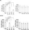
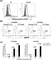
Cells transfected with a dominant-negative PI3K mutant decrease expression of targeted proteins mediated by poxvirus infection. Cl 41 cells and the Cl 41-derived cell line stably transfected with a dominant-negative PI3K mutant (Cl 41Δp85) were exposed with poxvirus, either parental VV, EGFP-VV, parental ALVAC, vCP1540, or vCP1452 at an MOI of 10 for 1 h at 37°C in a 5% CO2 incubator. After washing, cells were equally split into two aliquots: one aliquot of cells was directly used for intracellular staining with a specific Ab for the phospho-PI3K to measure activation of PI3K pathway, and the other aliquot of cells in each condition was resuspended in complete RPMI 1640 medium and then continuously cultured for an additional 1 to 12 h. Cells were then harvested at the checked points of 1, 6, and 12h postinfection for detecting EGFP expression (VV-EGFP or vCP1540 infection) by flow cytometric analysis (Fig. 4B and 4C), or detecting HIV-1 p24 expression (vCP1452 infection) by ELISA (Fig. 4D) to determine protein expression, or RNA and DNA extraction for real-time qPCR or qRT-PCR to determine the viral mRNA level and DNA copies (Fig. 4E). Fig. 4A shows a representative experiment of VV- or ALVAC-induced PI3K pathway activation. Gray histogram represents background without staining. Histogram in a thick line represents positive staining with a specific Ab for the phospho-PI3K, whereas histogram in a thin line represents negative background staining with an isotype-matched control Ab and histogram in a dotted line represents PI3K staining from the cells pre-treated with PI3K inhibitor LY294002. Fig. 4B: number in each plot shows the percentage of EGFP-positive cells checked at 6 h postinfection. Fig 4C: pooled data are mean ± SD of percentage of EGFP-positive cells presenting three independent experiments. Fig. 4D: pooled data are mean ± SD of HIV-1 p24 (pg/106 cells), presenting three independent experiments. Fig. 4E: viral cDNA or DNA copies (copies/106 cells), presenting three independent experiments. Black bar: virus-infected Cl 41 cells; open bar: virus-infected Cl 41Δp85.
Figure 4
Figure 4
Cells transfected with a dominant-negative PI3K mutant decrease expression of targeted proteins mediated by poxvirus infection. Cl 41 cells and the Cl 41-derived cell line stably transfected with a dominant-negative PI3K mutant (Cl 41Δp85) were exposed with poxvirus, either parental VV, EGFP-VV, parental ALVAC, vCP1540, or vCP1452 at an MOI of 10 for 1 h at 37°C in a 5% CO2 incubator. After washing, cells were equally split into two aliquots: one aliquot of cells was directly used for intracellular staining with a specific Ab for the phospho-PI3K to measure activation of PI3K pathway, and the other aliquot of cells in each condition was resuspended in complete RPMI 1640 medium and then continuously cultured for an additional 1 to 12 h. Cells were then harvested at the checked points of 1, 6, and 12h postinfection for detecting EGFP expression (VV-EGFP or vCP1540 infection) by flow cytometric analysis (Fig. 4B and 4C), or detecting HIV-1 p24 expression (vCP1452 infection) by ELISA (Fig. 4D) to determine protein expression, or RNA and DNA extraction for real-time qPCR or qRT-PCR to determine the viral mRNA level and DNA copies (Fig. 4E). Fig. 4A shows a representative experiment of VV- or ALVAC-induced PI3K pathway activation. Gray histogram represents background without staining. Histogram in a thick line represents positive staining with a specific Ab for the phospho-PI3K, whereas histogram in a thin line represents negative background staining with an isotype-matched control Ab and histogram in a dotted line represents PI3K staining from the cells pre-treated with PI3K inhibitor LY294002. Fig. 4B: number in each plot shows the percentage of EGFP-positive cells checked at 6 h postinfection. Fig 4C: pooled data are mean ± SD of percentage of EGFP-positive cells presenting three independent experiments. Fig. 4D: pooled data are mean ± SD of HIV-1 p24 (pg/106 cells), presenting three independent experiments. Fig. 4E: viral cDNA or DNA copies (copies/106 cells), presenting three independent experiments. Black bar: virus-infected Cl 41 cells; open bar: virus-infected Cl 41Δp85.
Figure 5
Figure 5
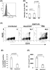
HIV-1 protein inhibitor suppresses poxvirus-mediated protein expression by targeting PI3K/Akt pathway. Monocytes were pretreated with atazanavir at 10 μM for 15 min at 37°C. Cells were then exposed with poxvirus, either ALVAC or vCP1452 at 10 MOI for various time intervals. (A) Cells exposed with ALVAC were harvested at 1 h post-exposure for flow cytometric analysis of PI3K activation with a specific Ab for the phospho-PI3K. (B) Pooled data are mean ± SD of phospho-PI3K fluorescence intensity presenting three independent experiments with different blood donors. (C) Monocytes pretreated with atazanavir at 10 uM for 15 min were infected with vCP1540 at an MOI of 10 for 6 h at 37°C in a 5% CO2 incubator. After infection, cells were harvested for surface staining with anti-human CD14PE Ab, and then subjected to flow cytometric analysis to determine EGFP expression on CD14-positive monocytes. The number in each dot plot shows the percentage of EGFP-positive monocytes in a representative experiment from three independent experiments with different donors. (D) Pooled data are mean ± SD of percentage of EGFP-positive monocytes presenting three independent experiments with different donors. (E) Monocytes infected with vCP1452 for 6 h were harvested and washed 2 times with ice-chilled PBS. Cells were resuspended in 50 μl PBS, and then lysed with 50 μl of lysis buffer appended in the HIV p24 ELISA kit for measuring HIV-1 Gag p24 expression. Pooled data are mean ± SD of HIV-1 p24 (pg/106 monocytes) presenting three independent experiments with different donors.

Similar articles

Cited by

References

    1. Moss B. Vaccinia and other poxvirus expression vectors. Curr Opin Biotechnol. 1992 Oct;3(5):518–22. - PubMed
    1. Moss B, Fuerst TR, Flexner C, Hugin A. Roles of vaccinia virus in the development of new vaccines. Vaccine. 1988 Apr;6(2):161–3. - PubMed
    1. Moss B, Flexner C. Vaccinia virus expression vectors. Annu Rev Immunol. 1987;5:305–24. - PubMed
    1. Poulet H, Minke J, Pardo MC, Juillard V, Nordgren B, Audonnet JC. Development and registration of recombinant veterinary vaccines. The example of the canarypox vector platform. Vaccine. 2007 Jul 26;25(30):5606–12. - PubMed
    1. Perkus ME, Tartaglia J, Paoletti E. Poxvirus-based vaccine candidates for cancer, AIDS, and other infectious diseases. J Leukoc Biol. 1995 Jul;58(1):1–13. - PubMed

Publication types

MeSH terms