. 2009 Jul;1793(7):1199-209.
doi: 10.1016/j.bbamcr.2009.04.012. Epub 2009 May 3.

Regulation of the formation and trafficking of vesicles from Golgi by PCH family proteins during chemotaxis

Affiliations

Regulation of the formation and trafficking of vesicles from Golgi by PCH family proteins during chemotaxis

S Lee et al. Biochim Biophys Acta. 2009 Jul.

Abstract

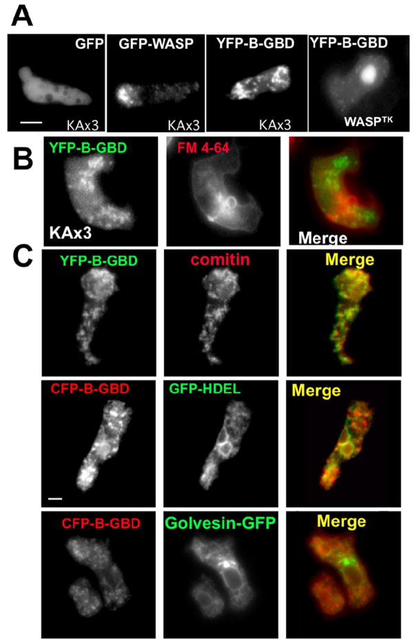
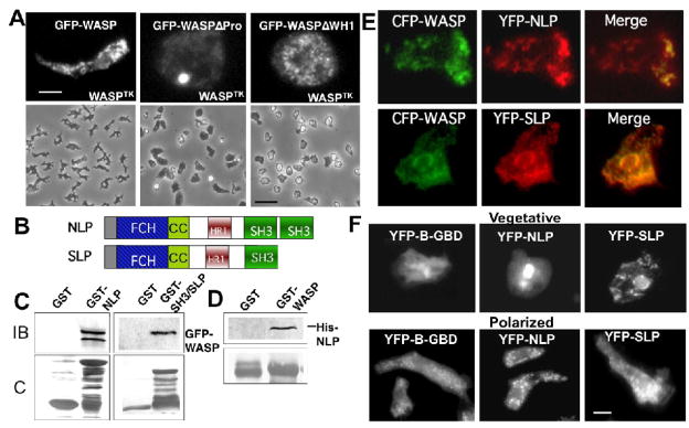
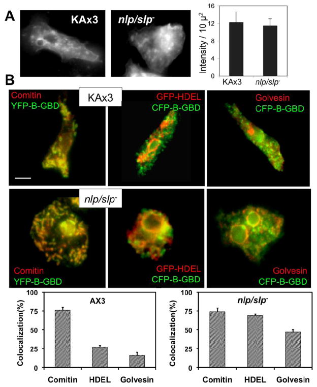
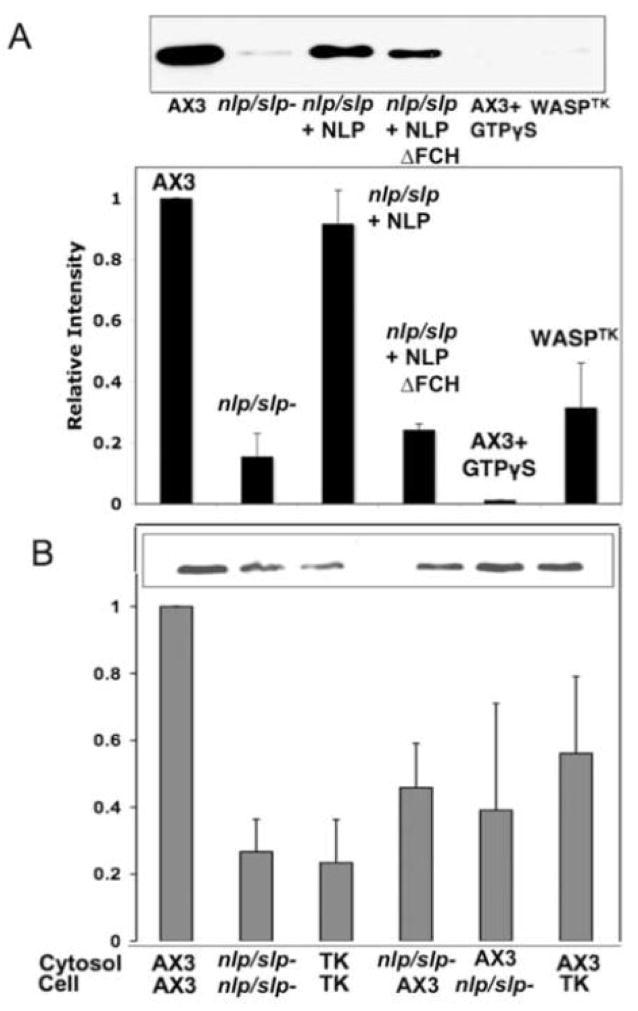
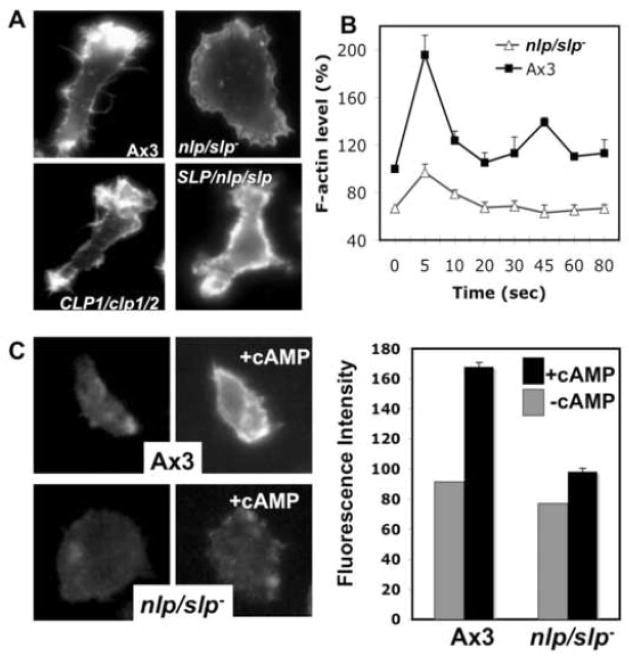
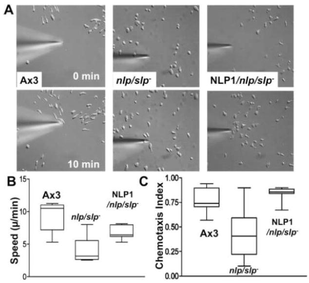
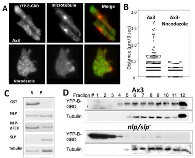
Previous study demonstrated that WASP localizes on vesicles during Dictyostelium chemotaxis and these vesicles appear to be preferentially distributed at the leading and trailing edge of migrating cells. In this study, we have examined the role of PCH family proteins, Nwk/Bzz1p-like protein (NLP) and Syndapin-like protein (SLP), in the regulation of the formation and trafficking of WASP-vesicles during chemotaxis. NLP and SLP appear to be functionally redundant and deletion of both nlp and slp genes causes the loss of polarized F-actin organization and significant defects in chemotaxis. WASP and NLP are colocalized on vesicles and interactions between two molecules via the SH3 domain of NLP/SLP and the proline-rich repeats of WASP are required for vesicle formation from Golgi. Microtubules are required for polarized trafficking of these vesicles as vesicles showing high directed mobility are absent in cells treated with nocodazole. Our results suggest that interaction of WASP with NLP/SLP is required for the formation and trafficking of vesicles from Golgi to the membrane, which might play a central role in the establishment of cell polarity during chemotaxis.


Figures

Figure 1
Figure 1
(A) Localization of GFP-WASP or YFP-B-GBD expressed in Dictyostelium cells migrating toward a cAMP gradient. Cells were pulsed for 5 hours and localizations of GFP-WASP or YFP-B-GBD in aggregation-competent KAx3 cells, or YFP-B-GBD in WASPTK cells were examined. Bar = 5 μm. (B) KAx3 cells were stained with FM4-64, a marker for internalized endosomal compartments, for 2 min and washed. After 10 min delay, cells were imaged to determine the colocalization. (C) KAx3 cells expressing YFP-B-GBD were stained with an antibody against comitin (a marker for Golgi and Golgi-derived cesicles). In bottom panels, fluorescence images of living cells co-expressing GFP-HDEL(an ER-specific retrieval signal in Dictyostelium) or golvesin-c-GFP (a marker for Golgi but not for Golgi-derived vesicles) and CFP-B-GBD are shown. Bar = 5 μm.
Figure 2
Figure 2
(A) Top panels show localization of GFP-WASP, GFP-WASPΔPro and GFP-WASPΔWH1 expressed in WASPTK cells (bar = 5 μm). Bottom panels show phase contrast pictures of these cells at lower magnification (bar = 50 μm). (B) Schematic diagram of NLP/SLP domain structure. FCH: FER/CIP4-homology; CC: coiled-coil; HR1: homology region 1; SH3: Src homology 3. (C) GST pull-down assay showing the interaction between WASP and NLP/SLP. GST-NLP- or GST-SH3 (SH3 domain of SLP)-bound agarose beads were incubated with the lysate of GFP-WASP expressing cells. Bound GFP-WASP was detected by immunoblot with the anti-GFP antibody. IB: immunoblot; C: Coomassie Blue staining. (D) Direct interaction between purified 6X His-tagged NLP and GST-WASP was also confirmed by pull down assay. (E) Fluorescence images of aggregation-competent cells co-expressing CFP-WASP and YFP-NLP or -SLP are shown. CFP-WASP showed punctate vesicle labeling which appears to overlap with YFP-NLP or 2 signal and these vesicles were enriched at the leading edge of migrating cells. (F) Change of subcellular localization of NLP/SLP upon polarization of cells. Images of YFP-NLP, YFP-SLP, or YFP-B-GBD were taken in cells at growth stage and in polarized cells at aggregation-competent stage. NLP/SLP are localized at the perinuclear region in non-polarized cells at growth stage, but with vesicles in polarized cells.
Figure 3
Figure 3
(A) Membrane endocytosis by aggregation-competent cells determined using FM1-43X. Ax3 or nlp/slp- cells were incubated with FM1-43X for 2 min and washed, followed by fixation for image collection. Cells were fixed after 2 min incubation with FM 1-43 followed by a 10 min delay to allow for endocytosis. Fluorescence intensity of FM1-43X dye in cells was measured and shown in the graph. Error bars represent SEM (n=10). (B) Colocalization of CFP-B-GBD with GFP-HDEL and golvesin-c-GFP in nlp/slp- cells. GFP-HDEL and golvesin-c-GFP overlaps with CFP-B-GBD in nlp/slp- cells, but not in KAx3 cells. At least 15 cells from two independent experiments were analyzed for colocalization as decribed in Methods section. The percent colocalization calculated among areas is shown for the percentage of YFP-B-GBD that colocalized with comitin, GFP-HDEL, or golvesin-c-GFP respectively.
Figure 4
Figure 4
Vesicle formation reconstitution assay. The role of NLP/SLP for vesicle formation from Golgi was examined using an assay that reconstitutes the formation of vesicles in a cell-free system. (A) Immunoblot of comitin on secretory vesicles formed in permeabilized cells reconstituted with the cytosol fraction prepared from the cell lysate. nlp/slp and WASPTK cells showed significantly reduced formation of vesicles, which can be rescued by GST-NLP, but not with NLP lacking FCH domain. Graph shows quantification of band intensities from four separate immunoblots. (B) Vesicle formation was measured in different combinations of the cytosols and permeabilized cells of wild type, nlp/slp-, or WASPTK cells.
Figure 5
Figure 5
F-actin organization and chemotaxis of nlp/slp- cells. (A) Aggregation-competent cells were fixed and stained with phallioidin. nlp/slp- cells exhibited severe defects in F-actin organization as they exhibit neither a prominent F-actin-enriched lamellipod nor cell polarity. This defect can be rescued by the expression of NLP or SLP. (B) In vivo actin polymerization assay measuring F-actin assembled in response to the chemoattractant (cAMP) stimulation. Basal F-actin level before cAMP stimulation is lower in nlp/slp- cells and F-actin polymerization upon cAMP stimulation is also defective in nlp/slp- cells. Error bars represent SEM (n=4) (C) Barbed-end staining of KAx3 or nlp/slp- cells. Barbed-ends were stained with Alexa595-labeled G-actin before and 5 seconds after cAMP stimulation. nlp/slp- cells show a greatly decreased number of free barbed ends at the cortical membrane upon cAMP stimulation relative to wild-type cells. A linescan analysis was performed to measure the pixel intensity at cortical membrane after background subtraction. Average fluorescence intensities measured at the cortical membrane (4 linescans per cell and 10–12 cells total) before and after cAMP stimulation are presented in the graph. Error bars represent SEM (n=8)
Figure 6
Figure 6
Abnormal chemotactic movement of nlp/slp- cells. (A) DIC images of cells migrating toward a cAMP gradient at 0 or 10 min. Wild-type cells are usually well polarized, move quickly and linearly toward the cAMP source. nlp/slp- cells appear to have difficulties developing a psuedopod in the direction of the chemoattractant gradient. The expression of YFP-NLP alone mostly rescues motility defects. (B) The speed and chemotaxis index of nlp/slp- cells is significantly decreased compared to KAx3 cells, but recovered by the expression of NLP. Chemotaxis of at least 7 cells from two independent experiments was measured.
Figure 7
Figure 7
(A) Distribution of vesicles decorated with YFP-B-GBD in chemotaxing or nocodazole-treated cells. Cells expressing YFP-B-GBD in aggregation stage were fixed and stained with rabbit anti-tubulin antibody. The YFP-B-GBD and microtubule images were superimposed for assessment of localization of YFP-B-GBD vesicles in a close proximity to microtubules. (B) Tracking of YFP-B-GBD vesicle movements in KAx3 or nocodazole-treated cells by live cell imaging. Speed of vesicle movement in wild type or cells treated with nocodazole was measured and plotted. Wild type cells showed brief periods of rapid directed movement leading to high speed (>0.25 μm/sec) that is absent in nocodazole-treated cells. (C) Binding of NLP/SLP and a NLP mutant to microtubule. GST-NLP or SLP were incubated with paclitaxel stabilized-microtubules. Supernatants (s) and pellets (p) were collected after ultracentrifugation over a 15% sucrose cushion and analyzed by SDS-PAGE for presence of GST-NLP or SLP by SDS-PAGE. (D) Cosedimentation of YFP-B-GBD with MT on a sucrose gradient. Lysates of wild type or nlp/slp- cells expressing YFP-B-GBD were applied onto 15–40% sucrose gradient and fractions were collected. YFP-B-GBD in fractions was detected by western blot with anti-GFP antibody.

Similar articles

Cited by

References

    1. Locker J, Goldblatt PJ, Leighton J. Ultrastructural features of invasion in chick embryo liver metastasis of Yoshida ascites hepatoma. Cancer Res. 1970;30:1632–1644. - PubMed
    1. Lahrtz F, Piali L, Spanaus KS, Seebach J, Fontana A. Chemokines and chemotaxis of leukocytes in infectious meningitis. J Neuroimmunol. 1998;85:33–43. - PubMed
    1. Downey GP. Mechanisms of leukocyte motility and chemotaxis. Curr Opin Immunol. 1994;6:113–124. - PubMed
    1. Devreotes PN, Zigmond SH. Chemotaxis in eukaryotic cells: A focus on leukocytes and Dictyostelium. Annu Rev Cell Biol. 1988;4:649–686. - PubMed
    1. Symons M, Derry JM, Karlak B, Jiang S, Lemahieu V, Mccormick F, Francke U, Abo A. Wiskott-Aldrich syndrome protein, a novel effector for the GTPase CDC42Hs, is implicated in actin polymerization. Cell. 1996;84:723–734. - PubMed

Publication types

MeSH terms

LinkOut - more resources