. 2009;11(3):R25.
doi: 10.1186/bcr2252. Epub 2009 May 1.

QLT0267, a small molecule inhibitor targeting integrin-linked kinase (ILK), and docetaxel can combine to produce synergistic interactions linked to enhanced cytotoxicity, reductions in P-AKT levels, altered F-actin architecture and improved treatment outcomes in an orthotopic breast cancer model

Affiliations

QLT0267, a small molecule inhibitor targeting integrin-linked kinase (ILK), and docetaxel can combine to produce synergistic interactions linked to enhanced cytotoxicity, reductions in P-AKT levels, altered F-actin architecture and improved treatment outcomes in an orthotopic breast cancer model

Jessica Kalra et al. Breast Cancer Res. 2009.

Abstract

Introduction: Substantial preclinical evidence has indicated that inhibition of integrin linked-kinase (ILK) correlates with cytotoxic/cytostatic cellular effects, delayed tumor growth in animal models of cancer, and inhibition of angiogenesis. Widely anticipated to represent a very promising therapeutic target in several cancer indications, it is increasingly evident that optimal therapeutic benefits obtained using ILK targeting strategies will only be achieved in combination settings. The purpose of this study was to investigate the therapeutic potential of the ILK small molecule inhibitor, QLT0267 (267), alone or in combination with chemotherapies commonly used to treat breast cancer patients.

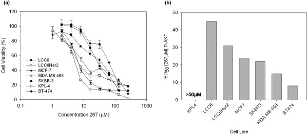
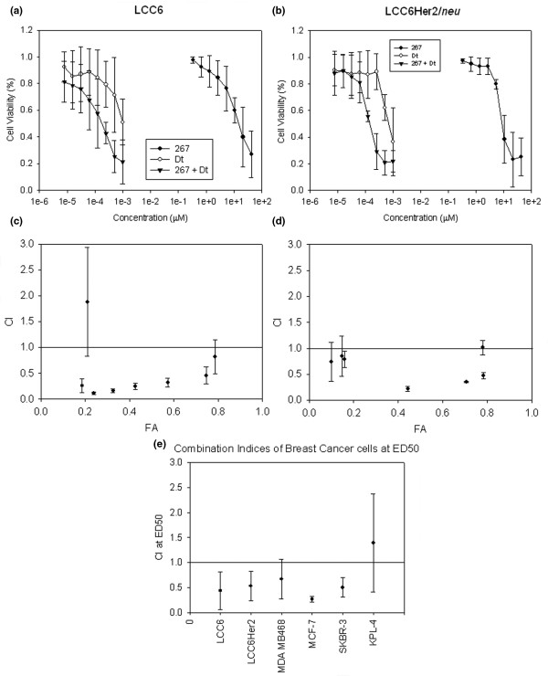
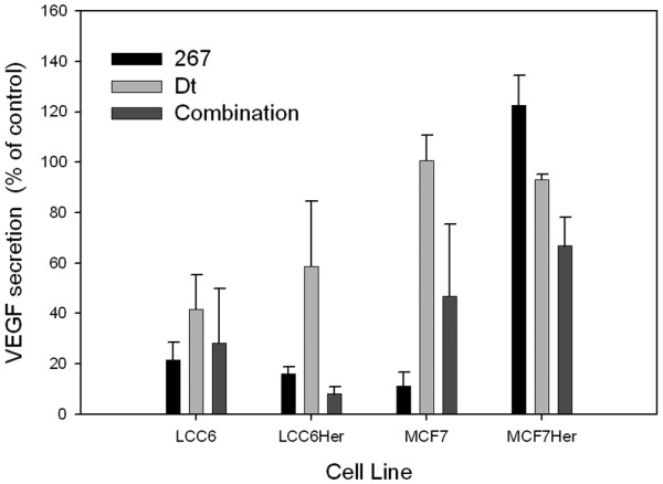
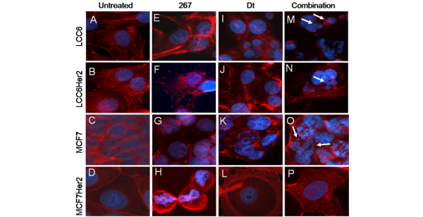
Methods: A single end-point metabolic assay was used as an initial screen for 267 interactions with selected chemotherapeutic agents. These in vitro assays were completed with seven breast cancer cell lines including several which over-expressed human epidermal growth factor receptor 2 (Her2). One agent, docetaxel (Dt), consistently produced synergistic interactions when combined with 267. Dt/267 interactions were further characterized by measuring therapeutic endpoints linked to phosphorylated protein kinase B (P-AKT) suppression, inhibition of vascular endothelial growth factor (VEGF) secretion and changes in cytoarchitecture. In vivo efficacy studies were completed in mice bearing orthotopic xenografts where tumor growth was assessed by bioluminescence and calliper methods.

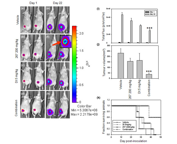
Results: The combination of 267 and Dt resulted in increased cytotoxic activity, as determined using an assay of metabolic activity. Combinations of cisplatin, doxorubicin, vinorelbine, paclitaxel, and trastuzumab produced antagonistic interactions. Further endpoint analysis in cell lines with low Her2 levels revealed that the 267/Dt combinations resulted in: a three-fold decrease in concentration (dose) of 267 required to achieve 50% inhibition of P-AKT; and a dramatic disruption of normal filamentous-actin cellular architecture. In contrast to Her2-positive cell lines, three-fold higher concentrations of 267 were required to achieve 50% inhibition of P-AKT when the drug was used in combination with Dt. In vivo studies focusing on low Her2-expressing breast cancer cells (LCC6) implanted orthotopically demonstrated that treatment with 267/Dt engendered improved therapeutic effects when compared with mice treated with either agent alone.

Conclusions: The findings indicate that the 267/Dt drug combination confers increased (synergistic) therapeutic efficacy towards human breast cancer cells that express low levels of Her2.


Figures

Figure 1
Figure 1
Breast cancer cells exhibit dose-dependent decrease in (a) cell viability and (b) P-AKT in response to increasing concentrations of the ILK small molecule inhibitor, QLT0267. (a) Seven breast cancer cell lines (LCC6, LCC6Her2, SKBR-3, KPL-4, BT-474, MBA-MB-468, and MCF-7) were treated with increasing doses (1 μm to 256 μM) of 267 for 72 hours and cell viability was evaluated using the AlamarBlue® assay. Percentage cell viability relative to control (untreated) cells are shown. Each data point represents the mean (± standard deviation) determined from three experiments done in triplicate. Treated cells were assessed for phosphorylated protein kinase B (P-AKT) using western blot analysis of protein lysates collected eight hours after treatment. (b) Western blot were analyzed using densitometry, dose-response curves were generated and analyzed using Calcusyn to determine the ED50 of 267 for PAKT suppression for each cell line. 267 = QLT0267; ILK = integrin-linked kinase.
Figure 2
Figure 2
Breast cancer cells treated with 267 and docetaxel combined at a fixed ratio and added at various concentrations exhibit synergistic effects based on a measured cell viability endpoint. (a) LCC6 and (b) LCC6Her2 cells were treated with increasing concentrations of 267, docetaxel (Dt) or 267 and Dt combined at a fixed ratio (50 μm:1 nm). Percentage cell viability relative to control cells not treated with drugs (100% viable) are shown and each data point is the average (± standard deviation) of triplicate samples. Combination index (CI) values determined by Calcusyn from dose-response curves of (c) LCC6 and (d) LCC6Her2 cells treated with 267/Dt combinations. Data points represent the average (± standard deviation) from triplicate experiments. CI values less than one are indicative of synergistic effects; CI values greater than one are indicative of antagonistic effects; and CI values equal to one are indicative of additive effects. Fraction affected (FA) is a measure of the determined effect (cytotoxicity as measured by an Alamar blue assay) and amount of drug to achieve a FA of 0.5 is referred as the ED50. (e) Mean CI values at ED50 for LCC6, LCC6Her2, MCF-7, MDA MB468, KPL-4, and SKBR-3 cells treated with 267 and Dt combined are shown and each data point represents the average (± standard deviation) from triplicate experiments.
Figure 3
Figure 3
The dose reduction index (DRI) calculated using the Calcusyn program was used to estimate the ED50 of drugs (267 and/or docetaxel) against the indicated cell lines. The DRI estimates the extent to which the dose of one or more agents in the combination can be reduced to achieve effect levels that are comparable with those achieved with single agents. Black bars indicate the mean ED50 calculated for the drugs when added alone and the grey bars indicate the mean ED50 calculated for the drugs when used in combination. (a) ED50s for QLT0267 (267); when used in combination to treat LCC6 cells the ED50 of 267 can be reduced by 3.6-fold. (b) ED50s for docetaxel; the dose reduction index assessment for docetaxel (Dt) indicated that dose reductions of up to 27.5 fold (LCC6 cells) can be obtained.
Figure 4
Figure 4
Dose response curves were generated for changes in P-AKT in LCC6 and LCC6Her2 cells after treatment with QLT0267, docetaxel, or a fixed ratio combination of QLT0267 and docetaxel. LCC6 and LCC6Her2 cells were treated for eight hours with increasing concentrations of QLT0267, docetaxel, or a fixed ratio combination of QLT0267 and docetaxel (50 μm:1 nm) to establish dose response curves based on an endpoint measuring suppression of P-AKT levels as determined by western blot analysis. Representative western blot images were collected using a fluorescence based imaging system (odyssey, Licor) where the colours (red or green) represent the secondary antibody used, show that increasing concentrations of (b) docetaxel (Dt) exerted no significant effect on the expression of integrin-linked kinase (ILK), protein kinase B (AKT), and phosphorylated protein kinase B (P-AKT) in either cell line. Treatment with increasing doses of (a) QLT0267 (267) alone or (c) in combination with Dt showed dose-dependent decrease in P-AKT. Densitometry assessment of western blots (n = 3) were used to estimate treatment response relative to controls (taken to be 100% P-AKT levels) and the resulting data was then analyzed by Calcusyn to determine estimated DRI. The DRI was then used to estimate the dose of 267 when used alone (black bars) or in combination with Dt (grey bars) needed to achieve a defined fraction affected (FA). The dose of drug required to achieve an FA of 0.5 is defined as the ED50 for the measured P-AKT suppression endpoint. The ED50 of 267 was about 30 μM in the (d) LCC6 cell line, while in the presence of (e) Dt the 267 ED50 was about 11 μM. In LCC6Her2 cells, more 267 was required when used in combination to achieve FA similar to that of single agents. For example in the LCC6Her2 cells, the 267 ED50 when used alone was about 30 μM, and this increased to 130 μM when 267 was used in combination with Dt.
Figure 5
Figure 5
Levels of P-AKT were measured in LCC6, LCC6Her2, MCF-7, and MCF-7Her2 cells after treatment with a singles dose of QLT0267, docetaxel, or a combination of QLT0267 and docetaxel. (a) LCC6, LCC6Her2, MCF-7, and MCF-7Her2 cells express baseline levels of integrin-linked kinase (ILK), protein kinase B (AKT), and phosphorylated AKT (P-AKT). (b) LCC6, (c) LCC6Her2, (d) MCF-7, and (e) MCF-7Her2 cells were treated for eight hours QLT0267 (267) (42 μM), docetaxel (Dt) (1 nM), or the combination of 267 and Dt. Western blot analysis using a fluorescence based imaging system (odyssey, Licor) shows that treatment with 267 and 267/Dt elicit considerable reductions in the level of P-AKT relative to controls (untreated cells). Treatment with Dt had no effect on P-AKT levels. Reductions in P-AKT were not attributable to changes in the level of ILK or AKT. Band intensities for P-AKT were normalized to actin then to untreated controls and changes in levels are indicated in the values provided just below the P-AKT band (n = 3).
Figure 6
Figure 6
Levels of secreted VEGF were measured in LCC6, LCC6Her2, MCF-7, and MCF-7Her2 cells after treatment with a singles dose of QLT0267, docetaxel, or a combination of QLT0267 and docetaxel. LCC6, LCC6Her2, MCF-7, and MCF-7Her2 were treated with 267 (10 μM), Dt (0.25 nM) or a combination of both for 72 hours. Conditioned media was collected subsequently assessed for vascular endothelial growth factor (VEGF) secretions using ELISA as described in the Materials and methods. All four cell lines assessed showed a decrease in VEGF secretion when treated with low doses of QLT0267 (267) alone or with the combination 267/docetaxel (Dt). Dt also moderately attenuated VEGF secretion in each cell line (n = 3).
Figure 7
Figure 7
267/Dt treatment of LCC6, LCC6Her2, MCF-7, and MCF-7Her2 causes disruption of normal F-actin cytoarchitecture and abnormal nuclear morphology. LCC6, LCC6Her2, MCF-7, and MCF-7Her2 were treated for eight hours with 267 (42 μM), Dt (1 nM) or the combination of 267 and Dt. Subsequently, cells were fixed using paraformaldehyde, permeabilized, and stained for nuclear material using Hoechst and F-actin using Texas red conjugated phalloidin. Representative photomicrographs of (a to d) untreated cells or cells treated with (e to h) QLT0267 (267), (i to l) docetaxel (Dt) and (m to p) the combination of 267/Dt are shown, where blue represents the nucleus and red the F-actin microfilaments. Combination treatment resulted in a distinct decrease in total F-actin staining, a change in actin organization, the appearance of apoptotic nuclear bodies (white arrows), as well as metaphase chromosomes suggestive of a cell cycle block in these cells.
Figure 8
Figure 8
267/Dt combination therapy in vivo correlates with reduced tumor burden and extended survival in orthotopic LCC6 breast cancer tumor model. Bioluminescent imaging of orthotopic LCC6 tumors are shown one day after treatment initiation and on day 22 after treatment initiation. The treatment groups included (a and e) vehicle controls, (b and f) QLT0267 (267), (c and g) docetaxel (Dt), and the (d and h) combination of 267/Dt treated animals (doses indicated on graph). Total light emission from tumors in animals was visualized and quantified. (i) Animals treated with the combination showed lower total light flux than all other treatment groups. Animals treated with 267 exhibited tumors with darkened areas in the core (see inset in f as an example). Tumor size in treated animals was measured by callipers and these data were used to estimate the (j) tumor volumes. The combination of 267/Dt was significantly lower (***P < 0.005) then all other treatment groups analyzed. (k) Kaplan-Meier survival analysis of data defining survival endpoints based on tumor ulceration and/or tumors more than 0.5 g were used to determine median survival times. For animals treated with 267 (200 mg/kg) the median survival time was 33 days (26 days post-treatment initiation), animals treated with Dt (5 mg/kg) exhibited a median survival time of 31 days (24 days post-treatment initiation); and animals treated with the 267/Dt combination exhibited a median survival time of more than 90 days (83 days post-treatment initiation). In this group three out of five animals were alive at day 90, while no animals were alive for any other treatment group.

Similar articles

Cited by

References

    1. Wu C, Dedhar S. Integrin-linked kinase (ILK) and its interactors: a new paradigm for the coupling of extracellular matrix to actin cytoskeleton and signaling complexes. J Cell Biol. 2001;155:505–510. doi: 10.1083/jcb.200108077. - DOI - PMC - PubMed
    1. Yau CY, Wheeler JJ, Sutton KL, Hedley DW. Inhibition of integrin-linked kinase by a selective small molecule inhibitor, QLT inhibits the PI3K/PKB/mTOR, Stat3, and FKHR pathways and tumor growth, and enhances gemcitabine-induced apoptosis in human orthotopic primary pancreatic cancer xenografts. Cancer Res. 2005;65:1497–1504. doi: 10.1158/0008-5472.CAN-04-2940. - DOI - PubMed
    1. Yoganathan TN, Costello P, Chen X, Jabali M, Yan J, Leung D, Zhang Z, Yee A, Dedhar S, Sanghera J. Integrin-linked kinase (ILK): a "hot" therapeutic target. Biochem Pharmacol. 2000;60:1115–1119. doi: 10.1016/S0006-2952(00)00444-5. - DOI - PubMed
    1. Hannigan G, Troussard AA, Dedhar S. Integrin-linked kinase: a cancer therapeutic target unique among its ILK. Nat Rev Cancer. 2005;5:51–63. doi: 10.1038/nrc1524. - DOI - PubMed
    1. Wu C. Integrin-linked kinase and PINCH: partners in regulation of cell-extracellular matrix interaction and signal transduction. J Cell Sci. 1999;112(Pt 24):4485–4489. - PubMed

Publication types

MeSH terms