Comparative Study
. 2009 Apr 29;29(17):5546-57.
doi: 10.1523/JNEUROSCI.0759-09.2009.

Alpha9 integrin promotes neurite outgrowth on tenascin-C and enhances sensory axon regeneration

Affiliations
Comparative Study

Alpha9 integrin promotes neurite outgrowth on tenascin-C and enhances sensory axon regeneration

Melissa R Andrews et al. J Neurosci. .

Abstract

Damaged CNS axons are prevented from regenerating by an environment containing many inhibitory factors. They also lack an integrin that interacts with tenascin-C, the main extracellular matrix glycoprotein of the CNS, which is upregulated after injury. The alpha9beta1 integrin heterodimer is a receptor for the nonalternatively spliced region of tenascin-C, but the alpha9 subunit is absent in adult neurons. In this study, we show that PC12 cells and adult rat dorsal root ganglion (DRG) neurons do not extend neurites on tenascin-C. However, after forced expression of alpha9 integrin, extensive neurite outgrowth from PC12 cells and adult rat DRG neurons occurs. Moreover, both DRG neurons and PC12 cells secrete tenascin-C, enabling alpha9-transfected cells to grow axons on tissue culture plastic. Using adeno-associated viruses to express alpha9 integrin in vivo in DRGs, we examined axonal regeneration after cervical dorsal rhizotomy or dorsal column crush in the adult rat. After rhizotomy, significantly more dorsal root axons regrew into the dorsal root entry zone at 6 weeks after injury in alpha9 integrin-expressing animals than in green fluorescent protein (GFP) controls. Similarly, after a dorsal column crush injury, there was significantly more axonal growth into the lesion site compared with GFP controls at 6 weeks after injury. Behavioral analysis after spinal cord injury revealed that both experimental and control groups had an increased withdrawal latency in response to mechanical stimulation when compared with sham controls; however, in response to heat stimulation, normal withdrawal latencies returned after alpha9 integrin treatment but remained elevated in control groups.


Figures

Figure 1.
Figure 1.
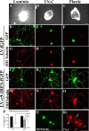
PC12 cells overexpressing α9 integrin extend neurites on TN-C, independent of the cytoplasmic domain. A, Quantification of neurite length from differentiated PC12 cells at 48 h, mock transfected or transfected with α4, α5, or α9 integrin. The asterisk (*) indicates significantly (p < 0.05) greater neurite outgrowth in α9-expressing PC12 cells on TN-C and plastic compared with mock transfected, α4, and α5-expressing PC12 cells. B–G, Immunofluorescent labeling of neurofilament in PC12 cells transduced with a lentivirus expressing fGFP (B–D) or α9 integrin-IRES-fGFP (E–G) grown on laminin (B, E), TN-C (C, F), or plastic (D, G). H, Quantification of neurite length from differentiated PC12 cells at 48 h, mock transfected or transfected with α9 integrin constructs (α5/α9, α9/α4, α9/α5, α9NOCYT). The asterisk (*) indicates significantly (p < 0.05) greater neurite outgrowth on TN-C or plastic in the presence of the α9 extracellular domain. I, Quantification of neurite length from differentiated PC12 cells at 24 h, mock transfected or transfected with α9/α9, α9/α4, or α9/α5 incubated in the presence or absence of anti-human α9β1 blocking antibody (“a9BLK”). All neurite outgrowth assays were repeated three times. The length of at least 50 randomly selected neurons was analyzed. The asterisk (*) indicates significantly (p < 0.05) less neurite outgrowth with α9β1 blocking antibody treatment of α9 integrin-expressing PC12 cells on TN-C or plastic. Error bars indicate SEM. Scale bar, 50 μm.
Figure 2.
Figure 2.
α9 integrin induces neurite outgrowth on adult rat cortical cryosections or on nitrocellulose filter implants from cortical lesions. Differentiated control (A, C) or α9/α9 (B, D) integrin-expressing PC12 cells (labeled with Cell Tracker Green) grown on cortical cryosections for 24 h, in the presence of anti-human α9β1 antibody, a9BLK (C, D). Quantification of neurite outgrowth of PC12 cells expressing human α9/α9, α9/α4, or α9/α5 grown on cortical cryosections for 24 h (E) or grown on filter implants (24 h) from 14-d-old cortical lesions (F) incubated in the presence or absence of anti-human α9β1 blocking antibody. Neurite outgrowth assays were repeated three times. The length of at least 50 randomly selected neurons was analyzed. The asterisk (*) indicates significantly (p < 0.05) greater neurite outgrowth of α9 chimeras on TN-C and plastic. Error bars indicate SEM. Scale bar, 100 μm.
Figure 3.
Figure 3.
PC12 cells express TN-C. A, Western blot analysis of cell extracts (left panels) and conditioned media (right panels) from differentiated PC12 cells transfected with the human integrins α4, α5, α5/α9, α9/α9, α9/α4, α9/α5, or α9 lacking the entire cytoplasmic domain (α9*) cultured for 48 h on PDL and examined for expression of osteopontin and full-length TN-C. Western blots were probed with rabbit IgG as a control. B, Full-length TN-C immunostaining in differentiated PC12 cells. Scale bar, 50 μm.
Figure 4.
Figure 4.
α9 integrin promotes adult sensory neurite outgrowth on TN-C. Adult DRG explants (A–C) or dissociated DRG neurons transduced with a lentivirus expressing fGFP (D–I) or a lentivirus expressing α9-IRES-fGFP (J–O) were cultured for 72 h on 10 μg/ml laminin (A, D, G, J, M), TN-C (B, E, H, K, N), or plastic (C, F, I, L, O). P, Quantification of dissociated DRG neurons transduced with an adenovirus expressing α9 integrin or LacZ plated on laminin, TN-C, or plastic. All neurite outgrowth assays were repeated three times. The length of at least 50 randomly selected neurons was analyzed. The asterisk (*) indicates significantly (p < 0.05) greater neurite outgrowth of dissociated DRGs expressing α9 integrin on TN-C and plastic. Dissociated DRG neurons were immunostained for βIII tubulin (Q) or TN-C (R). Error bars indicate SEM. Scale bars, 100 μm.
Figure 5.
Figure 5.
α9 integrin mRNA is expressed in neonate P0 DRG neurons, but not in adult sensory neurons, and is not upregulated after nerve injury. Postnatal day 0 (A–D) and adult dorsal root ganglia (E–H) were hybridized with antisense (A, E) and sense (D, H) probes against rat α9 integrin. Samples were immunolabeled with anti-neurofilament antibodies (3A10) (B, F) and Hoechst (C, G). α7 integrin (I) or α9 integrin (J) mRNA expression evaluated by RT-PCR from adult rat lumbar DRGs (L4, L5) 4 d after sciatic nerve crush from injured (i) and control (c) DRGs. K, α9 integrin mRNA expression evaluated by RT-PCR from adult rat lumbar DRGs (L4, L5) assessed after sciatic nerve crush in both injured (i) and control (c) DRGs over time (1, 4, 7 d after injury). Densitometry of integrin mRNA was performed and the relative changes in α7 integrin (I) or α9 integrin (J, K) expression after injury compared with control/uninjured levels is shown. Scale bar, 100 μm.
Figure 6.
Figure 6.
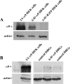
α9 integrin expression after viral transduction in vitro and in vivo. A, Western blot analysis of lysates from LV-α9-IRES-fGFP, AAV-α9, or AAV-fGFP transduced HEK cells. B, Western blot analysis of cell lysates of LV-α9-IRES-fGFP transduced HEK cells, or tissue lysates from AAV-α9 transduced DRGs, or control DRGs (in vivo) examined for the presence of human α9 integrin or actin (loading control).
Figure 7.
Figure 7.
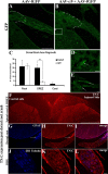
α9 integrin promotes dorsal root axon regrowth into the DREZ. Shown are horizontal sections of injured dorsal root with attached spinal cord, showing GFP-expressing axons 6 weeks after treatment transduced with AAV-fGFP alone (A) or with AAV-α9 integrin plus AAV-fGFP (B). The DREZ boundary is approximated with a dashed line. C, Quantification of GFP-expressing axons in the root, proximal to the crush, in the DREZ, and in the spinal cord. The asterisk (*) indicates significantly (p < 0.05) more axons were found in the DREZ in the α9 integrin group. High-magnification image of a GFP-labeled axon (D, arrow) located in the spinal cord after α9 integrin treatment from a region approximated by the box in B. High-magnification image of a DREZ after α9 integrin treatment showing a GFP-labeled axon entering the cord from the entry zone along with other axons within the DREZ (E, arrows). TN-C immunoreactivity is shown 3 weeks after dorsal root injury in the spinal cord (F), in the injured dorsal root (G–I), and in the DRG (J–L). Error bars indicate SEM. Scale bars: A, 50 μm; (in D) D, E, 10 μm; F, 500 μm; (in L) G–L, 100 μm.
Figure 8.
Figure 8.
α9 integrin enhances sensory axon growth into a dorsal column crush lesion. Shown are longitudinal sections through the dorsal column-injured spinal cord 6 weeks after treatment with AAV-α9 integrin plus AAV-fGFP (A–C, G–I), or AAV-fGFP alone (D–F, J). K, Quantification of GFP-labeled axons within the lesion boundaries in the α9 integrin-treated and the control-treated groups. The asterisk (*) indicates significantly (p < 0.05) more axons were present within the lesion site in the α9 group. L, M, Longitudinal section through the lesion site after α9 integrin-treatment immunostained for TN-C, showing a GFP-labeled axon growing along a TN-C-immunopositive fiber. N, O, Quantification of behavioral data for the pressure test (N) and the hot-plate test (O), before injury and at 5 weeks after injury. Error bars indicate SEM. Scale bars: A, D, L, 500 μm; (in F) B, C, E, F, 100 μm; (in G) G–J, M, 100 μm.

Similar articles

Cited by

References

    1. Baker KA, Hagg T. Developmental and injury-induced expression of alpha1beta1 and alpha6beta1 integrins in the rat spinal cord. Brain Res. 2007;1130:54–66. - PMC - PubMed
    1. Bartsch U. The extracellular matrix molecule tenascin-C: expression in vivo and functional characterization in vitro. Prog Neurobiol. 1996;49:145–168. - PubMed
    1. Carulli D, Rhodes KE, Brown DJ, Bonnert TP, Pollack SJ, Oliver K, Strata P, Fawcett JW. Composition of perineuronal nets in the adult rat cerebellum and the cellular origin of their components. J Comp Neurol. 2006;494:559–577. - PubMed
    1. Condic ML. Adult neuronal regeneration induced by transgenic integrin expression. J Neurosci. 2001;21:4782–4788. - PMC - PubMed
    1. Condic ML, Snow DM, Letourneau PC. Embryonic neurons adapt to the inhibitory proteoglycan aggrecan by increasing integrin expression. J Neurosci. 1999;19:10036–10043. - PMC - PubMed

Publication types

MeSH terms

LinkOut - more resources