. 2009 Jul;83(14):7252-60.
doi: 10.1128/JVI.00153-09. Epub 2009 Apr 29.

A shared interface mediates paramyxovirus interference with antiviral RNA helicases MDA5 and LGP2

Affiliations

A shared interface mediates paramyxovirus interference with antiviral RNA helicases MDA5 and LGP2

Jean-Patrick Parisien et al. J Virol. 2009 Jul.

Abstract

Diverse members of the Paramyxovirus family of negative-strand RNA viruses effectively suppress host innate immune responses through the actions of their V proteins. The V protein mediates interference with the interferon regulatory RNA helicase MDA5 to avoid cellular antiviral responses. Analysis of the interaction interface revealed the MDA5 helicase C domain as necessary and sufficient for association with V proteins from human parainfluenza virus type 2, parainfluenza virus type 5, measles virus, mumps virus, Hendra virus, and Nipah virus. The identified approximately 130-residue region is highly homologous between MDA5 and the related antiviral helicase LGP2, but not RIG-I. Results indicate that the paramyxovirus V proteins can also associate with LGP2. The V protein interaction was found to disrupt ATP hydrolysis mediated by both MDA5 and LGP2. These findings provide a potential mechanistic basis for V protein-mediated helicase interference and identify LGP2 as a second cellular RNA helicase targeted by paramyxovirus V proteins.


Figures
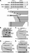
FIG. 1.
FIG. 1.
Map of V protein interaction regions of MDA5 and LGP2. (A) Diagrammatic representations of the LGP2, MDA5, and RIG-I proteins. (Top) Box diagrams illustrate the key features of the proteins, with CARD and helicase regions highlighted. Roman numerals depict the conserved helicase motifs, and boundaries of domain 1 and domain 2 are indicated. (Bottom) Illustration of MDA5 helicase domain protein fragments used for mapping the minimum V protein binding region. The black box depicts the N-terminal FLAG epitope tag, numbers refer to amino acid residues encompassed by the construct in parentheses, and + or − indicates V protein coprecipitation. (B) V protein associates with MDA5, the MDA5 helicase domain, and LGP2, but not RIG-I. FLAG-tagged helicase proteins were expressed in HEK293T cells along with HA-tagged measles virus V protein and lysates subject to FLAG immunoaffinity purification and anti-HA immunoblotting to detect protein coimmunoprecipitation. (C and D) Mapping the N-terminal boundary of the MDA5 V protein binding region. The experiment was similar to that shown in panel B, but we used serially truncated proteins as indicated. (E) Map of the C-terminal boundary of the MDA5 V protein binding region. The experiment was similar to those shown in panels B to D, but we used C-terminal truncation series as indicated. Vec, empty HA expression vector; Con, empty FLAG expression vector; FL, full-length protein; N, N-terminal CARD fragment; C, C-terminal helicase domain.
FIG. 2.
FIG. 2.
The MDA5 and LGP2 helicase C domains mediate the V protein interaction. (A) V protein association with defined MDA5 and LGP2 minimal targets. (Left) Full-length MDA5 (residues 1 to 1025), the MDA5 helicase domain (residues 327 to 1025), or MVBR fragment (residues 701 to 830) coprecipitate with the measles virus V protein. (Right) Full-length LGP2 (residues 1 to 678) or MVBR fragment (residues 351 to 479) coprecipitate with the measles virus V protein. (B) V protein interaction requires structural integrity of the helicase C domain. A coimmunoprecipitation assay was conducted using full-length, wild-type MDA5 or LGP2 (FL), or full-length proteins engineered with mutations to the indicated helicase conserved motifs (roman numerals).
FIG. 3.
FIG. 3.
Targeting of MDA5 and LGP2 via the MVBR is a property of diverse paramyxovirus V proteins and is mediated by the conserved CTD. (A) Diverse paramyxovirus V proteins coprecipitate with the minimal MDA5 or LGP2 target fragments. Another coprecipitation experiment was done using V proteins from PIV5 (P5), human parainfluenza virus type 2 (H2), mumps virus (Mu), measles virus (Me), Hendra virus (He), or Nipah virus (Ni). Expression of FLAG-tagged green fluorescent protein (GFP) combined with measles virus V protein served as a control for nonspecific interactions. (B) The V protein cysteine-rich C-terminal domain is sufficient to mediate association with the LGP2 and MDA5 MVBR fragments. FLAG-tagged full-length or target fragment of MDA5 (left) or LGP2 (right) was expressed along with glutathione S-transferase fusion proteins containing the measles virus (Me) or mumps virus (Mu) C-terminal domain, or as a control the measles virus N-terminal domain. Following FLAG or GST affinity purification, samples were probed for coprecipitation with antiserum specific to GST or FLAG, as indicated.
FIG. 4.
FIG. 4.
MVBR reacts with V protein from infected cells. Parallel plates of HEK293T cells were transfected with expression vectors for the indicated FLAG-tagged helicase proteins or fragments and then infected with 1 PFU/cell of PIV5 (+) or mock infected (−). Cell lysates were subjected to FLAG purification and immunoblotting with antiserum that recognizes the PIV5 V protein and P protein. Only V protein coprecipitated.
FIG. 5.
FIG. 5.
V protein interferes with ATP hydrolysis by MDA5 and LGP2. FLAG-tagged MDA5, LGP2, and RIG-I were immunoaffinity purified from HEK293T extracts with or without coexpressed V protein, and eluates were subjected to ATP hydrolysis analysis in the presence of poly(I:C). Top panels demonstrate copurification of V protein (PIV5V) with LGP2 and MDA5 but not RIG-I by immunoblotting. Middle panels demonstrate ATP hydrolysis activities by autoradiography, with positions of the origin, ATP, and free phosphate indicated. Bottom panels contain graphical representations of ATP hydrolysis activities, quantified by phosphorimaging analysis. Graphs plot the ratio of Pi to ATP, expressed as the percentage of the ATP hydrolysis activity without V protein. (A) Plasmids coding for FLAG-tagged LGP2, MDA5, or RIG-I RNA helicases were cotransfected with plasmids coding for HA-tagged PIV5 V protein or empty vector plasmid at a 1:1 ratio into HEK293T cells. Con, no-protein control; WT, wild type. (B) Four micrograms of cDNA plasmids coding for FLAG-tagged LGP2 helicase were cotransfected with empty vector (−) or 2 μg, 8 μg, or 16 μg of plasmid coding for HA-tagged PIV5 V protein. Total amounts of transfected DNA were equalized with empty vector for each reaction. (C) Plasmids coding for FLAG-tagged LGP2, MDA5, or RIG-I RNA helicases were cotransfected with empty vector control or plasmids coding for HA-tagged measles virus, mumps virus, or Nipah viurs V protein at a 1:3 ratio into HEK293T cells. *NS, nonspecific band.
FIG. 6.
FIG. 6.
Interference with ATP hydrolysis in infected cells. Results are for an experiment similar to that shown in Fig. 5, but FLAG-tagged MDA5, LGP2, RIG-I, or control GFP were immunoaffinity purified from HEK293T cells that were also infected with PIV5 and eluates were subjected to ATP hydrolysis analysis in the presence of poly(I:C). Top panels demonstrate copurification of V protein (PIV5V) with MDA5 (A) or LGP2 (B). Middle panels demonstrate ATP hydrolysis activities by autoradiography, and bottom panels contain graphical representations of ATP hydrolysis activities, quantified by phosphorimaging analysis. Graphs plot the ratio of Pi to ATP, expressed as a percentage of the ATP hydrolysis activity without V protein.

Similar articles

Cited by

References

    1. Andrejeva, J., K. S. Childs, D. F. Young, T. S. Carlos, N. Stock, S. Goodbourn, and R. E. Randall. 2004. The V proteins of paramyxoviruses bind the IFN-inducible RNA helicase, mda-5, and inhibit its activation of the IFN-beta promoter. Proc. Natl. Acad. Sci. USA 10117264-17269. - PMC - PubMed
    1. Bamming, D., and C. M. Horvath. 2009. Regulation of signal transduction by enzymatically inactive antiviral RNA helicase proteins MDA5, RIG-I and LGP2. J. Biol. Chem. 2849700-9712. - PMC - PubMed
    1. Childs, K., N. Stock, C. Ross, J. Andrejeva, L. Hilton, M. Skinner, R. Randall, and S. Goodbourn. 2007. mda-5, but not RIG-I, is a common target for paramyxovirus V proteins. Virology 359190-200. - PubMed
    1. Childs, K. S., J. Andrejeva, R. E. Randall, and S. Goodbourn. 2009. Mechanism of mda-5 inhibition by paramyxovirus V proteins. J. Virol. 831465-1473. - PMC - PubMed
    1. Cordin, O., J. Banroques, N. K. Tanner, and P. Linder. 2006. The DEAD-box protein family of RNA helicases. Gene 36717-37. - PubMed

Publication types

MeSH terms