

Loss of Wip1 sensitizes cells to stress- and DNA damage-induced apoptosis
- PMID: 19395378
- PMCID: PMC2719383
- DOI: 10.1074/jbc.M109.007823
Loss of Wip1 sensitizes cells to stress- and DNA damage-induced apoptosis
Abstract
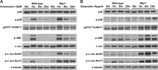
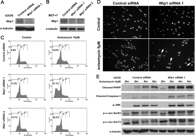
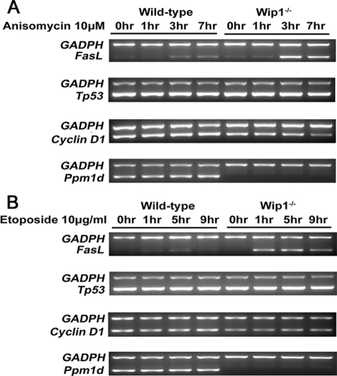
In response to various environmental stresses, the stress-responsive MAPKs p38 and JNK are activated and phosphorylate ATF2 and c-Jun transcription factors, thereby affecting cell-fate decision. Targeted gene disruption studies have established that JNK-c-Jun signaling plays a vital role in stress-induced apoptosis. The oncogenic phosphatase Wip1 acts as an important regulator in DNA damage pathway by dephosphorylating a spectrum of proteins including p53, p38, Chk1, Chk2, and ATM. In this study we show that Wip1 negatively regulates the activation of MKK4-JNK-c-Jun signaling during stress-induced apoptosis. The loss of Wip1 function sensitizes mouse embryonic fibroblasts to stress-induced apoptosis via the activation of both p38-ATF2 and JNK-c-Jun signaling. Here we reveal that Wip1 has dual roles in alternatively regulating stress- and DNA damage-induced apoptosis through p38/JNK MAPKs and p38/p53-dependent pathways, respectively. Our results point to Wip1 as a general regulator of apoptosis, which further supports its role in tumorigenesis.
Figures


Similar articles
-
Expression of a homeostatic regulator, Wip1 (wild-type p53-induced phosphatase), is temporally induced by c-Jun and p53 in response to UV irradiation.J Biol Chem. 2010 Mar 19;285(12):9067-76. doi: 10.1074/jbc.M109.070003. Epub 2010 Jan 21. J Biol Chem. 2010. PMID: 20093361 Free PMC article.
-
Arsenic trioxide augments Chk2/p53-mediated apoptosis by inhibiting oncogenic Wip1 phosphatase.J Biol Chem. 2008 Jul 4;283(27):18969-79. doi: 10.1074/jbc.M800560200. Epub 2008 May 15. J Biol Chem. 2008. PMID: 18482988
-
p53-inducible wip1 phosphatase mediates a negative feedback regulation of p38 MAPK-p53 signaling in response to UV radiation.EMBO J. 2000 Dec 1;19(23):6517-26. doi: 10.1093/emboj/19.23.6517. EMBO J. 2000. PMID: 11101524 Free PMC article.
-
The type 2C phosphatase Wip1: an oncogenic regulator of tumor suppressor and DNA damage response pathways.Cancer Metastasis Rev. 2008 Jun;27(2):123-35. doi: 10.1007/s10555-008-9127-x. Cancer Metastasis Rev. 2008. PMID: 18265945 Free PMC article. Review.
-
Regulation of the Wip1 phosphatase and its effects on the stress response.Front Biosci (Landmark Ed). 2012 Jan 1;17(4):1480-98. doi: 10.2741/3999. Front Biosci (Landmark Ed). 2012. PMID: 22201816 Free PMC article. Review.
Cited by
-
A novel mathematical model of ATM/p53/NF- κB pathways points to the importance of the DDR switch-off mechanisms.BMC Syst Biol. 2016 Aug 15;10(1):75. doi: 10.1186/s12918-016-0293-0. BMC Syst Biol. 2016. PMID: 27526774 Free PMC article.
-
Wip1 inhibitor GSK2830371 inhibits neuroblastoma growth by inducing Chk2/p53-mediated apoptosis.Sci Rep. 2016 Dec 19;6:38011. doi: 10.1038/srep38011. Sci Rep. 2016. PMID: 27991505 Free PMC article.
-
Expression of a homeostatic regulator, Wip1 (wild-type p53-induced phosphatase), is temporally induced by c-Jun and p53 in response to UV irradiation.J Biol Chem. 2010 Mar 19;285(12):9067-76. doi: 10.1074/jbc.M109.070003. Epub 2010 Jan 21. J Biol Chem. 2010. PMID: 20093361 Free PMC article.
-
LZAP inhibits p38 MAPK (p38) phosphorylation and activity by facilitating p38 association with the wild-type p53 induced phosphatase 1 (WIP1).PLoS One. 2011 Jan 24;6(1):e16427. doi: 10.1371/journal.pone.0016427. PLoS One. 2011. PMID: 21283629 Free PMC article.
-
Human MutY homolog induces apoptosis in etoposide-treated HEK293 cells.Oncol Lett. 2012 Dec;4(6):1203-1208. doi: 10.3892/ol.2012.921. Epub 2012 Sep 19. Oncol Lett. 2012. PMID: 23226797 Free PMC article.
References
Publication types
MeSH terms
Substances
LinkOut - more resources
Full Text Sources
Molecular Biology Databases
Research Materials
Miscellaneous
