. 2009 Apr 24;34(2):155-67.
doi: 10.1016/j.molcel.2009.02.032.

An alpha motif at Tas3 C terminus mediates RITS cis spreading and promotes heterochromatic gene silencing

Affiliations

An alpha motif at Tas3 C terminus mediates RITS cis spreading and promotes heterochromatic gene silencing

Haitao Li et al. Mol Cell. .

Abstract

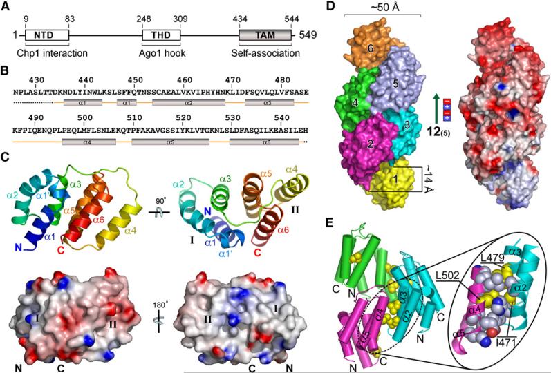
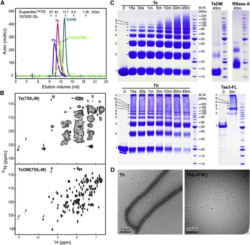
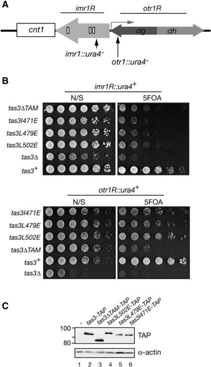
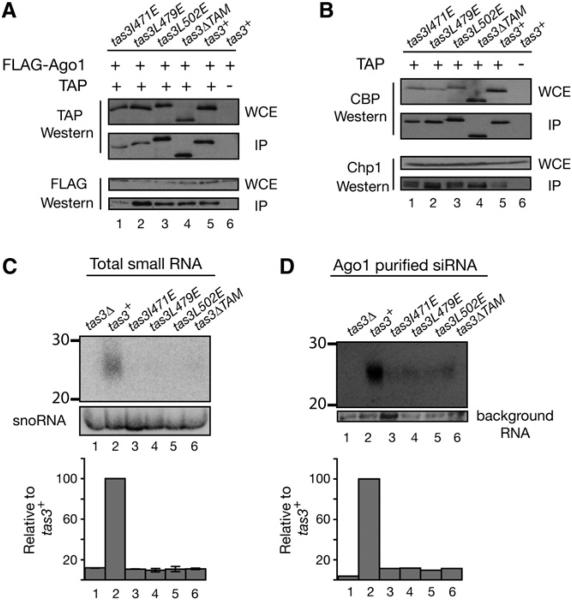
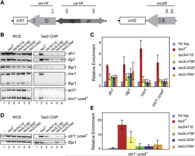
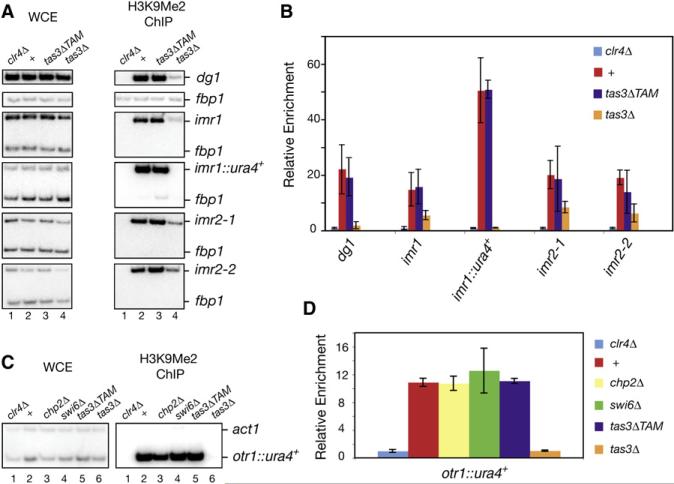
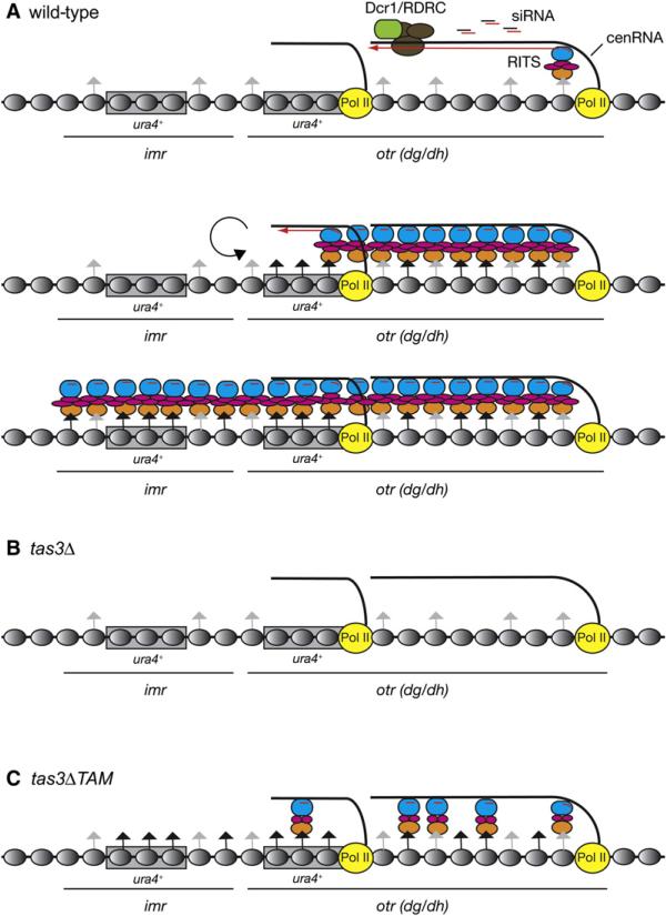
RNA interference (RNAi) plays a pivotal role in the formation of heterochromatin at the fission yeast centromeres. The RNA-induced transcriptional silencing (RITS) complex, composed of heterochromatic small interfering RNAs (siRNAs), the siRNA-binding protein Ago1, the chromodomain protein Chp1, and the Ago1/Chp1-interacting protein Tas3, provides a physical tether between the RNAi and heterochromatin assembly pathways. Here, we report the structural and functional characterization of a C-terminal Tas3 alpha-helical motif (TAM), which self-associates into a helical polymer and is required for cis spreading of RITS in centromeric DNA regions. Site-directed mutations of key residues within the hydrophobic monomer-monomer interface disrupt Tas3-TAM polymeric self-association in vitro and result in loss of gene silencing, spreading of RITS, and a dramatic reduction in centromeric siRNAs in vivo. These results demonstrate that, in addition to the chromodomain of Chp1 and siRNA-loaded Ago1, Tas3 self-association is required for RITS spreading and efficient heterochromatic gene silencing at centromeric repeat regions.


Figures

Figure 1
Figure 1. Structure of Tas3 C-Terminal TAM Domain and Its Self-Associated Polymer
(A) Domain architecture of full-length Tas3. The N-terminal domain (NTD) and the Tas3-homology domain (THD) are responsible for Chp1 and Ago1 interaction, respectively. The self-association C-terminal Tas3 α-helical motif (TAM) described in this paper is highlighted in gradient gray. (B) Secondary structure alignment of the TAM-containing Te1 construct (426−545) used for the crystallographic study. Dotted line denotes the unmodeled parts of Te1 in the solved crystal structure due to their high flexibility. (C) Ribbon and surface representations of the TAM monomer. The ribbon (top) is colored in ramp from blue (N terminus) to red (C terminus), and the surface (bottom) is colored by its electrostatic potential distribution ranging from −10 kT (red) to +10 kT (blue) as calculated by the program GRASP (Nicholls et al., 1991). The first subdomain (α1–α3) and the second subdomain (α4–α6) are denoted as I and II. The ribbon and surface drawings on the left are of the same orientation, with the bottom left and bottom right panels representing the largely exposed and buried surfaces upon self-association, respectively. (D) Surface view of the TAM polymer. The polymer obeys approximately 125 screw symmetry with a diameter of ∼50 Å and a rise of ∼14 Å per monomer. For clarity, only half cycle of the polymer is displayed. The right panel shows the electrostatic potential distribution along the polymer with the same coloring strategy as described for (C). Note the dipole feature of the polymer with negative charges and positive charges enriched on the top and at the bottom, respectively. (E) Details of the major polymerization interface. Key interface residue side chains are depicted in CPK with nitrogen atoms colored in blue, oxygen in red, and carbon in gray or yellow. Three critical hydrophobic residues (I471, L479, and L502) that are used for mutagenesis study are highlighted in yellow.
Figure 2
Figure 2. Tas3-TAM Domain Self-Associates in Solution
(A) Analytical gel filtration profile of wild-type Te (426−549), Te L479E/L502E double mutant (TeDM), wild-type Th (380−549), and Th L479E single mutant. (B) 1H-15N-HSQC NMR spectroscopy of 15N-labeled Te and TeDM samples at 750 μM; characteristic cross peaks that have similar chemical shifts in both samples are highlighted by block arrows. (C) In vitro crosslinking assays of Te, TeDM, RNase A control, Th, and full-length Tas3 (Tas3-FL). Crosslinking was performed at a protein concentration of 1.5 mg/ml for Tas3-FL and 4 mg/ml for all other samples using amine-reactive BS3 crosslinker. Detailed conditions are outlined in the Experimental Procedures. Bovine RNase A was selected as a monomeric protein control due to its similar lysine composition and molecular weight compared to Te. (D) Negative-stain electron micrograph of Th and Th(L479E).
Figure 3
Figure 3. tas3-TAM Mutations Cause a Dramatic Loss of ura4+ Silencing at imr1 but Only a Modest Loss at otr1
(A) Schematic diagram of S. pombe centromere (cen) 1. The locations of ura4+ inserts in imr1R and otr1R are shown with vertical arrows. cnt1, central core; imr1, innermost repeat 1; otr1, outer repeat 1; dg and dh, repeat elements in otr; white boxes in imr1R, tRNA genes. (B) Silencing assays showing that tas3-TAM mutations cause a dramatic loss of ura4+ silencing at imr1R, but not at otr1R. Note that tas3Δ cells have a more dramatic growth defect than tas3-TAM mutations. This is more apparent in the bottom panel, showing growth on N/S medium, in which the plate was photographed after a shorter period of growth. (C) TCA western blot showing that the level of mutant Tas3 protein is similar to wild-type.
Figure 4
Figure 4. Mutant Tas3-TAM Proteins Incorporate into RITS but Cause a Dramatic Reduction in cen siRNA Levels
(A and B) Western blots showing that Tas3-TAP coprecipitates with FLAG-Ago1 (A) or Chp1 (B). (C and D) End-labeled DNA oligos complementary to cen siRNAs (Verdel et al., 2004; Reinhart and Bartel, 2002) were used to probe 25 μg of total RNA (C) or 1 μg of FLAG-Ago1-associated RNA (D) from the indicated strains. snoR69 and a background RNA were used to normalize the abundance of total siRNAs (C) and Ago1-associated siRNAs (D) relative to wild-type, respectively.
Figure 5
Figure 5. Tas3-TAM Domain Is Required for RITS Spreading
(A) Schematic diagram showing the location of primers in imr1, dg1, dh1, imr2−1, and imr2−2 used in ChIP experiments. Abbreviations are described in the legend to Figure 3A. (B–E) ChIP experiments showing Tas3-TAP localization to imr1R or imr1R::ura4+ is abolished in tas3-TAM mutants. In contrast, a minor (2- to 3-fold; see bar graphs) decrease in Tas3-TAP occupancy at dh1, dg1, and otr1R::ura4+ is detected in the tas3-TAM mutants compared to wild-type. Fold enrichment from three independent immunoprecipitations were used to generate the bar graphs (C and E). Error bars represent standard deviations. Enrichment was normalized to act1 (for imr1R::ura4+) or fbp1 (for imr1, dg1, dh1, and otr1R::ura4+) controls. Value for the “No tag” control was set to 1.0.
Figure 6
Figure 6. Tas3-TAM Is Not Required for H3K9me Spreading
(A) H3K9me ChIP experiments were performed in wild-type or mutant (clr4Δ, tas3-TAM, or tas3Δ) cells, and primers corresponding to pericentromeric otr (dg1), imr (imr1, imr2−1, and imr2−2), and the imr1R::ura4+ insert were used to assess H3K9me enrichment compared to clr4Δ cells. The location of primers is shown in Figure 5A. (B) Graph showing the average H3K9me levels and deviation from the mean from two independent experiments. (C) H3K9me ChIP experiments for the otr1-R::ura4+ transgene were performed in wild-type or mutant (clr4Δ, tas3-TAM, chp2Δ, swi6Δ, or tas3Δ) cells. (D) Fold enrichment and deviation from the mean values from two independent experiments were used to generate the graphs. Enrichment was normalized to fbp1+. Values for the clr4Δ control was set to 1.0.
Figure 7
Figure 7. Model for Tas3-Mediated RITS Spreading at S. pombe Centromeres
(A) In wild-type cells, RITS recruitment to the centromeric otr region is mediated via siRNA base-pairing interactions with complementary nascent cen transcripts (cenRNA) and Chp1 binding to H3K9me (gray arrows). RITS recruitment targets Dcr1/RDRC to its substrate and leads to dsRNA synthesis and siRNA generation across the transcribed region. siRNA-programmed RITS mediates additional H3K9me within the otr region, whereas Tas3 self-association promotes the spreading of RITS and H3K9me (black arrows) across imr and ura4+ reporter transgenes (rectangles). The Chp1, Tas3, and Ago1 subunits of RITS are presented in orange, magenta, and blue colors, respectively. Gray ovals, nucleosomes; yellow circles, RNA polymerase II (Pol II). (B) In tas3Δ cells, RNAi-independent H3K9me is still present across imr and otr regions, but is absent from ura4+ reporter transgenes due to the absence of siRNA-mediated spreading. (C) In tas3ΔTAM cells, RITS is recruited to otr via Ago1 siRNAs and H3K9me binding to Chp1 chromodomain, but its spreading to the imr region is impaired. Even though H3K9me at imr and ura4+ inserted at imr is unaffected, RITS spreading is required for efficient silencing of the ura4+ transgene. Reduced levels of centromeric siRNAs in tas3ΔTAM cells may result from a defect in Dcr1/RDRC recruitment.

Similar articles

Cited by

References

    1. Bannister AJ, Zegerman P, Partridge JF, Miska EA, Thomas JO, Allshire RC, Kouzarides T. Selective recognition of methylated lysine 9 on histone H3 by the HP1 chromo domain. Nature. 2001;410:120–124. - PubMed
    1. Buhler M, Verdel A, Moazed D. Tethering RITS to a nascent transcript initiates RNAi- and heterochromatin-dependent gene silencing. Cell. 2006;125:873–886. - PubMed
    1. Buhler M, Haas W, Gygi SP, Moazed D. RNAi-dependent and -independent RNA turnover mechanisms contribute to heterochromatic gene silencing. Cell. 2007;129:707–721. - PubMed
    1. Buhler M, Spies N, Bartel D, Moazed D. TRAMP-mediated RNA surveillance prevents spurious entry of RNAs into the Schizosaccharomyces pombe siRNA pathway. Nat. Struct. Mol. Biol. 2008;15:1015–1023. - PMC - PubMed
    1. Cam HP, Sugiyama T, Chen ES, Chen X, FitzGerald PC, Grewal SI. Comprehensive analysis of heterochromatin- and RNAi-mediated epigenetic control of the fission yeast genome. Nat. Genet. 2005;37:809–819. - PubMed

Publication types

MeSH terms

LinkOut - more resources