. 2009 Jun 26;284(26):17731-41.
doi: 10.1074/jbc.M808506200. Epub 2009 Apr 22.

Identification and characterization of a novel lysophosphatidic acid receptor, p2y5/LPA6

Affiliations

Identification and characterization of a novel lysophosphatidic acid receptor, p2y5/LPA6

Keisuke Yanagida et al. J Biol Chem. .

Abstract

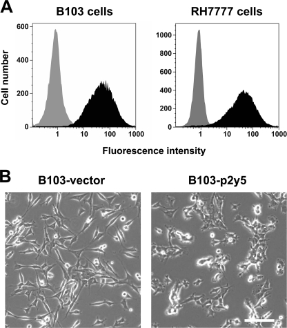
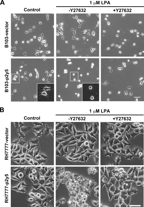
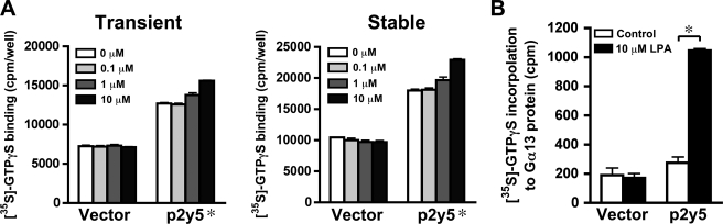
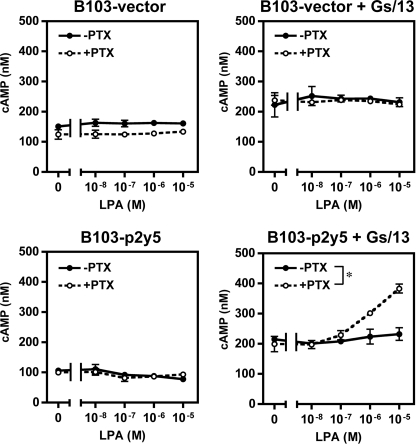
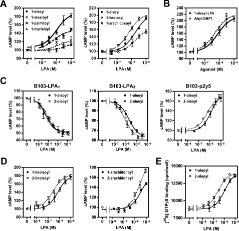
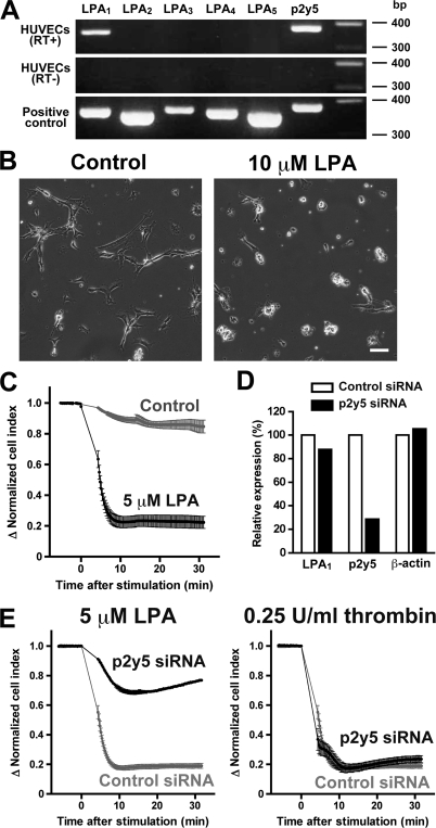
p2y5 is an orphan G protein-coupled receptor that is closely related to the fourth lysophosphatidic acid (LPA) receptor, LPA4. Here we report that p2y5 is a novel LPA receptor coupling to the G13-Rho signaling pathway. "LPA receptor-null" RH7777 and B103 cells exogenously expressing p2y5 showed [3H]LPA binding, LPA-induced [35S]guanosine 5'-3-O-(thio)triphosphate binding, Rho-dependent alternation of cellular morphology, and Gs/13 chimeric protein-mediated cAMP accumulation. LPA-induced contraction of human umbilical vein endothelial cells was suppressed by small interfering RNA knockdown of endogenously expressed p2y5. We also found that 2-acyl-LPA had higher activity to p2y5 than 1-acyl-LPA. A recent study has suggested that p2y5 is an LPA receptor essential for human hair growth. We confirmed that p2y5 is a functional LPA receptor and propose to designate this receptor LPA6.


Figures

FIGURE 1.
FIGURE 1.
Stable expression of p2y5 in B103 and RH7777 cells. A, flow cytometry analysis. B103 and RH7777 cells were stably transfected with the expression vector for p2y5 tagged with the HA epitope at the N terminus. After staining with an anti-HA antibody and a phycoerythrin-conjugated secondary antibody, HA-positive cells were sorted by the automated magnetic cell sorter and then subcultured. Data shown are the surface expression levels of HA epitope in subcultured polyclonal cells (closed area). Empty vector-transfected polyclonal cells served as a negative control (gray area). B, morphologies of B103-vector and B103-p2y5 cells in serum-containing medium. The cells were photographed 24 h after seeding. Bar, 50 μm.
FIGURE 2.
FIGURE 2.
LPA-induced neurite retraction in B103-p2y5 cells and membrane blebbing in RH7777-p2y5 cells. A, induction of neurite retraction in B103-p2y5 cells. The cells cultured in collagen-coated 24-well plates were serum-starved for 12 h, stimulated with 1 μm 1-oleoyl-LPA for 15 min, and fixed with 2% paraformaldehyde. Where indicated, the cells were pretreated with 5 μm Y27632 for 10 min (middle panels). Bar, 50 μm. The boxed areas are magnified in the inset in the same panels. B, induction of membrane blebbing in RH7777-p2y5 cells. Cell culture, stimulation, and fixation were performed in the same manner as in A. Bar, 50 μm.
FIGURE 3.
FIGURE 3.
[3H]LPA binding to RH7777-p2y5 cell membrane. A, Western blotting of HA-tagged LPA receptors (top) and [3H]-1-oleoyl-LPA binding to transiently expressed LPA receptors (bottom). Membrane fractions of RH7777 cells transiently expressing each receptor were used to confirm the receptor expression by anti-HA antibody (top). Membrane fractions were incubated with 10 nm [3H]LPA in the presence (for detecting nonspecific binding) or absence (for total binding) of 10 μm unlabeled 1-oleoyl-LPA for 70 min at 4 °C (bottom). Data are means ± S.E. (n = 3) of a representative of three independent experiments with similar results. *, p < 0.01 (using unpaired two-tailed t test). B, Scatchard analysis data of the specific binding of [3H]LPA to LPA1, LPA2, LPA4, and LPA5. Data shown are representative of at least two independent experiments with similar results. C, [3H]1-oleoyl-LPA binding to the membrane fractions from RH7777 cells stably expressing p2y5. Membrane fractions of RH7777-p2y5 cells were incubated with 30 nm [3H]LPA in the presence (for detecting nonspecific binding) or absence (for total binding) of 10 μm unlabeled 1-oleoyl-LPA for 70 min at 4 °C. Data are means ± S.E. (n = 3) of a representative of three independent experiments with similar results. *, p < 0.05 (using unpaired two-tailed t test). D, sensitivity to GTPγS. Membrane fractions of RH7777-p2y5 cells were incubated with or without 100 μm GTPγS for 30 min at room temperature, and a [3H]LPA binding assay was performed in the same manner as in A. Data are means ± S.E. (n = 3) of a representative of three independent experiments with similar results. *, p < 0.05 (using unpaired two-tailed t test).
FIGURE 4.
FIGURE 4.
LPA-induced [35S]GTPγS binding via p2y5. A, membrane fractions from RH7777 cells transiently (left) or stably (right) expressing p2y5 were incubated with 0.5 nm [35S]GTPγS and the indicated concentrations of LPA for 30 min at 30 °C in the presence of 0.3 μm GDP. Data are means ± S.E. (n = 3) of a representative of three independent experiments with similar results; *, p < 0.001 for vector versus p2y5 using two-way analysis of variance. B, LPA-induced [35S]GTPγS incorporation to Gα13 protein. The membrane fraction from RH7777 cells transiently expressing p2y5 was incubated with 0.5 nm [35S]GTPγS and 10 μm 1-oleoyl-LPA for 30 min at 30 °C in the presence of 0.3 μm GDP. Then membrane protein was solubilized, and Gα13 protein was immunoprecipitated to detect [35S]GTPγS incorporated. Data are means ± S.E. (n = 3) of a representative of two independent experiments with similar results. *, p < 0.001 (using unpaired two-tailed t test).
FIGURE 5.
FIGURE 5.
Adenylyl cyclase activation by p2y5 through a Gs/13 chimeric protein. B103-vector and B103-p2y5 cells were transfected with Gs/13 expression plasmid (right) or control plasmid (left). Serum-starved cells were pretreated with or without 100 ng/ml PTX for 12 h and stimulated with the indicated concentrations of 1-oleoyl-LPA in the presence of 0.5 mm 3-isobutyl-1-methylxanthine and 10 μm forskolin. After a 30-min incubation at room temperature, the cells were solubilized. cAMP concentrations in the cell lysates are shown. Data are means ± S.E. (n = 3) of a representative of three independent experiments with similar results. *, p < 0.001 (using two-way analysis of variance).
FIGURE 6.
FIGURE 6.
Ligand selectivity of p2y5. Activities of various LPA species and alkyl-OMPT were examined by the cAMP assay using B103-p2y5 cells transfected with Gs/13 after pretreatment with PTX (A–D). Agonistic activities of 1-acyl-LPA species with saturated fatty acid (A, left) or unsaturated fatty acid (A, right), alkyl-OMPT (B), and 2-acyl-LPA species (C and D) are presented as a percentage of forskolin-induced cAMP accumulation in the absence of LPA. 2-Acyl-LPA species (C–E) were prepared as described under “Experimental Procedures.” The preference of p2y5 for 2-acyl-LPA was consistently observed in the GTPγS binding assay using membrane fractions from RH7777- p2y5 cells (E). Data are means ± S.E. (n = 3) of a representative of three independent experiments with similar results.
FIGURE 7.
FIGURE 7.
p2y5-dependent contraction of HUVECs. A, mRNA expression profile of LPA receptors in HUVECs. RNA was isolated from HUVECs and reverse-transcribed. cDNA was amplified using primers described under “Experimental Procedures.” PCR templates for “positive controls” were cDNA from HEK293 cells (for LPA1 and LPA3), cDNA from HL-60 cells (for LPA4 and p2y5), and expression plasmids for LPA2 and LPA5 in pCXN2.1. B, contraction of HUVECs by LPA stimulation. HUVECs cultured in poly-l-lysine-coated glass-bottomed 35-mm dishes were serum-starved, stimulated with 10 μm 1-oleoyl-LPA for 30 min, and fixed with 2% paraformaldehyde. Bar, 50 μm. C, quantification of HUVEC contraction using the xCELLigence system. The serum-starved cells were stimulated with 5 μm LPA. A decrease in cell index was observed upon LPA stimulation. Data are means ± S.D. (n = 6) of a representative of two independent experiments with similar results. D, quantitative real time PCR analysis in HUVECs transfected with siRNAs. Expression levels of LPA1, p2y5, and β-actin mRNA in the cells transfected with p2y5 siRNA (Silencer Select siRNA, ID numbers; s19798) are presented as a percentage of those with control siRNA. Data are means (n = 2) of a representative of four independent experiments with similar results. E, effect of p2y5 on contraction of HUVECs. The cells transfected with siRNAs were serum-starved. The morphological changes upon the stimulation with 5 μm LPA (left panel) or 0.25 units/ml thrombin (right panel) were evaluated in the same manner as C. Data are means ± S.D. (n = 6) of a representative of four (LPA) and two (thrombin) independent experiments with similar results. Another p2y5 siRNA with different target sequence gave consistent results (Fig. S3).

Similar articles

Cited by

References

    1. Tokumura A. ( 1995) Prog. Lipid Res. 34, 151– 184 - PubMed
    1. Ishii I., Fukushima N., Ye X., Chun J. ( 2004) Annu. Rev. Biochem. 73, 321– 354 - PubMed
    1. Herr D. R., Chun J. ( 2007) Curr. Drug Targets 8, 155– 167 - PubMed
    1. Murph M., Tanaka T., Liu S., Mills G. B. ( 2006) Clin. Cancer Res. 12, 6598– 6602 - PubMed
    1. Watterson K. R., Lanning D. A., Diegelmann R. F., Spiegel S. ( 2007) Wound Repair Regen. 15, 607– 616 - PubMed

Publication types

MeSH terms

LinkOut - more resources